|
Retroperitoneal
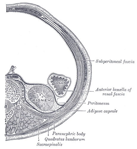
The retroperitoneal space (retroperitoneum) is the anatomical space (sometimes a potential space) behind (''retro'') the peritoneum. It has no specific delineating anatomical structures. Organs are retroperitoneal if they have peritoneum on their anterior side only. Structures that are not suspended by mesentery in the abdominal cavity and that lie between the parietal peritoneum and abdominal wall are classified as retroperitoneal. This is different from organs that are not retroperitoneal, which have peritoneum on their posterior side and are suspended by mesentery in the abdominal cavity. The retroperitoneum can be further subdivided into the following: *Perirenal (or perinephric) space *Anterior pararenal (or paranephric) space *Posterior pararenal (or paranephric) space Retroperitoneal structures Structures that lie behind the peritoneum are termed "retroperitoneal". Organs that were once suspended within the abdominal cavity by mesentery but migrated posterior to the ... [...More Info...] [...Related Items...] OR: [Wikipedia] [Google] [Baidu] |
Retroperitoneal Fibrosis
Retroperitoneal fibrosis or Ormond's disease is a disease featuring the proliferation of fibrous tissue in the retroperitoneum, the compartment of the body containing the kidneys, aorta, renal tract, and various other structures. It may present with lower back pain, kidney failure, hypertension, deep vein thrombosis, and other obstructive symptoms. It is named after John Kelso Ormond, who rediscovered the condition in 1948. Causes The association of idiopathic retroperitoneal fibrosis with various immune-related conditions and response to immunosuppression led to a search for an autoimmune cause of idiopathic RPF. Many of these previously idiopathic cases can now be attributed to IgG4-related disease, an autoimmune disorder proposed in 2003. Otherwise, one-third of cases are secondary to malignancy, medication (methysergide, hydralazine, beta blockers), prior radiotherapy, or certain infections. Other associations include: * connective tissue disease * Riedel's thyroiditis * sc ... [...More Info...] [...Related Items...] OR: [Wikipedia] [Google] [Baidu] |
Retroperitoneal Hemorrhage
Retroperitoneal bleeding is an accumulation of blood in the retroperitoneal space. Signs and symptoms may include abdominal or upper leg pain, hematuria, and shock. It can be caused by major trauma or by non-traumatic mechanisms. Signs and symptoms Signs and symptoms may include: * abdominal pain. * upper leg pain. * hematuria. * shock. Causes Retroperitoneal bleeds are most often caused by major trauma, such as from a traffic collisions or a fall. Less common non-traumatic causes including: * anticoagulation. * a ruptured aortic aneurysm. * a ruptured renal aneurysm. * acute pancreatitis. * malignancy. Retroperitoneal bleeds may also be iatrogenic, caused accidentally during medical procedures. Such procedures include cannulating the femoral artery for cardiac catheterization or for interventional radiology, and the administration of a psoas compartment nerve block. Diagnosis Neurology As well as initial symptoms, the accumulation of blood in the retroperitoneal space ... [...More Info...] [...Related Items...] OR: [Wikipedia] [Google] [Baidu] |
Perirenal Fat
The retroperitoneal space (retroperitoneum) is the anatomical space (sometimes a potential space) behind (''retro'') the peritoneum. It has no specific delineating anatomical structures. Organs are retroperitoneal if they have peritoneum on their anterior side only. Structures that are not suspended by mesentery in the abdominal cavity and that lie between the parietal peritoneum and abdominal wall are classified as retroperitoneal. This is different from organs that are not retroperitoneal, which have peritoneum on their posterior side and are suspended by mesentery in the abdominal cavity. The retroperitoneum can be further subdivided into the following: *Perirenal (or perinephric) space *Anterior pararenal (or paranephric) space *Posterior pararenal (or paranephric) space Retroperitoneal structures Structures that lie behind the peritoneum are termed "retroperitoneal". Organs that were once suspended within the abdominal cavity by mesentery but migrated posterior to the p ... [...More Info...] [...Related Items...] OR: [Wikipedia] [Google] [Baidu] |
Retroperitoneal Lymph Node Dissection
Retroperitoneal lymph node dissection (RPLND) is a surgical procedure to remove abdominal lymph nodes. It is used to treat testicular cancer, as well as to help establish the exact stage and type of the cancer. Indications Testicular cancer metastasizes in a predictable pattern, and lymph nodes in the retroperitoneum are typically the first place it lands. By examining the removed lymphatic tissue, a pathologist can determine whether the disease has spread. If no malignant tissue is found, the cancer can be labeled Stage I, limited to the testicle. The procedure is common in the treatment of Stage I and II non-seminomatous germ cell tumors. In seminomas, another form of testicular cancer, radiation therapy is generally preferred to the invasive RPLND procedure. Whether RPLND is needed after orchiectomy depends on the type of tumor and its stage. RPLND may be performed to remove tumor remnants that persist after chemotherapy, because these remnants might otherwise spread and b ... [...More Info...] [...Related Items...] OR: [Wikipedia] [Google] [Baidu] |
Peritoneum
The peritoneum is the serous membrane forming the lining of the abdominal cavity or coelom in amniotes and some invertebrates, such as annelids. It covers most of the intra-abdominal (or coelomic) organs, and is composed of a layer of mesothelium supported by a thin layer of connective tissue. This peritoneal lining of the cavity supports many of the abdominal organs and serves as a conduit for their blood vessels, lymphatic vessels, and nerves. The abdominal cavity (the space bounded by the vertebrae, abdominal muscles, diaphragm, and pelvic floor) is different from the intraperitoneal space (located within the abdominal cavity but wrapped in peritoneum). The structures within the intraperitoneal space are called "intraperitoneal" (e.g., the stomach and intestines), the structures in the abdominal cavity that are located behind the intraperitoneal space are called "retroperitoneal" (e.g., the kidneys), and those structures below the intraperitoneal space are called "subp ... [...More Info...] [...Related Items...] OR: [Wikipedia] [Google] [Baidu] |
Spatium
{{set index article In anatomy, a spatium or anatomic space is a space (cavity or gap). Anatomic spaces are often landmarks to find other important structures. When they fill with gases (such as air) or liquids (such as blood) in pathological ways, they can suffer conditions such as pneumothorax, edema, or pericardial effusion. Many anatomic spaces are potential spaces, which means that they are potential rather than realized (with their realization being dynamic according to physiologic or pathophysiologic events). In other words, they are like an empty plastic bag that has not been opened (two walls collapsed against each other; no interior volume until opened) or a balloon that has not been inflated. Examples of anatomic spaces (or potential spaces) include: *Axillary space *Buccal space * Canine space *Cystohepatic triangle * Deep perineal space * Deep temporal space *Epidural space * Extraperitoneal space *Fascial spaces of the head and neck * Infratemporal space *Intercosta ... [...More Info...] [...Related Items...] OR: [Wikipedia] [Google] [Baidu] |
Colon (anatomy)
The large intestine, also known as the large bowel, is the last part of the gastrointestinal tract and of the digestive system in tetrapods. Water is absorbed here and the remaining waste material is stored in the rectum as feces before being removed by defecation. The colon is the longest portion of the large intestine, and the terms are often used interchangeably but most sources define the large intestine as the combination of the cecum, colon, rectum, and anal canal. Some other sources exclude the anal canal. In humans, the large intestine begins in the right iliac region of the pelvis, just at or below the waist, where it is joined to the end of the small intestine at the cecum, via the ileocecal valve. It then continues as the colon ascending the abdomen, across the width of the abdominal cavity as the transverse colon, and then descending to the rectum and its endpoint at the anal canal. Overall, in humans, the large intestine is about long, which is about one-fifth o ... [...More Info...] [...Related Items...] OR: [Wikipedia] [Google] [Baidu] |
Adrenal Gland
The adrenal glands (also known as suprarenal glands) are endocrine glands that produce a variety of hormones including adrenaline and the steroids aldosterone and cortisol. They are found above the kidneys. Each gland has an outer cortex which produces steroid hormones and an inner medulla. The adrenal cortex itself is divided into three main zones: the zona glomerulosa, the zona fasciculata and the zona reticularis. The adrenal cortex produces three main types of steroid hormones: mineralocorticoids, glucocorticoids, and androgens. Mineralocorticoids (such as aldosterone) produced in the zona glomerulosa help in the regulation of blood pressure and electrolyte balance. The glucocorticoids cortisol and cortisone are synthesized in the zona fasciculata; their functions include the regulation of metabolism and immune system suppression. The innermost layer of the cortex, the zona reticularis, produces androgens that are converted to fully functional sex hormones in the gonads ... [...More Info...] [...Related Items...] OR: [Wikipedia] [Google] [Baidu] |
Renal Fascia
The renal fascia is a layer of connective tissue encapsulating the kidneys and the adrenal glands. It can be divided into: *The anterior renal fascia, also called Gerota's fascia (after Dimitrie Gerota) *The posterior renal fascia, also called Zuckerkandl's fascia or fascia retrorenalis The renal fascia separates the adipose capsule of kidney from the overlying pararenal fat. The deeper layers below the renal fascia are, in order, the adipose capsule (or perirenal fat), the renal capsule and finally the parenchyma of the renal cortex. The spaces about the kidney are typically divided into three compartments: the perinephric space and the anterior and posterior pararenal spaces. Anterior renal fascia * Medial attachment: Passes anterior to the kidney, renal vessels, abdominal aorta and inferior vena cava and fuses with the anterior layer of the renal fascia of the opposite kidney. * Lateral attachment: Fuses with the psoas fascia and side of the body of the vertebrae. * Superior at ... [...More Info...] [...Related Items...] OR: [Wikipedia] [Google] [Baidu] |
Adrenal Gland
The adrenal glands (also known as suprarenal glands) are endocrine glands that produce a variety of hormones including adrenaline and the steroids aldosterone and cortisol. They are found above the kidneys. Each gland has an outer cortex which produces steroid hormones and an inner medulla. The adrenal cortex itself is divided into three main zones: the zona glomerulosa, the zona fasciculata and the zona reticularis. The adrenal cortex produces three main types of steroid hormones: mineralocorticoids, glucocorticoids, and androgens. Mineralocorticoids (such as aldosterone) produced in the zona glomerulosa help in the regulation of blood pressure and electrolyte balance. The glucocorticoids cortisol and cortisone are synthesized in the zona fasciculata; their functions include the regulation of metabolism and immune system suppression. The innermost layer of the cortex, the zona reticularis, produces androgens that are converted to fully functional sex hormones in the gonads ... [...More Info...] [...Related Items...] OR: [Wikipedia] [Google] [Baidu] |
Sarcoma
A sarcoma is a malignant tumor, a type of cancer that arises from transformed cells of mesenchymal (connective tissue) origin. Connective tissue is a broad term that includes bone, cartilage, fat, vascular, or hematopoietic tissues, and sarcomas can arise in any of these types of tissues. As a result, there are many subtypes of sarcoma, which are classified based on the specific tissue and type of cell from which the tumor originates. Sarcomas are ''primary'' connective tissue tumors, meaning that they arise in connective tissues. This is in contrast to ''secondary'' (or "metastatic") connective tissue tumors, which occur when a cancer from elsewhere in the body (such as the lungs, breast tissue or prostate) spreads to the connective tissue. The word ''sarcoma'' is derived from the Greek σάρκωμα ''sarkōma'' "fleshy excrescence or substance", itself from σάρξ ''sarx'' meaning "flesh". Classification Sarcomas are typically divided into two major groups: bone sarcom ... [...More Info...] [...Related Items...] OR: [Wikipedia] [Google] [Baidu] |
Duodenum
The duodenum is the first section of the small intestine in most higher vertebrates, including mammals, reptiles, and birds. In fish, the divisions of the small intestine are not as clear, and the terms anterior intestine or proximal intestine may be used instead of duodenum. In mammals the duodenum may be the principal site for iron absorption. The duodenum precedes the jejunum and ileum and is the shortest part of the small intestine. In humans, the duodenum is a hollow jointed tube about 25–38 cm (10–15 inches) long connecting the stomach to the middle part of the small intestine. It begins with the duodenal bulb and ends at the suspensory muscle of duodenum. Duodenum can be divided into four parts: the first (superior), the second (descending), the third (horizontal) and the fourth (ascending) parts. Structure The duodenum is a C-shaped structure lying adjacent to the stomach. It is divided anatomically into four sections. The first part of the duodenum lies ... [...More Info...] [...Related Items...] OR: [Wikipedia] [Google] [Baidu] |


