|
Retinal Disease
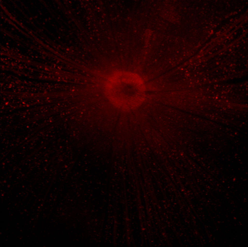
The retina (from la, rete "net") is the innermost, light-sensitive layer of tissue of the eye of most vertebrates and some molluscs. The optics of the eye create a focused two-dimensional image of the visual world on the retina, which then processes that image within the retina and sends nerve impulses along the optic nerve to the visual cortex to create visual perception. The retina serves a function which is in many ways analogous to that of the film or image sensor in a camera. The neural retina consists of several layers of neurons interconnected by synapses and is supported by an outer layer of pigmented epithelial cells. The primary light-sensing cells in the retina are the photoreceptor cells, which are of two types: rods and cones. Rods function mainly in dim light and provide monochromatic vision. Cones function in well-lit conditions and are responsible for the perception of colour through the use of a range of opsins, as well as high-acuity vision used for tas ... [...More Info...] [...Related Items...] OR: [Wikipedia] [Google] [Baidu] |
Plural
The plural (sometimes abbreviated pl., pl, or ), in many languages, is one of the values of the grammatical category of number. The plural of a noun typically denotes a quantity greater than the default quantity represented by that noun. This default quantity is most commonly one (a form that represents this default quantity of one is said to be of ''singular'' number). Therefore, plurals most typically denote two or more of something, although they may also denote fractional, zero or negative amounts. An example of a plural is the English word ''cats'', which corresponds to the singular ''cat''. Words of other types, such as verbs, adjectives and pronouns, also frequently have distinct plural forms, which are used in agreement with the number of their associated nouns. Some languages also have a dual (denoting exactly two of something) or other systems of number categories. However, in English and many other languages, singular and plural are the only grammatical numbers, exce ... [...More Info...] [...Related Items...] OR: [Wikipedia] [Google] [Baidu] |
Cone Cell
Cone cells, or cones, are photoreceptor cells in the retinas of vertebrate eyes including the human eye. They respond differently to light of different wavelengths, and the combination of their responses is responsible for color vision. Cones function best in relatively bright light, called the photopic region, as opposed to rod cells, which work better in dim light, or the scotopic region. Cone cells are densely packed in the fovea centralis, a 0.3 mm diameter rod-free area with very thin, densely packed cones which quickly reduce in number towards the periphery of the retina. Conversely, they are absent from the optic disc, contributing to the blind spot. There are about six to seven million cones in a human eye (vs ~92 million rods), with the highest concentration being towards the macula. Cones are less sensitive to light than the rod cells in the retina (which support vision at low light levels), but allow the perception of color. They are also able to perceive ... [...More Info...] [...Related Items...] OR: [Wikipedia] [Google] [Baidu] |
University Of Pennsylvania Health System
The University of Pennsylvania Health System (UPHS) is a major multi-hospital health system headquartered in Philadelphia, Pennsylvania. UPHS and the Perelman School of Medicine at the University of Pennsylvania together comprise Penn Medicine, a clinical and research entity of the University of Pennsylvania. UPHS hospitals include the Hospital of the University of Pennsylvania, Penn Presbyterian Medical Center, Pennsylvania Hospital, Chester County Hospital, Lancaster General Hospital, and Princeton Medical Center. The UPHS is home to the first hospital in the United States, the Pennsylvania Hospital. History The Chester County Hospital and Health System joined UPHS in 2013. Two years later, Lancaster General Health (LG Health) joined the UPHS family. Princeton Health officially merged into UPHS in January 2018. Phoenixville Hospital in Phoenixville, Pennsylvania, was previously part of the UPHS network but was sold to Community Health Systems in 2004. Phoenixville Hospi ... [...More Info...] [...Related Items...] OR: [Wikipedia] [Google] [Baidu] |
Central Nervous System
The central nervous system (CNS) is the part of the nervous system consisting primarily of the brain and spinal cord. The CNS is so named because the brain integrates the received information and coordinates and influences the activity of all parts of the bodies of bilaterally symmetric and triploblastic animals—that is, all multicellular animals except sponges and diploblasts. It is a structure composed of nervous tissue positioned along the rostral (nose end) to caudal (tail end) axis of the body and may have an enlarged section at the rostral end which is a brain. Only arthropods, cephalopods and vertebrates have a true brain (precursor structures exist in onychophorans, gastropods and lancelets). The rest of this article exclusively discusses the vertebrate central nervous system, which is radically distinct from all other animals. Overview In vertebrates, the brain and spinal cord are both enclosed in the meninges. The meninges provide a barrier to chemicals ... [...More Info...] [...Related Items...] OR: [Wikipedia] [Google] [Baidu] |
Diencephalon
The diencephalon (or interbrain) is a division of the forebrain (embryonic ''prosencephalon''). It is situated between the telencephalon and the midbrain (embryonic ''mesencephalon''). The diencephalon has also been known as the 'tweenbrain in older literature. It consists of structures that are on either side of the third ventricle, including the thalamus, the hypothalamus, the epithalamus and the subthalamus. The diencephalon is one of the main vesicles of the brain formed during embryogenesis. During the third week of development a neural tube is created from the ectoderm, one of the three primary germ layers. The tube forms three main vesicles during the third week of development: the prosencephalon, the mesencephalon and the rhombencephalon. The prosencephalon gradually divides into the telencephalon and the diencephalon. Structure The diencephalon consists of the following structures: *Thalamus *Hypothalamus including the posterior pituitary * Epithalamus which cons ... [...More Info...] [...Related Items...] OR: [Wikipedia] [Google] [Baidu] |
Embryonic Development
An embryo is an initial stage of development of a multicellular organism. In organisms that reproduce sexually, embryonic development is the part of the life cycle that begins just after fertilization of the female egg cell by the male sperm cell. The resulting fusion of these two cells produces a single-celled zygote that undergoes many cell divisions that produce cells known as blastomeres. The blastomeres are arranged as a solid ball that when reaching a certain size, called a morula, takes in fluid to create a cavity called a blastocoel. The structure is then termed a blastula, or a blastocyst in mammals. The mammalian blastocyst hatches before implantating into the endometrial lining of the womb. Once implanted the embryo will continue its development through the next stages of gastrulation, neurulation, and organogenesis. Gastrulation is the formation of the three germ layers that will form all of the different parts of the body. Neurulation forms the nervous s ... [...More Info...] [...Related Items...] OR: [Wikipedia] [Google] [Baidu] |
Axon
An axon (from Greek ἄξων ''áxōn'', axis), or nerve fiber (or nerve fibre: see spelling differences), is a long, slender projection of a nerve cell, or neuron, in vertebrates, that typically conducts electrical impulses known as action potentials away from the nerve cell body. The function of the axon is to transmit information to different neurons, muscles, and glands. In certain sensory neurons ( pseudounipolar neurons), such as those for touch and warmth, the axons are called afferent nerve fibers and the electrical impulse travels along these from the periphery to the cell body and from the cell body to the spinal cord along another branch of the same axon. Axon dysfunction can be the cause of many inherited and acquired neurological disorders that affect both the peripheral and central neurons. Nerve fibers are classed into three types group A nerve fibers, group B nerve fibers, and group C nerve fibers. Groups A and B are myelinated, and group C are unmyelinated. ... [...More Info...] [...Related Items...] OR: [Wikipedia] [Google] [Baidu] |
Retinal Ganglion Cell
A retinal ganglion cell (RGC) is a type of neuron located near the inner surface (the ganglion cell layer) of the retina of the eye. It receives visual information from photoreceptors via two intermediate neuron types: bipolar cells and retina amacrine cells. Retina amacrine cells, particularly narrow field cells, are important for creating functional subunits within the ganglion cell layer and making it so that ganglion cells can observe a small dot moving a small distance. Retinal ganglion cells collectively transmit image-forming and non-image forming visual information from the retina in the form of action potential to several regions in the thalamus, hypothalamus, and mesencephalon, or midbrain. Retinal ganglion cells vary significantly in terms of their size, connections, and responses to visual stimulation but they all share the defining property of having a long axon that extends into the brain. These axons form the optic nerve, optic chiasm, and optic tract. A s ... [...More Info...] [...Related Items...] OR: [Wikipedia] [Google] [Baidu] |
Action Potential
An action potential occurs when the membrane potential of a specific cell location rapidly rises and falls. This depolarization then causes adjacent locations to similarly depolarize. Action potentials occur in several types of animal cells, called excitable cells, which include neurons, muscle cells, and in some plant cells. Certain endocrine cells such as pancreatic beta cells, and certain cells of the anterior pituitary gland are also excitable cells. In neurons, action potentials play a central role in cell-cell communication by providing for—or with regard to saltatory conduction, assisting—the propagation of signals along the neuron's axon toward synaptic boutons situated at the ends of an axon; these signals can then connect with other neurons at synapses, or to motor cells or glands. In other types of cells, their main function is to activate intracellular processes. In muscle cells, for example, an action potential is the first step in the chain of events l ... [...More Info...] [...Related Items...] OR: [Wikipedia] [Google] [Baidu] |
Brain
A brain is an organ that serves as the center of the nervous system in all vertebrate and most invertebrate animals. It is located in the head, usually close to the sensory organs for senses such as vision. It is the most complex organ in a vertebrate's body. In a human, the cerebral cortex contains approximately 14–16 billion neurons, and the estimated number of neurons in the cerebellum is 55–70 billion. Each neuron is connected by synapses to several thousand other neurons. These neurons typically communicate with one another by means of long fibers called axons, which carry trains of signal pulses called action potentials to distant parts of the brain or body targeting specific recipient cells. Physiologically, brains exert centralized control over a body's other organs. They act on the rest of the body both by generating patterns of muscle activity and by driving the secretion of chemicals called hormones. This centralized control allows rapid and coordinated responses ... [...More Info...] [...Related Items...] OR: [Wikipedia] [Google] [Baidu] |
Pupillary Light Reflex
The pupillary light reflex (PLR) or photopupillary reflex is a reflex that controls the diameter of the pupil, in response to the intensity (luminance) of light that falls on the retinal ganglion cells of the retina in the back of the eye, thereby assisting in adaptation of vision to various levels of lightness/darkness. A greater intensity of light causes the pupil to constrict ( miosis/myosis; thereby allowing less light in), whereas a lower intensity of light causes the pupil to dilate ( mydriasis, expansion; thereby allowing more light in). Thus, the pupillary light reflex regulates the intensity of light entering the eye. Light shone into one eye will cause both pupils to constrict. Terminology The pupil is the dark circular opening in the center of the iris and is where light enters the eye. By analogy with a camera, the pupil is equivalent to aperture, whereas the iris is equivalent to the diaphragm. It may be helpful to consider the ''Pupillary reflex'' as an Iris' ref ... [...More Info...] [...Related Items...] OR: [Wikipedia] [Google] [Baidu] |
Circadian Rhythm
A circadian rhythm (), or circadian cycle, is a natural, internal process that regulates the sleep–wake cycle and repeats roughly every 24 hours. It can refer to any process that originates within an organism (i.e., endogenous) and responds to the environment ( entrained by the environment). These 24-hour rhythms are driven by a circadian clock, and they have been widely observed in animals, plants, fungi and cyanobacteria. The term ''circadian'' comes from the Latin ''circa'', meaning "approximately", and ''dies'', meaning "day". Processes with 24-hour cycles are more generally called diurnal rhythms; diurnal rhythms should not be called circadian rhythms unless they can be confirmed as endogenous, and not environmental. Although circadian rhythms are endogenous, they are adjusted to the local environment by external cues called zeitgebers (German for "time givers"), which include light, temperature and redox cycles. In clinical settings, an abnormal circadian rhythm in h ... [...More Info...] [...Related Items...] OR: [Wikipedia] [Google] [Baidu] |


