|
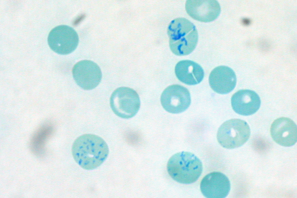
Reticulocytopenia
Reticulocytopenia is the medical term for an abnormal decrease of reticulocytes in the body. Reticulocytes are new, immature red blood cells. Causes Reticulocytopenia is usually a result of viral parvovirus B19 infection, which invades and destroys red blood cell precursors and halts the red cell production. If infection occurs in individuals with sickle cell anemia,"Sickle Cell Disease - Basic Principles and Clinical Practice" Edited by Stephen H. Embury, Robert P. Hebbel, Narla Mohandas and Martin Steinberg. Copyright 1996 by Lippincott-Raven. Section IV p352. spherocytosis, or beta thalassemia, it will lead to incorporation of two anemia-induced mechanisms: decreased red cell production and hemolysis. The result is a rapid and severe anemia (aplastic crisis) which may require blood transfusion. Diagnosis See also * Erythropoiesis – process of creating red blood cells * Hemolytic anemia – reduced number of red blood cells due to destruction of the cells after they were ... [...More Info...] [...Related Items...] OR: [Wikipedia] [Google] [Baidu] |
Sickle Cell Anemia
Sickle cell disease (SCD) is a group of blood disorders typically inherited from a person's parents. The most common type is known as sickle cell anaemia. It results in an abnormality in the oxygen-carrying protein haemoglobin found in red blood cells. This leads to a rigid, sickle-like shape under certain circumstances. Problems in sickle cell disease typically begin around 5 to 6 months of age. A number of health problems may develop, such as attacks of pain (known as a sickle cell crisis), anemia, swelling in the hands and feet, bacterial infections and stroke. Long-term pain may develop as people get older. The average life expectancy in the developed world is 40 to 60 years. Sickle cell disease occurs when a person inherits two abnormal copies of the β-globin gene (''HBB'') that makes haemoglobin, one from each parent. This gene occurs in chromosome 11. Several subtypes exist, depending on the exact mutation in each haemoglobin gene. An attack can be set off by tempera ... [...More Info...] [...Related Items...] OR: [Wikipedia] [Google] [Baidu] |
Reticulocytes
Reticulocytes are immature red blood cells (RBCs). In the process of erythropoiesis (red blood cell formation), reticulocytes develop and mature in the bone marrow and then circulate for about a day in the blood stream before developing into mature red blood cells. Like mature red blood cells, in mammals, reticulocytes do not have a cell nucleus. They are called reticulocytes because of a reticular (mesh-like) network of ribosomal RNA that becomes visible under a microscope with certain stains such as new methylene blue and Romanowsky stain. Clinical significance To accurately measure reticulocyte counts, automated counters use a combination of laser excitation, detectors and a fluorescent dye that marks RNA and DNA (such as titan yellow or polymethine). Reticulocytes appear slightly bluer than other red cells when looked at with the normal Romanowsky stain. Reticulocytes are also relatively large, a characteristic that is described by the mean corpuscular volume. The normal ... [...More Info...] [...Related Items...] OR: [Wikipedia] [Google] [Baidu] |
Red Blood Cells
Red blood cells (RBCs), also referred to as red cells, red blood corpuscles (in humans or other animals not having nucleus in red blood cells), haematids, erythroid cells or erythrocytes (from Greek language, Greek ''erythros'' for "red" and ''kytos'' for "hollow vessel", with ''-cyte'' translated as "cell" in modern usage), are the most common type of blood cell and the vertebrate's principal means of delivering oxygen (O2) to the body tissue (biology), tissues—via blood flow through the circulatory system. RBCs take up oxygen in the lungs, or in fish the gills, and release it into tissues while squeezing through the body's capillary, capillaries. The cytoplasm of a red blood cell is rich in hemoglobin, an iron-containing biomolecule that can bind oxygen and is responsible for the red color of the cells and the blood. Each human red blood cell contains approximately 270 million hemoglobin molecules. The cell membrane is composed of proteins and lipids, and this structure ... [...More Info...] [...Related Items...] OR: [Wikipedia] [Google] [Baidu] |
Parvovirus B19
Primate erythroparvovirus 1, generally referred to as B19 virus (B19V), parvovirus B19 or sometimes erythrovirus B19, is the first (and until 2005 the only) known human virus in the family ''Parvoviridae'', genus ''Erythroparvovirus''; it measures only 23–26 nm in diameter. The name is derived from Latin, parvum meaning small, reflecting the fact that B19 ranks among the smallest DNA viruses. B19 virus is most known for causing disease in the pediatric population; however, it can also affect adults. It is the classic cause of the childhood rash called fifth disease or erythema infectiosum, or "slapped cheek syndrome". The virus was discovered by chance in 1975 by Australian virologist Yvonne Cossart. It gained the B19 name because it was discovered in well B19 of a large series of microtiter plates. Virology Erythroviruses belong to the ''Parvoviridae'' family of small DNA viruses. Human parvovirus B19 is a non-enveloped, icosahedral virus that contains a single-stranded ... [...More Info...] [...Related Items...] OR: [Wikipedia] [Google] [Baidu] |
Spherocytosis
Spherocytosis is the presence of spherocytes in the blood, i.e. erythrocytes (red blood cells) that are sphere-shaped rather than bi-concave disk shaped as normal. Spherocytes are found in all hemolytic anemias to some degree. Hereditary spherocytosis and autoimmune hemolytic anemia are characterized by having ''only'' spherocytes. Causes Spherocytes are found in immunologically-mediated hemolytic anemias and in hereditary spherocytosis, but the former would have a positive direct Coombs test and the latter would not. The misshapen but otherwise healthy red blood cells are mistaken by the spleen for old or damaged red blood cells and it thus constantly breaks them down, causing a cycle whereby the body destroys its own blood supply (auto-hemolysis). A complete blood count (CBC) may show increased reticulocytes, a sign of increased red blood cell production, and decreased hemoglobin and hematocrit. The term "non-hereditary spherocytosis" is occasionally used, albeit rarely. Lists ... [...More Info...] [...Related Items...] OR: [Wikipedia] [Google] [Baidu] |
Beta Thalassemia
Beta thalassemias (β thalassemias) are a group of inherited blood disorders. They are forms of thalassemia caused by reduced or absent synthesis of the beta chains of hemoglobin that result in variable outcomes ranging from severe anemia to clinically asymptomatic individuals. Global annual incidence is estimated at one in 100,000. Beta thalassemias occur due to malfunctions in the hemoglobin subunit beta or HBB. The severity of the disease depends on the nature of the mutation. HBB blockage over time leads to decreased beta-chain synthesis. The body's inability to construct new beta-chains leads to the underproduction of HbA (adult hemoglobin). Reductions in HbA available overall to fill the red blood cells in turn leads to microcytic anemia. Microcytic anemia ultimately develops in respect to inadequate HBB protein for sufficient red blood cell functioning. Due to this factor, the patient may require blood transfusions to make up for the blockage in the beta-chains. Repeated ... [...More Info...] [...Related Items...] OR: [Wikipedia] [Google] [Baidu] |
Hemolysis
Hemolysis or haemolysis (), also known by several other names, is the rupturing (lysis) of red blood cells (erythrocytes) and the release of their contents (cytoplasm) into surrounding fluid (e.g. blood plasma). Hemolysis may occur in vivo or in vitro. One cause of hemolysis is the action of hemolysins, toxins that are produced by certain pathogenic bacteria or fungi. Another cause is intense physical exercise. Hemolysins damage the red blood cell's cytoplasmic membrane, causing lysis and eventually cell death. Etymology From hemo- + -lysis, from , "blood") + , "loosening"). Inside the body Hemolysis inside the body can be caused by a large number of medical conditions, including some parasites (''e.g.'', ''Plasmodium''), some autoimmune disorders (''e.g.'', autoimmune haemolytic anaemia, drug-induced hemolytic anemia, atypical hemolytic uremic syndrome (aHUS)), some genetic disorders (''e.g.'', Sickle-cell disease or G6PD deficiency), or blood with too low a solute conc ... [...More Info...] [...Related Items...] OR: [Wikipedia] [Google] [Baidu] |
Erythropoiesis
Erythropoiesis (from Greek 'erythro' meaning "red" and 'poiesis' "to make") is the process which produces red blood cells (erythrocytes), which is the development from erythropoietic stem cell to mature red blood cell. It is stimulated by decreased O2 in circulation, which is detected by the kidneys, which then secrete the hormone erythropoietin.Sherwood, L, Klansman, H, Yancey, P: ''Animal Physiology'', Brooks/Cole, Cengage Learning, 2005. This hormone stimulates proliferation and differentiation of red cell precursors, which activates increased erythropoiesis in the hemopoietic tissues, ultimately producing red blood cells (erythrocytes). In postnatal birds and mammals (including humans), this usually occurs within the red bone marrow. In the early fetus, erythropoiesis takes place in the mesodermal cells of the yolk sac. By the third or fourth month, erythropoiesis moves to the liver. After seven months, erythropoiesis occurs in the bone marrow. Increased levels of physical ac ... [...More Info...] [...Related Items...] OR: [Wikipedia] [Google] [Baidu] |
Hemolytic Anemia
Hemolytic anemia or haemolytic anaemia is a form of anemia due to hemolysis, the abnormal breakdown of red blood cells (RBCs), either in the blood vessels (intravascular hemolysis) or elsewhere in the human body (extravascular). This most commonly occurs within the spleen, but also can occur in the reticuloendothelial system or mechanically (prosthetic valve damage). Hemolytic anemia accounts for 5% of all existing anemias. It has numerous possible consequences, ranging from general symptoms to life-threatening systemic effects. The general classification of hemolytic anemia is either intrinsic or extrinsic. Treatment depends on the type and cause of the hemolytic anemia. Symptoms of hemolytic anemia are similar to other forms of anemia ( fatigue and shortness of breath), but in addition, the breakdown of red cells leads to jaundice and increases the risk of particular long-term complications, such as gallstones and pulmonary hypertension. Signs and symptoms Symptoms of hemolytic ... [...More Info...] [...Related Items...] OR: [Wikipedia] [Google] [Baidu] |
Nutritional Anemia
Anemia is a deficiency in the size or number of red blood cells or in the amount of hemoglobin they contain.This deficiency limits the exchange of O2 and CO2 between the blood and the tissue cells.Globally, young children, women, and older adults are at the highest risk of developing anemia. Anemia can be classified based on different parameters, and one classification depends on whether it is related to nutrition or not so there are two types: nutritional anemia and non-nutritional anemia. Nutritional anemia refers to anemia that can be directly attributed to nutritional disorders or deficiencies. Examples include Iron deficiency anemia and pernicious anemia. It is often discussed in a pediatric context. According to the World Health Organization, a hemoglobin concentration below 110 g/L for children under 5 years of age and pregnant women, and below 130 g/L for men indicates anemia. Hemoglobin is a blood protein that transports oxygen to the cells of the body. Without oxygen ... [...More Info...] [...Related Items...] OR: [Wikipedia] [Google] [Baidu] |
Spherocytosis
Spherocytosis is the presence of spherocytes in the blood, i.e. erythrocytes (red blood cells) that are sphere-shaped rather than bi-concave disk shaped as normal. Spherocytes are found in all hemolytic anemias to some degree. Hereditary spherocytosis and autoimmune hemolytic anemia are characterized by having ''only'' spherocytes. Causes Spherocytes are found in immunologically-mediated hemolytic anemias and in hereditary spherocytosis, but the former would have a positive direct Coombs test and the latter would not. The misshapen but otherwise healthy red blood cells are mistaken by the spleen for old or damaged red blood cells and it thus constantly breaks them down, causing a cycle whereby the body destroys its own blood supply (auto-hemolysis). A complete blood count (CBC) may show increased reticulocytes, a sign of increased red blood cell production, and decreased hemoglobin and hematocrit. The term "non-hereditary spherocytosis" is occasionally used, albeit rarely. Lists ... [...More Info...] [...Related Items...] OR: [Wikipedia] [Google] [Baidu] |


