|
Rashkind Balloon Atrial Septostomy
Atrial septostomy is a surgical procedure in which a small hole is created between the upper two chambers of the heart, the atria. This procedure is primarily used to palliate dextro-Transposition of the great arteries or d-TGA (often imprecisely called transposition of the great arteries), a life-threatening cyanotic congenital heart defect seen in infants. It is performed prior to an arterial switch operation. Atrial septostomy has also seen limited use as a surgical treatment for pulmonary hypertension. The first atrial septostomy (then less precisely called a septectomy) was developed by Vivien Thomas in a canine model and performed in humans by Alfred Blalock. The Rashkind balloon procedure, a common atrial septostomy technique, was developed in 1966 by American cardiologist William Rashkind at the Children's Hospital of Philadelphia. There are two types of this procedure: balloon atrial septostomy (also called endovascular atrial septostomy, Rashkind atrial balloon septos ... [...More Info...] [...Related Items...] OR: [Wikipedia] [Google] [Baidu] |
Surgery
Surgery ''cheirourgikē'' (composed of χείρ, "hand", and ἔργον, "work"), via la, chirurgiae, meaning "hand work". is a medical specialty that uses operative manual and instrumental techniques on a person to investigate or treat a pathological condition such as a disease or injury, to help improve bodily function, appearance, or to repair unwanted ruptured areas. The act of performing surgery may be called a surgical procedure, operation, or simply "surgery". In this context, the verb "operate" means to perform surgery. The adjective surgical means pertaining to surgery; e.g. surgical instruments or surgical nurse. The person or subject on which the surgery is performed can be a person or an animal. A surgeon is a person who practices surgery and a surgeon's assistant is a person who practices surgical assistance. A surgical team is made up of the surgeon, the surgeon's assistant, an anaesthetist, a circulating nurse and a surgical technologist. Surgery usually spa ... [...More Info...] [...Related Items...] OR: [Wikipedia] [Google] [Baidu] |
Pulmonary Circulation
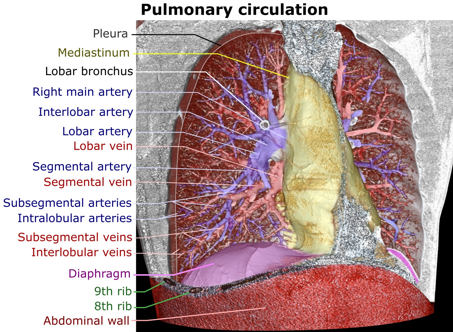
The pulmonary circulation is a division of the circulatory system in all vertebrates. The circuit begins with deoxygenated blood returned from the body to the right atrium of the heart where it is pumped out from the right ventricle to the lungs. In the lungs the blood is oxygenated and returned to the left atrium to complete the circuit. The other division of the circulatory system is the systemic circulation that begins with receiving the oxygenated blood from the pulmonary circulation into the left atrium. From the atrium the oxygenated blood enters the left ventricle where it is pumped out to the rest of the body, returning as deoxygenated blood back to the pulmonary circulation. The blood vessels of the pulmonary circulation are the pulmonary arteries and the pulmonary veins. A separate circulatory circuit known as the bronchial circulation supplies oxygenated blood to the tissue of the larger airways of the lung. Structure De-oxygenated blood leaves the heart, goe ... [...More Info...] [...Related Items...] OR: [Wikipedia] [Google] [Baidu] |
Right Atrium
The atrium ( la, ātrium, , entry hall) is one of two upper chambers in the heart that receives blood from the circulatory system. The blood in the atria is pumped into the heart ventricles through the atrioventricular valves. There are two atria in the human heart – the left atrium receives blood from the pulmonary circulation, and the right atrium receives blood from the venae cavae of the systemic circulation. During the cardiac cycle the atria receive blood while relaxed in diastole, then contract in systole to move blood to the ventricles. Each atrium is roughly cube-shaped except for an ear-shaped projection called an atrial appendage, sometimes known as an auricle. All animals with a closed circulatory system have at least one atrium. The atrium was formerly called the 'auricle'. That term is still used to describe this chamber in some other animals, such as the ''Mollusca''. They have thicker muscular walls than the atria do. Structure Humans have a four-chambered h ... [...More Info...] [...Related Items...] OR: [Wikipedia] [Google] [Baidu] |
Vein
Veins are blood vessels in humans and most other animals that carry blood towards the heart. Most veins carry deoxygenated blood from the tissues back to the heart; exceptions are the pulmonary and umbilical veins, both of which carry oxygenated blood to the heart. In contrast to veins, arteries carry blood away from the heart. Veins are less muscular than arteries and are often closer to the skin. There are valves (called ''pocket valves'') in most veins to prevent backflow. Structure Veins are present throughout the body as tubes that carry blood back to the heart. Veins are classified in a number of ways, including superficial vs. deep, pulmonary vs. systemic, and large vs. small. * Superficial veins are those closer to the surface of the body, and have no corresponding arteries. *Deep veins are deeper in the body and have corresponding arteries. *Perforator veins drain from the superficial to the deep veins. These are usually referred to in the lower limbs and feet. *Communic ... [...More Info...] [...Related Items...] OR: [Wikipedia] [Google] [Baidu] |
Balloon Catheter
A balloon catheter is a type of "soft" catheter with an inflatable "balloon" at its tip which is used during a catheterization procedure to enlarge a narrow opening or passage within the Human body, body. The deflated balloon catheter is positioned, then inflated to perform the necessary procedure, and deflated again in order to be removed. Some common uses include: *angioplasty or balloon septostomy, via cardiac catheterization (heart cath) *tuboplasty via uterus, uterine catheterization *pyeloplasty using a detachable inflatable balloon stent positioned via a cystoscopy, cystocopic transvesicular approach. Angioplasty balloon catheters Balloon catheters used in angioplasty are either of Over-the-Wire (OTW) or Rapid Exchange (Rx) design. Rx catheters nowadays are about 90% of the Coronary Intervention market. While OTW Catheters may still be useful in highly tortuous vascular pathways, they sacrifice deflation time and pushability. When a balloon catheter is used to compress p ... [...More Info...] [...Related Items...] OR: [Wikipedia] [Google] [Baidu] |
Cyanotic Heart Defect
A cyanotic heart defect is any congenital heart defect (CHD) that occurs due to deoxygenated blood bypassing the lungs and entering the systemic circulation, or a mixture of oxygenated and unoxygenated blood entering the systemic circulation. It is caused by structural defects of the heart such as right-to-left or bidirectional shunting, malposition of the great arteries, or any condition which increases pulmonary vascular resistance. The result may be the development of collateral circulation. Types * Tetralogy of Fallot (ToF) * Total anomalous pulmonary venous connection * Hypoplastic left heart syndrome (HLHS) * Transposition of the great arteries (d-TGA) * Truncus arteriosus (Persistent) * Tricuspid atresia * Interrupted aortic arch * Pulmonary atresia (PA) * Pulmonary stenosis (critical) * Eisenmenger syndrome (reversal of shunt due to pulmonary hypertension).Page Signs and symptoms Presentation includes the following: * Clubbing * The patient assuming a crouching posi ... [...More Info...] [...Related Items...] OR: [Wikipedia] [Google] [Baidu] |
Cor Pulmonale
Pulmonary heart disease, also known as cor pulmonale, is the enlargement and failure of the right ventricle of the heart as a response to increased vascular resistance (such as from pulmonic stenosis) or high blood pressure in the lungs. Chronic pulmonary heart disease usually results in right ventricular hypertrophy (RVH), whereas acute pulmonary heart disease usually results in dilatation. Hypertrophy is an adaptive response to a long-term increase in pressure. Individual muscle cells grow larger (in thickness) and change to drive the increased contractile force required to move the blood against greater resistance. Dilatation is a stretching (in length) of the ventricle in response to acute increased pressure. To be classified as pulmonary heart disease, the cause must originate in the pulmonary circulation system; RVH due to a systemic defect is not classified as pulmonary heart disease. Two causes are vascular changes as a result of tissue damage (e.g. disease, hypoxic in ... [...More Info...] [...Related Items...] OR: [Wikipedia] [Google] [Baidu] |
Hypoxia (medical)
Hypoxia is a condition in which the body or a region of the body is deprived of adequate oxygen supply at the tissue level. Hypoxia may be classified as either '' generalized'', affecting the whole body, or ''local'', affecting a region of the body. Although hypoxia is often a pathological condition, variations in arterial oxygen concentrations can be part of the normal physiology, for example, during strenuous physical exercise.. Hypoxia differs from hypoxemia and anoxemia, in that hypoxia refers to a state in which oxygen present in a tissue or the whole body is insufficient, whereas hypoxemia and anoxemia refer specifically to states that have low or no oxygen in the blood. Hypoxia in which there is complete absence of oxygen supply is referred to as anoxia. Hypoxia can be due to external causes, when the breathing gas is hypoxic, or internal causes, such as reduced effectiveness of gas transfer in the lungs, reduced capacity of the blood to carry oxygen, compromised general ... [...More Info...] [...Related Items...] OR: [Wikipedia] [Google] [Baidu] |
Pulmonary
The lungs are the primary organs of the respiratory system in humans and most other animals, including some snails and a small number of fish. In mammals and most other vertebrates, two lungs are located near the backbone on either side of the heart. Their function in the respiratory system is to extract oxygen from the air and transfer it into the bloodstream, and to release carbon dioxide from the bloodstream into the atmosphere, in a process of gas exchange. Respiration is driven by different muscular systems in different species. Mammals, reptiles and birds use their different muscles to support and foster breathing. In earlier tetrapods, air was driven into the lungs by the pharyngeal muscles via buccal pumping, a mechanism still seen in amphibians. In humans, the main muscle of respiration that drives breathing is the diaphragm. The lungs also provide airflow that makes vocal sounds including human speech possible. Humans have two lungs, one on the left and one on the ... [...More Info...] [...Related Items...] OR: [Wikipedia] [Google] [Baidu] |
Cardio
{{dab ...
Cardio (from Greek καρδίᾱ ''kardia'', 'heart') may refer to: * Of the Heart * Cardiology ** Cardiovascular system * Aerobic exercise, also known as cardio * ''Cardio'' (album), a 2010 album by Miguel Bosé See also * * * Physical exercise Exercise is a body activity that enhances or maintains physical fitness and overall health and wellness. It is performed for various reasons, to aid growth and improve strength, develop muscles and the cardiovascular system, hone athletic ... [...More Info...] [...Related Items...] OR: [Wikipedia] [Google] [Baidu] |
Systemic Circulation
The blood circulatory system is a system of organs that includes the heart, blood vessels, and blood which is circulated throughout the entire body of a human or other vertebrate. It includes the cardiovascular system, or vascular system, that consists of the heart and blood vessels (from Greek ''kardia'' meaning ''heart'', and from Latin ''vascula'' meaning ''vessels''). The circulatory system has two divisions, a systemic circulation or circuit, and a pulmonary circulation or circuit. Some sources use the terms ''cardiovascular system'' and ''vascular system'' interchangeably with the ''circulatory system''. The network of blood vessels are the great vessels of the heart including large elastic arteries, and large veins; other arteries, smaller arterioles, capillaries that join with venules (small veins), and other veins. The circulatory system is closed in vertebrates, which means that the blood never leaves the network of blood vessels. Some invertebrates such as arthro ... [...More Info...] [...Related Items...] OR: [Wikipedia] [Google] [Baidu] |
Muscle
Skeletal muscles (commonly referred to as muscles) are organs of the vertebrate muscular system and typically are attached by tendons to bones of a skeleton. The muscle cells of skeletal muscles are much longer than in the other types of muscle tissue, and are often known as muscle fibers. The muscle tissue of a skeletal muscle is striated – having a striped appearance due to the arrangement of the sarcomeres. Skeletal muscles are voluntary muscles under the control of the somatic nervous system. The other types of muscle are cardiac muscle which is also striated and smooth muscle which is non-striated; both of these types of muscle tissue are classified as involuntary, or, under the control of the autonomic nervous system. A skeletal muscle contains multiple fascicles – bundles of muscle fibers. Each individual fiber, and each muscle is surrounded by a type of connective tissue layer of fascia. Muscle fibers are formed from the fusion of developmental myoblasts in ... [...More Info...] [...Related Items...] OR: [Wikipedia] [Google] [Baidu] |
.jpg)


