|
Ramstedt's Operation
Pyloromyotomy is a surgical procedure in which a portion of the muscle fibers of the pyloric muscle are cut. This is typically done in cases where the contents from the stomach are inappropriately stopped by the pyloric muscle, causing the stomach contents to build up in the stomach and unable to be appropriately digested. The procedure is typically performed in cases of " hypertrophic pyloric stenosis" in young children. In most cases, the procedure can be performed with either an open approach or a laparoscopic approach and the patients typically have good outcomes with minimal complications. History and development The development of the procedure has attributed to Dr. Conrad Ramstedt in 1911, who originally named the procedure Ramstedt's Operation. However, the procedure was truly performed about 17 months earlier by Sir Harold Stiles in 1910 at the Royal Hospital for sick children. In 1991, the first laparoscopic pyloromyotomy was performed by Dr. Alain and Dr. Grousseau ... [...More Info...] [...Related Items...] OR: [Wikipedia] [Google] [Baidu] |
Pylorus
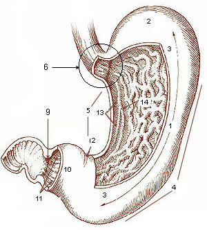
The pylorus ( or ), or pyloric part, connects the stomach to the duodenum. The pylorus is considered as having two parts, the ''pyloric antrum'' (opening to the body of the stomach) and the ''pyloric canal'' (opening to the duodenum). The ''pyloric canal'' ends as the ''pyloric orifice'', which marks the junction between the stomach and the duodenum. The orifice is surrounded by a sphincter, a band of muscle, called the ''pyloric sphincter''. The word ''pylorus'' comes from Greek πυλωρός, via Latin. The word ''pylorus'' in Greek means "gatekeeper", related to "gate" ( el, pyle) and is thus linguistically related to the word " pylon". Structure The pylorus is the furthest part of the stomach that connects to the duodenum. It is divided into two parts, the ''antrum'', which connects to the body of the stomach, and the ''pyloric canal'', which connects to the duodenum. Antrum The ''pyloric antrum'' is the initial portion of the pylorus. It is near the bottom of the stomach, ... [...More Info...] [...Related Items...] OR: [Wikipedia] [Google] [Baidu] |
Gastrointestinal Tract
The gastrointestinal tract (GI tract, digestive tract, alimentary canal) is the tract or passageway of the digestive system that leads from the mouth to the anus. The GI tract contains all the major organ (biology), organs of the digestive system, in humans and other animals, including the esophagus, stomach, and intestines. Food taken in through the mouth is digestion, digested to extract nutrients and absorb energy, and the waste expelled at the anus as feces. ''Gastrointestinal'' is an adjective meaning of or pertaining to the stomach and intestines. Nephrozoa, Most animals have a "through-gut" or complete digestive tract. Exceptions are more primitive ones: sponges have small pores (ostium (sponges), ostia) throughout their body for digestion and a larger dorsal pore (osculum) for excretion, comb jellies have both a ventral mouth and dorsal anal pores, while cnidarians and acoels have a single pore for both digestion and excretion. The human gastrointestinal tract consists o ... [...More Info...] [...Related Items...] OR: [Wikipedia] [Google] [Baidu] |
Incisional Hernia
An incisional hernia is a type of hernia caused by an incompletely-healed surgical wound. Since median incisions in the abdomen are frequent for abdominal exploratory surgery, ventral incisional hernias are often also classified as ventral hernias due to their location. Not all ventral hernias are from incisions, as some may be caused by other trauma or congenital problems. Signs and symptoms Clinically, incisional hernias present as a bulge or protrusion at or near the area of a surgical incision. Virtually any prior abdominal operation can develop an incisional hernia at the scar area (provided adequate healing does not occur due to infection), including large abdominal procedures such as intestinal or vascular surgery, and small incisions, such as ( appendix removal or abdominal exploratory surgery). While incisional hernias can occur at any incision, they tend to occur more commonly along a straight line from the xiphoid process of the sternum straight down to the pubis, ... [...More Info...] [...Related Items...] OR: [Wikipedia] [Google] [Baidu] |
Wound Dehiscence
Wound dehiscence is a surgical complication in which a wound ruptures along a surgical incision. Risk factors include age, collagen disorder such as Ehlers–Danlos syndrome, diabetes, obesity, poor knotting or grabbing of stitches, and trauma to the wound after surgery. Signs Signs of dehiscence can include bleeding, pain, inflammation, fever, or the wound opening spontaneously. An internal surgical wound dehiscence can occur internally, as a consequence of hysterectomy, at the site of the vaginal cuff. Cause A primary cause of wound dehiscence is sub-acute infection, resulting from inadequate or imperfect aseptic technique. Coated suture, such as Vicryl, generally breaks down at a rate predicted to correspond with tissue healing, but is hastened in the presence of bacteria. In the absence of other known metabolic factors which inhibit healing and may have contributed to suture dehiscence, subacute infection should be suspected, and the protocol for obtaining wound culture ... [...More Info...] [...Related Items...] OR: [Wikipedia] [Google] [Baidu] |
Fascia
A fascia (; plural fasciae or fascias; adjective fascial; from Latin: "band") is a band or sheet of connective tissue, primarily collagen, beneath the skin that attaches to, stabilizes, encloses, and separates muscles and other internal organs. Fascia is classified by layer, as superficial fascia, deep fascia, and ''visceral'' or ''parietal'' fascia, or by its function and anatomical location. Like ligaments, aponeuroses, and tendons, fascia is made up of fibrous connective tissue containing closely packed bundles of collagen fibers oriented in a wavy pattern parallel to the direction of pull. Fascia is consequently flexible and able to resist great unidirectional tension forces until the wavy pattern of fibers has been straightened out by the pulling force. These collagen fibers are produced by fibroblasts located within the fascia. Fasciae are similar to ligaments and tendons as they have collagen as their major component. They differ in their location and function: ligament ... [...More Info...] [...Related Items...] OR: [Wikipedia] [Google] [Baidu] |
Surgical Suture
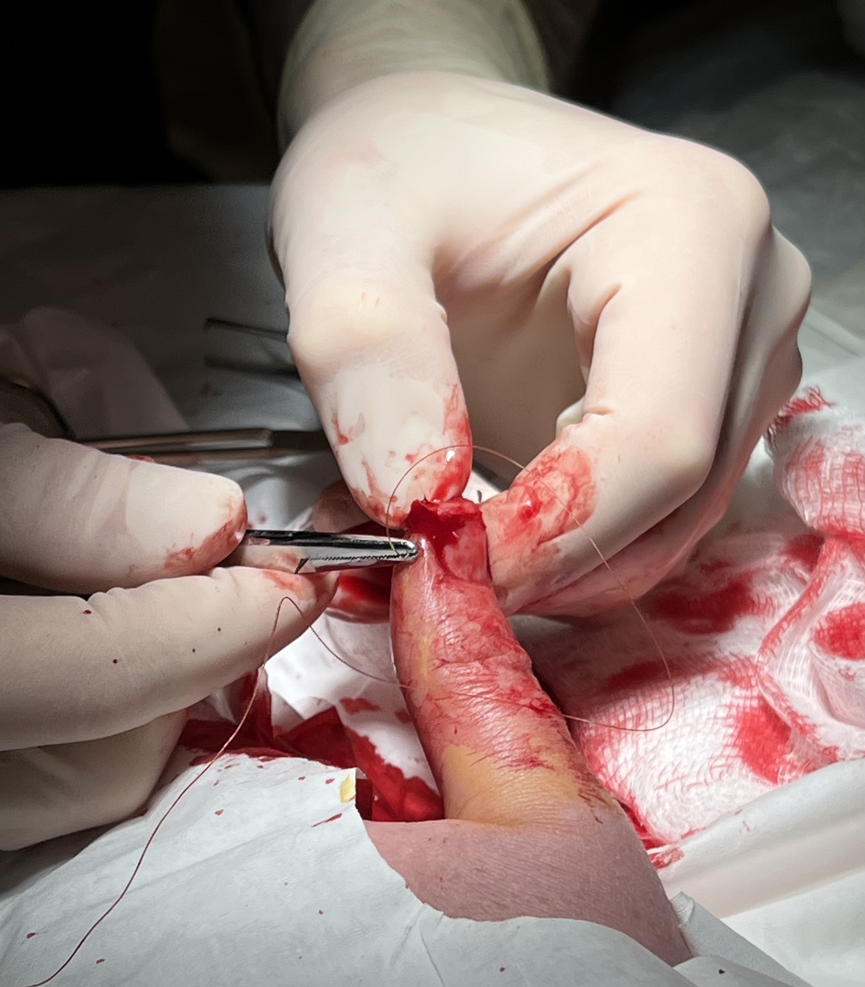
A surgical suture, also known as a stitch or stitches, is a medical device used to hold body tissues together and approximate wound edges after an injury or surgery. Application generally involves using a needle with an attached length of thread. There are numerous types of suture which differ by needle shape and size as well as thread material and characteristics. Selection of surgical suture should be determined by the characteristics and location of the wound or the specific body tissues being approximated. In selecting the needle, thread, and suturing technique to use for a specific patient, a medical care provider must consider the tensile strength of the specific suture thread needed to efficiently hold the tissues together depending on the mechanical and shear forces acting on the wound as well as the thickness of the tissue being approximated. One must also consider the elasticity of the thread and ability to adapt to different tissues, as well as the memory of the threa ... [...More Info...] [...Related Items...] OR: [Wikipedia] [Google] [Baidu] |
Trocar
A trocar (or trochar) is a medical or veterinary device that is made up of an awl (which may be a metal or plastic sharpened or non-bladed tip), a cannula (essentially a hollow tube), and a seal. Trocars are placed through the abdomen during laparoscopic surgery. The trocar functions as a portal for the subsequent placement of other instruments, such as graspers, scissors, staplers, etc. Trocars also allow the escape of gas or fluid from organs within the body. Etymology The word ''trocar'', less commonly ''trochar'', comes from French ''trocart'', ''trois-quarts'' (three-fourths), from ''trois'' 'three' and ''carre'' 'side, face of an instrument', first recorded in the ''Dictionnaire des Arts et des Sciences'', 1694, by Thomas Corneille, younger brother of Pierre Corneille. History Originally, doctors used trocars to relieve pressure build-up of fluids (edema) or gases (bloating). Patents for trocars appeared early in the 19th century, although their use dated back possibly ... [...More Info...] [...Related Items...] OR: [Wikipedia] [Google] [Baidu] |
Abdominal Cavity
The abdominal cavity is a large body cavity in humans and many other animals that contains many organs. It is a part of the abdominopelvic cavity. It is located below the thoracic cavity, and above the pelvic cavity. Its dome-shaped roof is the thoracic diaphragm, a thin sheet of muscle under the lungs, and its floor is the pelvic inlet, opening into the pelvis. Structure Organs Organs of the abdominal cavity include the stomach, liver, gallbladder, spleen, pancreas, small intestine, kidneys, large intestine, and adrenal glands. Peritoneum The abdominal cavity is lined with a protective membrane termed the peritoneum. The inside wall is covered by the parietal peritoneum. The kidneys are located behind the peritoneum, in the retroperitoneum, outside the abdominal cavity. The viscera are also covered by visceral peritoneum. Between the visceral and parietal peritoneum is the peritoneal cavity, which is a potential space. It contains a serous fluid called peritoneal fluid tha ... [...More Info...] [...Related Items...] OR: [Wikipedia] [Google] [Baidu] |
Mucosa
A mucous membrane or mucosa is a membrane that lines various cavities in the body of an organism and covers the surface of internal organs. It consists of one or more layers of epithelial cells overlying a layer of loose connective tissue. It is mostly of endodermal origin and is continuous with the skin at body openings such as the eyes, eyelids, ears, inside the nose, inside the mouth, lips, the genital areas, the urethral opening and the anus. Some mucous membranes secrete mucus, a thick protective fluid. The function of the membrane is to stop pathogens and dirt from entering the body and to prevent bodily tissues from becoming dehydrated. Structure The mucosa is composed of one or more layers of epithelial cells that secrete mucus, and an underlying lamina propria of loose connective tissue. The type of cells and type of mucus secreted vary from organ to organ and each can differ along a given tract. Mucous membranes line the digestive, respiratory and reproductive trac ... [...More Info...] [...Related Items...] OR: [Wikipedia] [Google] [Baidu] |
Pyloric Stenosis
Pyloric stenosis is a narrowing of the opening from the stomach to the first part of the small intestine (the pylorus). Symptoms include projectile vomiting without the presence of bile. This most often occurs after the baby is fed. The typical age that symptoms become obvious is two to twelve weeks old. The cause of pyloric stenosis is unclear. Risk factors in babies include birth by cesarean section, preterm birth, bottle feeding, and being first born. The diagnosis may be made by feeling an olive-shaped mass in the baby's abdomen. This is often confirmed with ultrasound. Treatment initially begins by correcting dehydration and electrolyte problems. This is then typically followed by surgery, although some treat the condition without surgery by using atropine. Results are generally good both in the short term and in the long term. About one to two per 1,000 babies are affected, and males are affected about four times more often than females. The condition is very rare in ... [...More Info...] [...Related Items...] OR: [Wikipedia] [Google] [Baidu] |
Hypertrophy
Hypertrophy is the increase in the volume of an organ or tissue due to the enlargement of its component cells. It is distinguished from hyperplasia, in which the cells remain approximately the same size but increase in number.Updated by Linda J. Vorvick. 8/14/1Hyperplasia/ref> Although hypertrophy and hyperplasia are two distinct processes, they frequently occur together, such as in the case of the hormonally-induced proliferation and enlargement of the cells of the uterus during pregnancy. Eccentric hypertrophy is a type of hypertrophy where the walls and chamber of a hollow organ undergo growth in which the overall size and volume are enlarged. It is applied especially to the left ventricle of heart. Sarcomeres are added in series, as for example in dilated cardiomyopathy (in contrast to hypertrophic cardiomyopathy, a type of concentric hypertrophy, where sarcomeres are added in parallel). Gallery File:*+ * Photographic documentation on sexual education - Hypertrophy of bre ... [...More Info...] [...Related Items...] OR: [Wikipedia] [Google] [Baidu] |


