|
Radiate Ligament Of Head Of Rib
The radiate ligament of head of rib is a ligament of the costovertebral joint that typically connects the anterior edge of the head of each rib, and the side of the bodies of two adjacent vertebrae Each vertebra (: vertebrae) is an irregular bone with a complex structure composed of bone and some hyaline cartilage, that make up the vertebral column or spine, of vertebrates. The proportions of the vertebrae differ according to their spinal ... and their intervertebral discs. The ligament is formed as a thickening of the anterior portion of the joint capsule of the costovertebral joint, and thus reinforces it anteriorly. Anatomy In the case of the first rib, the radiate ligament attaches to the bodies of the vertebrae C7 and T1. In the case of ribs X-XII - each of which articulates with a single vertebra - the radiate ligaments attach onto the corresponding vertebra and the above vertebra. Structure The ligament consists of three bands: * The superior band extends superi ... [...More Info...] [...Related Items...] OR: [Wikipedia] [Google] [Baidu] [Amazon] |
Costovertebral Articulations
The costovertebral joints are the joints that connect the ribs to the vertebral column. * The articulation of the head of rib connects the head of the rib and the bodies of vertebrae. * The costotransverse joint connects the rib with the transverse processes Each vertebra (: vertebrae) is an irregular bone with a complex structure composed of bone and some hyaline cartilage, that make up the vertebral column or spine, of vertebrates. The proportions of the vertebrae differ according to their spina ... of vertebrae. References External links Diagram at ithaca.edu via web.archive.org Joints {{musculoskeletal-stub ... [...More Info...] [...Related Items...] OR: [Wikipedia] [Google] [Baidu] [Amazon] |
Head Of Rib
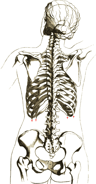
The rib cage or thoracic cage is an endoskeletal enclosure in the thorax of most vertebrates that comprises the ribs, vertebral column and sternum, which protect the vital organs of the thoracic cavity, such as the heart, lungs and great vessels and support the shoulder girdle to form the core part of the axial skeleton. A typical human thoracic cage consists of 12 pairs of ribs and the adjoining costal cartilages, the sternum (along with the manubrium and xiphoid process), and the 12 thoracic vertebrae articulating with the ribs. The thoracic cage also provides attachments for extrinsic skeletal muscles of the neck, upper limbs, upper abdomen and back, and together with the overlying skin and associated fascia and muscles, makes up the thoracic wall. In tetrapods, the rib cage intrinsically holds the muscles of respiration ( diaphragm, intercostal muscles, etc.) that are crucial for active inhalation and forced exhalation, and therefore has a major ventilatory function in th ... [...More Info...] [...Related Items...] OR: [Wikipedia] [Google] [Baidu] [Amazon] |
Vertebrae
Each vertebra (: vertebrae) is an irregular bone with a complex structure composed of bone and some hyaline cartilage, that make up the vertebral column or spine, of vertebrates. The proportions of the vertebrae differ according to their spinal segment and the particular species. The basic configuration of a vertebra varies; the vertebral body (also ''centrum'') is of bone and bears the load of the vertebral column. The upper and lower surfaces of the vertebra body give attachment to the intervertebral discs. The posterior part of a vertebra forms a vertebral arch, in eleven parts, consisting of two pedicles (pedicle of vertebral arch), two laminae, and seven process (anatomy), processes. The laminae give attachment to the ligamenta flava (ligaments of the spine). There are vertebral notches formed from the shape of the pedicles, which form the intervertebral foramina when the vertebrae articulation (anatomy), articulate. These foramina are the entry and exit conduits for the spi ... [...More Info...] [...Related Items...] OR: [Wikipedia] [Google] [Baidu] [Amazon] |
Intervertebral Disc
An intervertebral disc (British English), also spelled intervertebral disk (American English), lies between adjacent vertebrae in the vertebral column. Each disc forms a fibrocartilaginous joint (a symphysis), to allow slight movement of the vertebrae, to act as a ligament to hold the vertebrae together, and to function as a shock absorber for the spine. Structure Intervertebral discs consist of an outer fibrous ring, the ''anulus (or annulus) fibrosus disci intervertebralis'', which surrounds an inner gel-like center, the ''nucleus pulposus''. The ''anulus fibrosus'' consists of several layers (laminae) of fibrocartilage made up of both type I and type II collagen. Type I is concentrated toward the edge of the ring, where it provides greater strength. The stiff laminae can withstand compressive forces. The fibrous intervertebral disc contains the ''nucleus pulposus'' and this helps to distribute pressure evenly across the disc. This prevents the development of stress conc ... [...More Info...] [...Related Items...] OR: [Wikipedia] [Google] [Baidu] [Amazon] |
Anterior Longitudinal Ligament
The anterior longitudinal ligament is a ligament that extends across the anterior/ventral aspect of the vertebral bodies and intervertebral discs the spine. It may be partially cut to treat certain abnormal curvatures in the vertebral column, such as kyphosis. Anatomy The anterior longitudinal ligament extends superoinferiorly between the basiocciput of the skull and the anterior tubercle of the atlas (cervical vertebra C1) superiorly, and the superior part of the sacrum inferiorly; inferiorly, it ends at the sacral promontory. It broadens inferiorly. Inferiorly, it becomes continuous with the anterior sacrococcygeal ligament. Superiorly, between the skull and atlas, the ligament is continuous laterally with the anterior atlantooccipital membrane. The ligament is thick and slightly more narrow over the vertebral bodies and thinner but slightly wider over the intervertebral discs. It tends to be narrower and thicker around thoracic vertebrae, and wider and thinner around ... [...More Info...] [...Related Items...] OR: [Wikipedia] [Google] [Baidu] [Amazon] |
Thoracic Ganglia
The thoracic ganglia are paravertebral ganglia. The thoracic portion of the sympathetic trunk typically has 12 thoracic ganglia. Emerging from the ganglia are thoracic splanchnic nerves (the cardiopulmonary, the greater, lesser, and least splanchnic nerves) that help provide sympathetic innervation to thoracic and abdominal structures. The thoracic part of sympathetic trunk lies posterior to the costovertebral pleura and is hence not a content of the posterior mediastinum Also, the ganglia of the thoracic sympathetic trunk have both white and gray rami communicantes. The white rami communicantes carry sympathetic fibers arising in the spinal cord into the sympathetic trunk, while the gray rami communicantes carry postganglionic nerve fibers of the sympathetic nervous system back to the spinal nerves A spinal nerve is a mixed nerve, which carries motor, sensory, and autonomic signals between the spinal cord and the body. In the human body there are 31 pairs of spinal ne ... [...More Info...] [...Related Items...] OR: [Wikipedia] [Google] [Baidu] [Amazon] |
Sympathetic Trunk
The sympathetic trunk (sympathetic chain, gangliated cord) is a paired bundle of nerve fibers that run from the base of the skull to the coccyx. It is a major component of the sympathetic nervous system. Structure The sympathetic trunk lies just lateral to the vertebral bodies for the entire length of the vertebral column. It interacts with the anterior rami of spinal nerves by way of rami communicantes. The sympathetic trunk permits preganglionic fibers of the sympathetic nervous system to ascend to spinal levels superior to T1 and descend to spinal levels inferior to L2/3.Greenstein B., Greenstein A. (2002): Color atlas of neuroscience – Neuroanatomy and neurophysiology. Thieme, Stuttgart – New York, . The superior end of it is continued upward through the carotid canal into the skull, and forms a plexus on the internal carotid artery; the inferior part travels in front of the coccyx, where it converges with the other trunk at a structure known as the ganglion impar ... [...More Info...] [...Related Items...] OR: [Wikipedia] [Google] [Baidu] [Amazon] |
Pleura
The pleurae (: pleura) are the two flattened closed sacs filled with pleural fluid, each ensheathing each lung and lining their surrounding tissues, locally appearing as two opposing layers of serous membrane separating the lungs from the mediastinum, the inside surfaces of the surrounding chest walls and the diaphragm. Although wrapped onto itself resulting in an apparent double layer, each lung is surrounded by a single, continuous pleural membrane. The portion of the pleura that covers the surface of each lung is often called the visceral pleura. This can lead to some confusion, as the lung is not the only visceral organ covered by the pleura. The pleura typically dips between the lobes of the lung as ''fissures'', and is formed by the invagination of lung buds into each thoracic sac during embryonic development. The portion of the pleura seen as the outer layer covers the chest wall, the diaphragm and the mediastinum and is often also misleadingly called the parieta ... [...More Info...] [...Related Items...] OR: [Wikipedia] [Google] [Baidu] [Amazon] |
Azygos Vein
The azygos vein (from Ancient Greek ἄζυγος (ázugos), meaning 'unwedded' or 'unpaired') is a vein running up the right side of the thoracic vertebral column draining itself towards the superior vena cava. It connects the systems of superior vena cava and inferior vena cava and can provide an alternative path for blood to the right atrium when either of the venae cavae is blocked. Structure The azygos vein transports deoxygenated blood from the posterior walls of the thorax and abdomen into the superior vena cava. It is formed by the union of the ascending lumbar veins with the right subcostal veins at the level of the 12th thoracic vertebra, ascending to the right of the descending aorta and thoracic duct, passing behind the right crus of diaphragm, anterior to the vertebral bodies of T12 to T5 and right posterior intercostal arteries. At the level of T4 vertebrae, it arches over the root of the right lung from behind to the front to join the superior vena cava. The tra ... [...More Info...] [...Related Items...] OR: [Wikipedia] [Google] [Baidu] [Amazon] |
Interarticular Ligament
The costovertebral joints are the joints that connect the ribs to the vertebral column. * The articulation of the head of rib connects the head of the rib and the bodies of vertebrae. * The costotransverse joint connects the rib with the transverse processes Each vertebra (: vertebrae) is an irregular bone with a complex structure composed of bone and some hyaline cartilage, that make up the vertebral column or spine, of vertebrates. The proportions of the vertebrae differ according to their spina ... of vertebrae. References External links Diagram at ithaca.edu via web.archive.org Joints {{musculoskeletal-stub ... [...More Info...] [...Related Items...] OR: [Wikipedia] [Google] [Baidu] [Amazon] |
Synovial Membranes
{{disambiguation ...
Synovial () may refer to: * Synovial fluid * Synovial joint * Synovial membrane * Synovial bursa A synovial bursa, usually simply bursa (: bursae or bursas), is a small fluid-filled sac lined by synovial membrane with an inner capillary layer of viscous synovial fluid (similar in consistency to that of a raw egg white). It provides a cushi ... [...More Info...] [...Related Items...] OR: [Wikipedia] [Google] [Baidu] [Amazon] |

