|
Rubeosis Iridis
Rubeosis iridis is a medical condition of the iris of the eye in which new abnormal blood vessels (formed by neovascularization) are found on the surface of the iris. Causes This condition is often associated with diabetes in advanced proliferative diabetic retinopathy. Other conditions causing rubeosis iridis include central retinal vein occlusion, ocular ischemic syndrome, and chronic retinal detachment. Pathophysiology It is usually associated with disease processes in the retina, which involve the retina becoming starved of oxygen (ischaemic). The ischemic retina releases a variety of factors, the most important of which is vascular endothelial growth factor (VEGF). These factors stimulate the formation of new blood vessels ( angiogenesis). These new vessels do not have the same characteristics as the blood vessels originally formed in the eye. In addition, new blood vessels can form in areas that do not have them. Specifically, new blood vessels can be observed on the ... [...More Info...] [...Related Items...] OR: [Wikipedia] [Google] [Baidu] |
Medical Condition
A disease is a particular abnormal condition that negatively affects the structure or function (biology), function of all or part of an organism, and that is not immediately due to any external injury. Diseases are often known to be medical conditions that are associated with specific signs and symptoms. A disease may be caused by external factors such as pathogens or by internal dysfunctions. For example, internal dysfunctions of the immune system can produce a variety of different diseases, including various forms of immunodeficiency, hypersensitivity, allergy, allergies and autoimmune disorders. In humans, ''disease'' is often used more broadly to refer to any condition that causes pain, Abnormality (behavior), dysfunction, distress (medicine), distress, social problems, or death to the person affected, or similar problems for those in contact with the person. In this broader sense, it sometimes includes injury, injuries, disability, disabilities, #Disorder, disorders, s ... [...More Info...] [...Related Items...] OR: [Wikipedia] [Google] [Baidu] |
Vascular Endothelial Growth Factor
Vascular endothelial growth factor (VEGF, ), originally known as vascular permeability factor (VPF), is a signal protein produced by many cells that stimulates the formation of blood vessels. To be specific, VEGF is a sub-family of growth factors, the platelet-derived growth factor family of cystine-knot growth factors. They are important signaling proteins involved in both vasculogenesis (the '' de novo'' formation of the embryonic circulatory system) and angiogenesis (the growth of blood vessels from pre-existing vasculature). It is part of the system that restores the oxygen supply to tissues when blood circulation is inadequate such as in hypoxic conditions. Serum concentration of VEGF is high in bronchial asthma and diabetes mellitus. VEGF's normal function is to create new blood vessels during embryonic development, new blood vessels after injury, muscle following exercise, and new vessels (collateral circulation) to bypass blocked vessels. It can contribute to disease. S ... [...More Info...] [...Related Items...] OR: [Wikipedia] [Google] [Baidu] |
Corneal Neovascularization
Corneal neovascularization (CNV) is the in-growth of new blood vessels from the pericorneal plexus into avascular corneal tissue as a result of oxygen deprivation. Maintaining avascularity of the corneal stroma is an important aspect of corneal pathophysiology as it is required for corneal transparency and optimal vision. A decrease in corneal transparency causes visual acuity deterioration. Corneal tissue is avascular in nature and the presence of vascularization, which can be deep or superficial, is always pathologically related. Corneal neovascularization is a sight-threatening condition that can be caused by inflammation related to infection, chemical injury, autoimmune conditions, immune hypersensitivity, post-corneal transplantation, and traumatic conditions among other ocular pathologies. Common causes of CNV within the cornea include trachoma, corneal ulcers, phylctenular keratoconjunctivitisrosacea keratitis interstitial keratitissclerosing keratitis chemical burns, a ... [...More Info...] [...Related Items...] OR: [Wikipedia] [Google] [Baidu] |
Choroidal Neovascularization
Choroidal neovascularization (CNV) is the creation of new blood vessels in the choroid layer of the eye. Choroidal neovascularization is a common cause of neovascular degenerative maculopathy (i.e. 'wet' macular degeneration) commonly exacerbated by extreme myopia, malignant myopic degeneration, or age-related developments. Causes CNV can occur rapidly in individuals with defects in Bruch's membrane, the innermost layer of the choroid. It is also associated with excessive amounts of vascular endothelial growth factor (VEGF). As well as in wet macular degeneration, CNV can also occur frequently with the rare genetic disease pseudoxanthoma elasticum and rarely with the more common optic disc drusen. CNV has also been associated with extreme myopia or malignant myopic degeneration, where in choroidal neovascularization occurs primarily in the presence of cracks within the retinal (specifically) macular tissue known as lacquer cracks. Symptoms CNV can create a sudden deterioration ... [...More Info...] [...Related Items...] OR: [Wikipedia] [Google] [Baidu] |
Panretinal Photocoagulation
Diabetic retinopathy (also known as diabetic eye disease), is a medical condition in which damage occurs to the retina due to diabetes mellitus. It is a leading cause of blindness in developed countries. Diabetic retinopathy affects up to 80 percent of those who have had both type 1 and type 2 diabetes for 20 years or more. In at least 90% of new cases, progression to more aggressive forms of sight threatening retinopathy and maculopathy could be reduced with proper treatment and monitoring of the eyes The longer a person has diabetes, the higher his or her chances of developing diabetic retinopathy. Each year in the United States, diabetic retinopathy accounts for 12% of all new cases of blindness. It is also the leading cause of blindness in people aged 20 to 64. Signs and symptoms Nearly all people with diabetes develop some degree of retina damage ("retinopathy") over several decades with the disease. For many, that damage can only be detected by a retinal exam, and has ... [...More Info...] [...Related Items...] OR: [Wikipedia] [Google] [Baidu] |
Neovascular Glaucoma
Glaucoma is a group of eye diseases that result in damage to the optic nerve (or retina) and cause vision loss. The most common type is open-angle (wide angle, chronic simple) glaucoma, in which the drainage angle for fluid within the eye remains open, with less common types including closed-angle (narrow angle, acute congestive) glaucoma and normal-tension glaucoma. Open-angle glaucoma develops slowly over time and there is no pain. Peripheral vision may begin to decrease, followed by central vision, resulting in blindness if not treated. Closed-angle glaucoma can present gradually or suddenly. The sudden presentation may involve severe eye pain, blurred vision, mid-dilated pupil, redness of the eye, and nausea. Vision loss from glaucoma, once it has occurred, is permanent. Eyes affected by glaucoma are referred to as being glaucomatous. Risk factors for glaucoma include increasing age, high pressure in the eye, a family history of glaucoma, and use of steroid medication. F ... [...More Info...] [...Related Items...] OR: [Wikipedia] [Google] [Baidu] |
Intraocular Pressure
Intraocular pressure (IOP) is the fluid pressure inside the eye Eyes are organs of the visual system. They provide living organisms with vision, the ability to receive and process visual detail, as well as enabling several photo response functions that are independent of vision. Eyes detect light and conv .... Tonometry is the method eye care professionals use to determine this. IOP is an important aspect in the evaluation of patients at risk of glaucoma. Most tonometers are calibrated to measure pressure in millimeters of mercury (mmHg). Physiology Intraocular pressure is determined by the production and drainage of aqueous humour by the ciliary body and its drainage via the trabecular meshwork and uveoscleral outflow. The reason for this is because the Vitreous body, vitreous humour in the Posterior segment of eyeball, posterior segment has a relatively fixed volume and thus does not affect intraocular pressure regulation. An important quantitative relationship (Goldmann ... [...More Info...] [...Related Items...] OR: [Wikipedia] [Google] [Baidu] |
Fibrosis
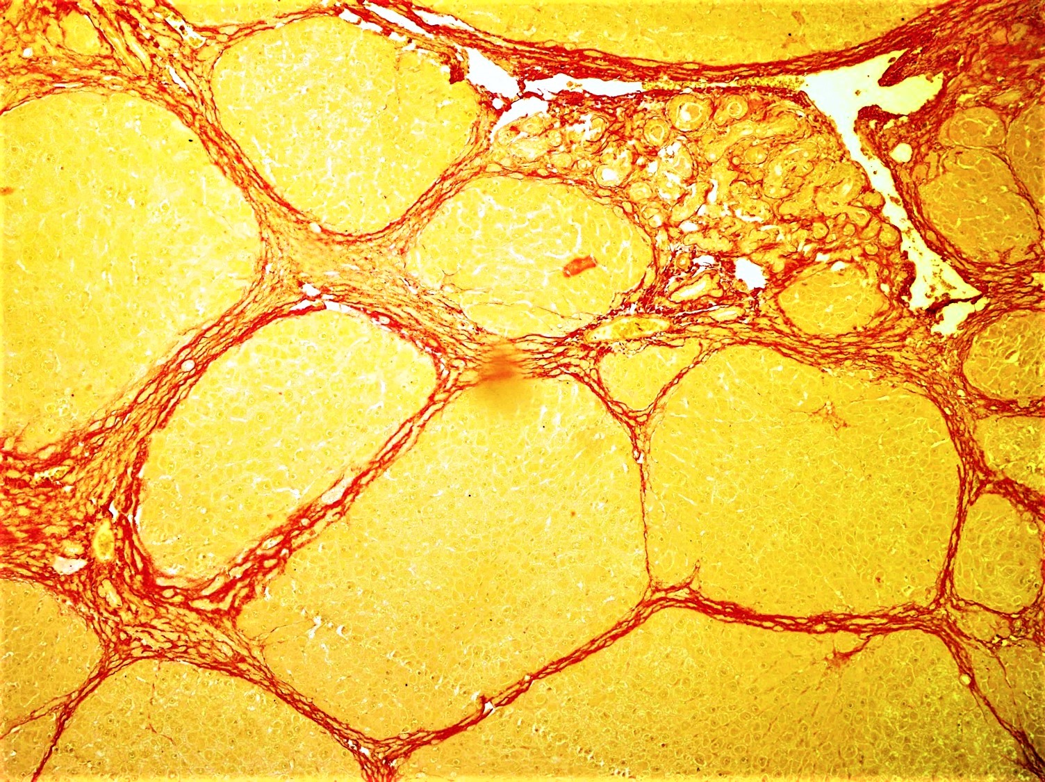
Fibrosis, also known as fibrotic scarring, is a pathological wound healing in which connective tissue replaces normal parenchymal tissue to the extent that it goes unchecked, leading to considerable tissue remodelling and the formation of permanent scar tissue. Repeated injuries, chronic inflammation and repair are susceptible to fibrosis where an accidental excessive accumulation of extracellular matrix components, such as the collagen is produced by fibroblasts, leading to the formation of a permanent fibrotic scar. In response to injury, this is called scarring, and if fibrosis arises from a single cell line, this is called a fibroma. Physiologically, fibrosis acts to deposit connective tissue, which can interfere with or totally inhibit the normal architecture and function of the underlying organ or tissue. Fibrosis can be used to describe the pathological state of excess deposition of fibrous tissue, as well as the process of connective tissue deposition in healing. Define ... [...More Info...] [...Related Items...] OR: [Wikipedia] [Google] [Baidu] |
Angiogenesis
Angiogenesis is the physiological process through which new blood vessels form from pre-existing vessels, formed in the earlier stage of vasculogenesis. Angiogenesis continues the growth of the vasculature by processes of sprouting and splitting. Vasculogenesis is the embryonic formation of endothelial cells from mesoderm cell precursors, and from neovascularization, although discussions are not always precise (especially in older texts). The first vessels in the developing embryo form through vasculogenesis, after which angiogenesis is responsible for most, if not all, blood vessel growth during development and in disease. Angiogenesis is a normal and vital process in growth and development, as well as in wound healing and in the formation of granulation tissue. However, it is also a fundamental step in the transition of tumors from a benign state to a malignant one, leading to the use of angiogenesis inhibitors in the treatment of cancer. The essential role of angi ... [...More Info...] [...Related Items...] OR: [Wikipedia] [Google] [Baidu] |
Ischaemic
Ischemia or ischaemia is a restriction in blood supply to any tissue, muscle group, or organ of the body, causing a shortage of oxygen that is needed for cellular metabolism (to keep tissue alive). Ischemia is generally caused by problems with blood vessels, with resultant damage to or dysfunction of tissue i.e. hypoxia and microvascular dysfunction. It also implies local hypoxia in a part of a body resulting from constriction (such as vasoconstriction, thrombosis, or embolism). Ischemia causes not only insufficiency of oxygen, but also reduced availability of nutrients and inadequate removal of metabolic wastes. Ischemia can be partial (poor perfusion) or total blockage. The inadequate delivery of oxygenated blood to the organs must be resolved either by treating the cause of the inadequate delivery or reducing the oxygen demand of the system that needs it. For example, patients with myocardial ischemia have a decreased blood flow to the heart and are prescribed with ... [...More Info...] [...Related Items...] OR: [Wikipedia] [Google] [Baidu] |
Iris (anatomy)
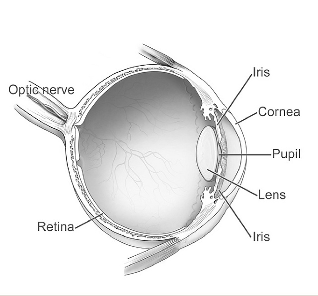
In humans and most mammals and birds, the iris (plural: ''irides'' or ''irises'') is a thin, annular structure in the eye, responsible for controlling the diameter and size of the pupil, and thus the amount of light reaching the retina. Eye color is defined by the iris. In optical terms, the pupil is the eye's aperture, while the iris is the diaphragm. Structure The iris consists of two layers: the front pigmented fibrovascular layer known as a stroma and, beneath the stroma, pigmented epithelial cells. The stroma is connected to a sphincter muscle ( sphincter pupillae), which contracts the pupil in a circular motion, and a set of dilator muscles (dilator pupillae), which pull the iris radially to enlarge the pupil, pulling it in folds. The sphincter pupillae is the opposing muscle of the dilator pupillae. The pupil's diameter, and thus the inner border of the iris, changes size when constricting or dilating. The outer border of the iris does not change size. The constr ... [...More Info...] [...Related Items...] OR: [Wikipedia] [Google] [Baidu] |


