|
Reinke Crystals
Reinke crystals are rod-like cytoplasmic inclusions which can be found in Leydig cells of the testes. Occurring only in adult humans and wild bush rats, their function is unknown. Ovarian stromal tumors having a predominant pattern of fibroma or thecoma but also containing cells typical of steroid hormone-secreting cells were reported. Some of the tumors were classified as luteinized thecomas because the steroid cells resembled lutein cells and lacked crystalloids of Reinke. But others were classified as stromal Leydig cell tumors as seen in tumors of the testes because Reinke crystalloids were identified in the steroid cells. The Stromal Leydig tumors occurred at an average age of 61 years and were associated with Ovarian hyperandrogenism which led to virilization in some cases, endometrial hyperplasia in other cases, and endometrial hyperplasia with carcinoma in the rest of the cases. Luteinized thecomas and stromal Leydig cell tumors are indistinguishable except for the presence ... [...More Info...] [...Related Items...] OR: [Wikipedia] [Google] [Baidu] |
Cytoplasmic Inclusions
In cellular biology, inclusions are diverse intracellularShively, J. M. (ed.). (2006). ''Microbiology Monographs Vol. 1: Inclusions in Prokaryotes''. Berlin, Heidelberg: Springer-Verlaglink non-living substances ( ergastic substances) that are not bound by membranes. Inclusions are stored nutrients/deutoplasmic substances, secretory products, and pigment granules. Examples of inclusions are glycogen granules in the liver and muscle cells, lipid droplets in fat cells, pigment granules in certain cells of skin and hair, and crystals of various types.Leslie P. Gartner and James L. Hiatt ; Text book of Histology; 3rd edition Cytoplasmic inclusions are an example of a biomolecular condensate arising by liquid-solid, liquid-gel or liquid-liquid phase separation. These structures were first observed by O. F. Müller in 1786. Examples Glycogen: Glycogen is the most common form of glucose in animals and is especially abundant in cells of muscles, and liver. It appears in electro ... [...More Info...] [...Related Items...] OR: [Wikipedia] [Google] [Baidu] |
Leydig Cell
Leydig cells, also known as interstitial cells of the testes and interstitial cells of Leydig, are found adjacent to the seminiferous tubules in the testicle and produce testosterone in the presence of luteinizing hormone (LH). They are polyhedral in shape and have a large, prominent nucleus, an eosinophilic cytoplasm, and numerous lipid-filled vesicles. Structure The mammalian Leydig cell is a polyhedral epithelioid cell with a single eccentrically located ovoid nucleus. The nucleus contains one to three prominent nucleoli and large amounts of dark-staining peripheral heterochromatin. The acidophilic cytoplasm usually contains numerous membrane-bound lipid droplets and large amounts of smooth endoplasmic reticulum (SER). Besides the abundance of SER with scattered patches of rough endoplasmic reticulum, several mitochondria are also prominent within the cytoplasm. Reinke crystals have lipofuscin pigment and rod-shaped crystal-like structures 3 to 20 micrometres in diameter. Adult ... [...More Info...] [...Related Items...] OR: [Wikipedia] [Google] [Baidu] |
Testes
A testicle or testis (plural testes) is the male reproductive gland or gonad in all bilaterians, including humans. It is homologous to the female ovary. The functions of the testes are to produce both sperm and androgens, primarily testosterone. Testosterone release is controlled by the anterior pituitary luteinizing hormone, whereas sperm production is controlled both by the anterior pituitary follicle-stimulating hormone and gonadal testosterone. Structure Appearance Males have two testicles of similar size contained within the scrotum, which is an extension of the abdominal wall. Scrotal asymmetry, in which one testicle extends farther down into the scrotum than the other, is common. This is because of the differences in the vasculature's anatomy. For 85% of men, the right testis hangs lower than the left one. Measurement and volume The volume of the testicle can be estimated by palpating it and comparing it to ellipsoids of known sizes. Another method is to use cali ... [...More Info...] [...Related Items...] OR: [Wikipedia] [Google] [Baidu] |
Bush Rat
The bush rat or Australian bush rat (''Rattus fuscipes'') is a small Australian Nocturnality, nocturnal animal. It is an omnivore and one of the most common indigenous species of rat on the continent, found in many heathland areas of Victoria (Australia), Victoria and New South Wales. Taxonomy The description of the species by G. R. Waterhouse was published in the second part of the series ''Zoology of the Voyage of H.M.S. Beagle'', edited by Charles Darwin. The species was assigned to the genus ''Mus (genus), Mus'', a once broader classification, and later placed with the genus ''Rattus''. The collection of the type specimen was made when HMS ''Beagle'' was anchored at King George Sound, a port at the southwest of the continent. The capture was noted by Darwin as "caught in a trap baited with cheese, amongst the bushes …". The type locality has been determined as Little Grove, Western Australia, south of Mount Melville in the city of Albany, Western Australia, Albany. The ... [...More Info...] [...Related Items...] OR: [Wikipedia] [Google] [Baidu] |
Brenner Tumour
Brenner tumors are an uncommon subtype of the surface epithelial-stromal tumor group of ovarian neoplasms. The majority are benign, but some can be malignant. They are most frequently found incidentally on pelvic examination or at laparotomy. Brenner tumours very rarely can occur in other locations, including the testes. Presentation On gross pathological examination, they are solid, sharply circumscribed and pale yellow-tan in colour. 90% are unilateral (arising in one ovary, the other is unaffected). The tumours can vary in size from less than to . Borderline and malignant Brenner tumours are possible but each are rare. Diagnosis Histologically, there are nests of transitional epithelial (urothelial) cells with longitudinal nuclear grooves (coffee bean nuclei) lying in abundant fibrous stroma. Also recall that the "coffee bean nuclei" are the nuclear grooves exceptionally pathognomonic to the sex cord stromal tumor, the ovarian granulosa cell tumor, with the fluid-fill ... [...More Info...] [...Related Items...] OR: [Wikipedia] [Google] [Baidu] |
Optical Microscope
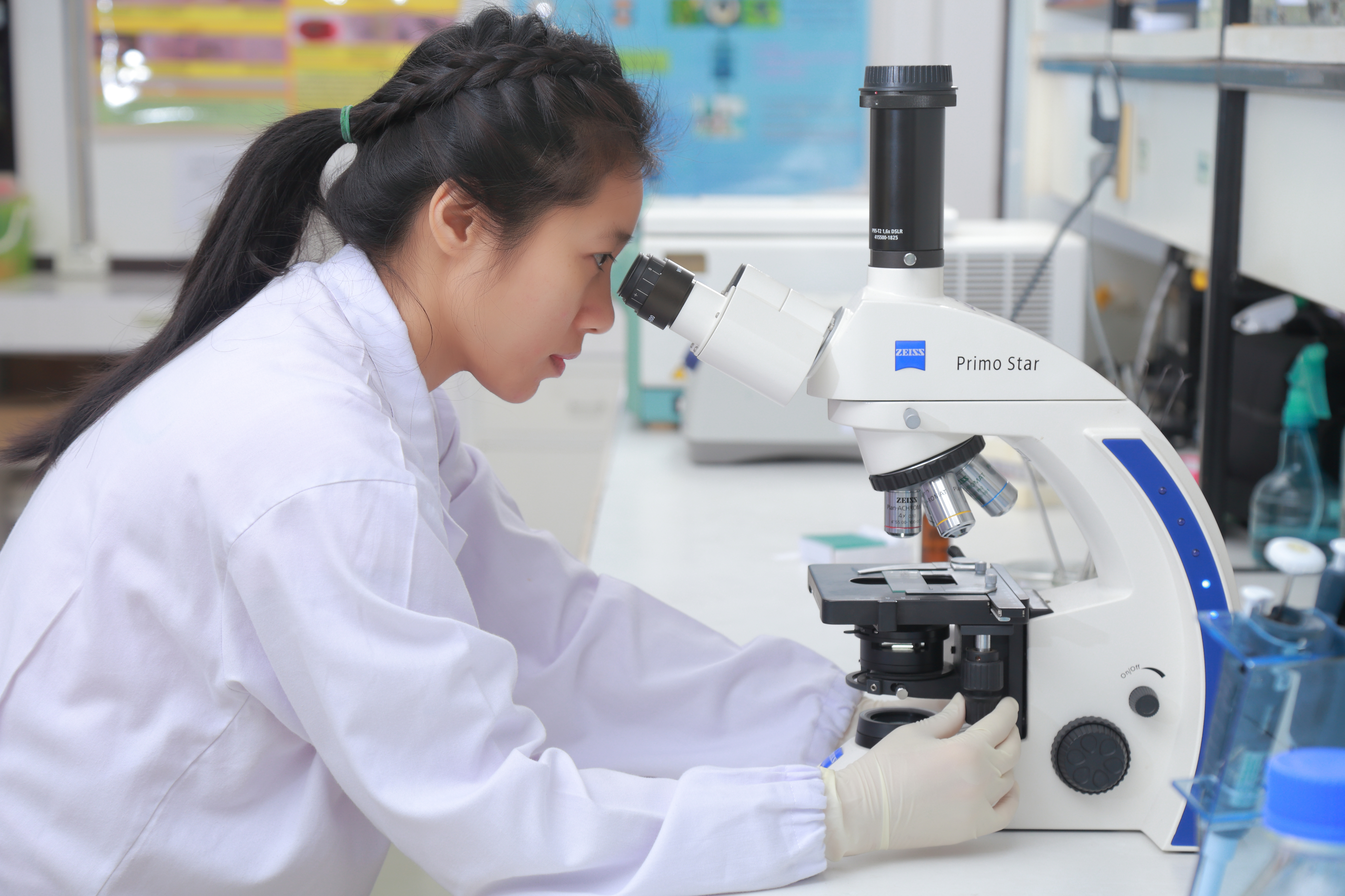
The optical microscope, also referred to as a light microscope, is a type of microscope that commonly uses visible light and a system of lenses to generate magnified images of small objects. Optical microscopes are the oldest design of microscope and were possibly invented in their present compound form in the 17th century. Basic optical microscopes can be very simple, although many complex designs aim to improve resolution and sample contrast. The object is placed on a stage and may be directly viewed through one or two eyepieces on the microscope. In high-power microscopes, both eyepieces typically show the same image, but with a stereo microscope, slightly different images are used to create a 3-D effect. A camera is typically used to capture the image (micrograph). The sample can be lit in a variety of ways. Transparent objects can be lit from below and solid objects can be lit with light coming through ( bright field) or around (dark field) the objective lens. Polarised ... [...More Info...] [...Related Items...] OR: [Wikipedia] [Google] [Baidu] |
Giemsa Stain
Giemsa stain (), named after German chemist and bacteriologist Gustav Giemsa, is a nucleic acid stain used in cytogenetics and for the histopathological diagnosis of malaria and other parasites. Uses It is specific for the phosphate groups of DNA and attaches itself to regions of DNA where there are high amounts of adenine-thymine bonding. Giemsa stain is used in Giemsa banding, commonly called G-banding, to stain chromosomes and often used to create a Karyotype, karyogram (chromosome map). It can identify chromosomal aberrations such as chromosomal translocation, translocations and chromosomal inversion, rearrangements. It stains the trophozoite ''Trichomonas vaginalis'', which presents with greenish discharge and motile cells on wet prep. Giemsa stain is also a Differential staining, differential stain, such as when it is combined with Wright's stain, Wright stain to form Wright-Giemsa stain. It can be used to study the adherence of pathogenic bacteria to human cells. It dif ... [...More Info...] [...Related Items...] OR: [Wikipedia] [Google] [Baidu] |
Trichrome Stain
Trichrome staining is a histological staining method that uses two or more acid dyes in conjunction with a polyacid. Staining differentiates tissues by tinting them in contrasting colours. It increases the contrast of microscopic features in cells and tissues, which makes them easier to see when viewed through a microscope. The word '' trichrome'' means "three colours". The first staining protocol that was described as "trichrome" was Mallory's trichrome stain, which differentially stained erythrocytes to a red colour, muscle tissue to a red colour, and collagen to a blue colour. Some other trichrome staining protocols are the Masson's trichrome stain, Lillie's trichrome, and the Gömöri trichrome stain. Purpose Without trichrome staining, discerning one feature from another can be extremely difficult. Smooth muscle tissue, for example, is hard to differentiate from collagen. A trichrome stain can colour the muscle tissue red, and the collagen fibres green or blue. Liver ... [...More Info...] [...Related Items...] OR: [Wikipedia] [Google] [Baidu] |
Gram Staining
In microbiology and bacteriology, Gram stain (Gram staining or Gram's method), is a method of staining used to classify bacterial species into two large groups: gram-positive bacteria and gram-negative bacteria. The name comes from the Danish bacteriologist Hans Christian Gram, who developed the technique in 1884. Gram staining differentiates bacteria by the chemical and physical properties of their cell walls. Gram-positive cells have a thick layer of peptidoglycan in the cell wall that retains the primary stain, crystal violet. Gram-negative cells have a thinner peptidoglycan layer that allows the crystal violet to wash out on addition of ethanol. They are stained pink or red by the counterstain, commonly safranin or fuchsine. Lugol's iodine solution is always added after addition of crystal violet to strengthen the bonds of the stain with the cell membrane. Gram staining is almost always the first step in the preliminary identification of a bacterial organism. While Gram stain ... [...More Info...] [...Related Items...] OR: [Wikipedia] [Google] [Baidu] |
PAS Stain
PAS or Pas may refer to: Companies and organizations * Pakistan Academy of Sciences * Pakistan Administrative Service * Pan Am Southern, a freight railroad owned by Norfolk Southern and Pan Am Railways * Pan American Silver, a mining company in Canada * Paradox Access Solutions, a construction company * Percussive Arts Society, percussion organization * Poetry Association of Scotland * Polish Academy of Sciences * Port Auxiliary Service, formerly the British Admiralty Yard Craft Service * Production Automotive Services, an American specialty vehicle manufacturer Political parties * Malaysian Islamic Party, Malaysia * Partido Alianza Social, Mexico * Party of Action and Solidarity, Moldova Places * The Pas (electoral district), in Manitoba, Canada * The Pas, town in Canada * Le Pas, commune in France * Sihanoukville Autonomous Port (Port Autonome de Sihanoukville), Cambodia Science * PAS diastase stain * PAS domain, a protein domain * Panic and Agoraphobia Scale, a psychologica ... [...More Info...] [...Related Items...] OR: [Wikipedia] [Google] [Baidu] |
Leydig Cell Tumour
Leydig cell tumour, also Leydig cell tumor (US spelling), (testicular) interstitial cell tumour and (testicular) interstitial cell tumor (US spelling), is a member of the sex cord-stromal tumour group of ovarian and testicular cancers. It arises from Leydig cells. While the tumour can occur at any age, it occurs most often in young adults. A Sertoli–Leydig cell tumour is a combination of a Leydig cell tumour and a Sertoli cell tumour from Sertoli cells. Presentation The majority of Leydig cell tumors are found in males, usually at 5–10 years of age or in middle adulthood (30–60 years). Children typically present with precocious puberty. Due to excess testosterone secreted by the tumour, one-third of female patients present with a recent history of progressive masculinization. Masculinization is preceded by anovulation, oligomenorrhea, amenorrhea and ''defeminization''. Additional signs include acne and hirsutism, voice deepening, clitoromegaly, temporal hair recession, a ... [...More Info...] [...Related Items...] OR: [Wikipedia] [Google] [Baidu] |


.jpg)

