|
Radionuclide Test
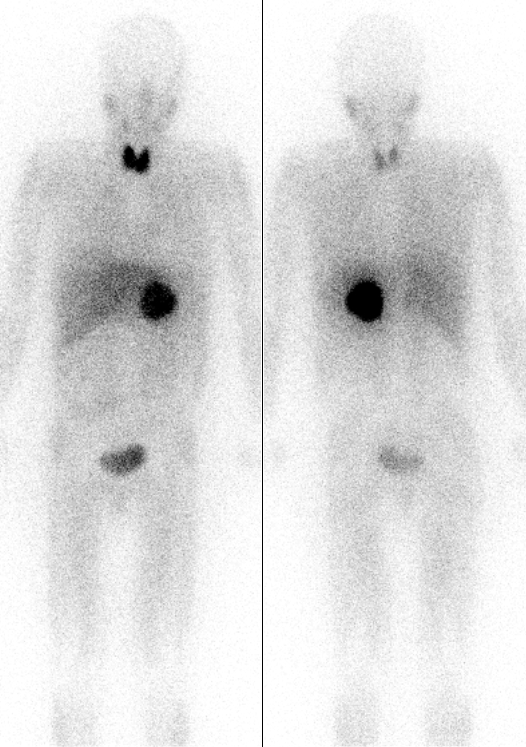
Nuclear medicine (nuclear radiology, nucleology), is a medical specialty involving the application of radioactive substances in the diagnosis and treatment of disease. Nuclear imaging is, in a sense, ''radiology done inside out'', because it records radiation emitted from within the body rather than radiation that is transmitted through the body from external sources like X-ray generators. In addition, nuclear medicine scans differ from radiology, as the emphasis is not on imaging anatomy, but on the function. For such reason, it is called a physiological imaging modality. Single photon emission computed tomography (SPECT) and positron emission tomography (PET) scans are the two most common imaging modalities in nuclear medicine. Diagnostic medical imaging Diagnostic In nuclear medicine imaging, radiopharmaceuticals are taken internally, for example, through inhalation, intravenously, or orally. Then, external detectors (gamma cameras) capture and form images from the radi ... [...More Info...] [...Related Items...] OR: [Wikipedia] [Google] [Baidu] |
PET Scan
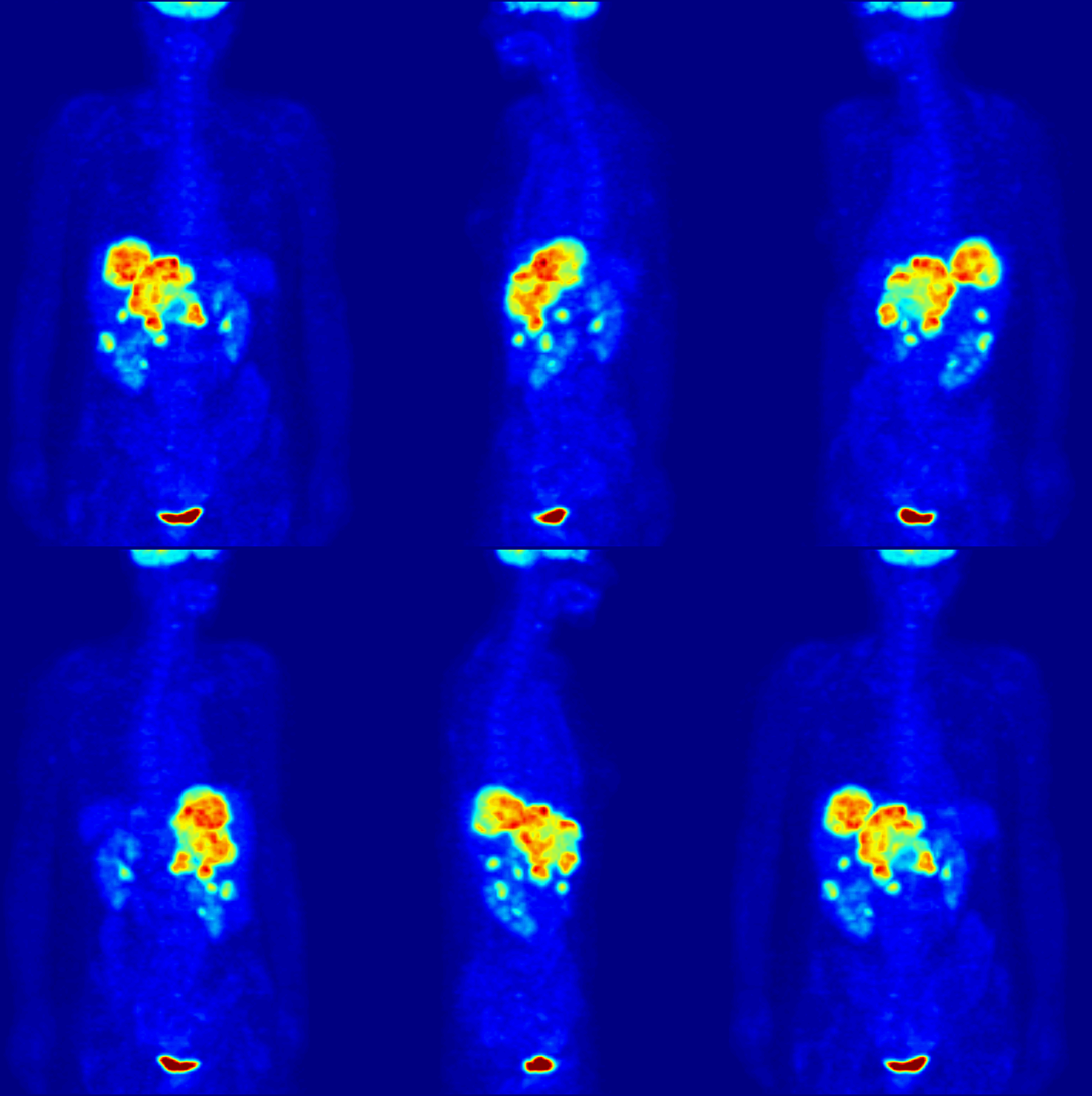
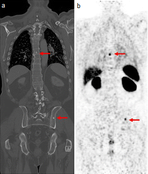
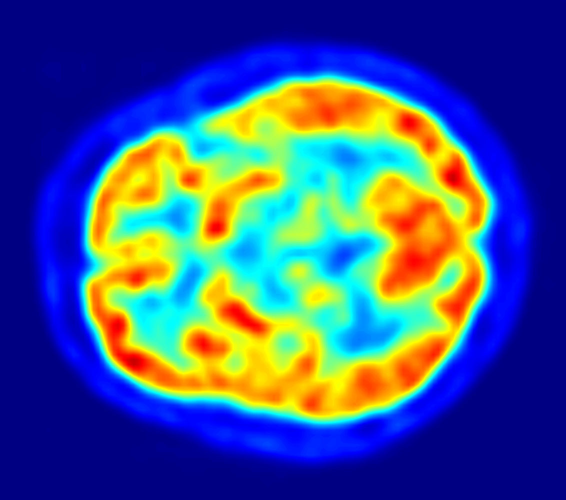
Positron emission tomography (PET) is a functional imaging technique that uses radioactive substances known as radiotracers to visualize and measure changes in Metabolism, metabolic processes, and in other physiological activities including blood flow, regional chemical composition, and absorption. Different tracers are used for various imaging purposes, depending on the target process within the body, such as: * Fluorodeoxyglucose (18F), Fluorodeoxyglucose ([18F]FDG or FDG) is commonly used to detect cancer; * Sodium fluoride#Medical imaging, [18F]Sodium fluoride (Na18F) is widely used for detecting bone formation; * Oxygen-15 (15O) is sometimes used to measure blood flow. PET is a common medical imaging, imaging technique, a Scintigraphy#Process, medical scintillography technique used in nuclear medicine. A radiopharmaceutical—a radioisotope attached to a drug—is injected into the body as a radioactive tracer, tracer. When the radiopharmaceutical undergoes beta plus decay ... [...More Info...] [...Related Items...] OR: [Wikipedia] [Google] [Baidu] |
Technetium (99mTc) Sestamibi
Technetium (99mTc) sestamibi ( INN; commonly sestamibi; USP: technetium Tc 99m sestamibi; trade name Cardiolite) is a pharmaceutical agent used in nuclear medicine imaging. The drug is a coordination complex consisting of the radioisotope technetium-99m bound to six (sesta=6) methoxyisobutylisonitrile (MIBI) ligands. The anion is not defined. The generic drug became available late September 2008. A scan of a patient using MIBI is commonly known as a "MIBI scan". Sestamibi is taken up by tissues with large numbers of mitochondria and negative plasma membrane potentials. Sestamibi is mainly used to image the myocardium (heart muscle). It is also used in the work-up of primary hyperparathyroidism to identify parathyroid adenomas, for radioguided surgery of the parathyroid and in the work-up of possible breast cancer. Cardiac imaging (MIBI scan) A ''MIBI scan'' or ''sestamibi scan'' is now a common method of cardiac imaging. Technetium (99mTc) sestamibi is a lipophilic cation ... [...More Info...] [...Related Items...] OR: [Wikipedia] [Google] [Baidu] |
MIBG
Iobenguane, or MIBG, is an aralkylguanidine analog of the adrenergic neurotransmitter norepinephrine (noradrenaline), typically used as a radiopharmaceutical. It acts as a blocking agent for adrenergic neurons. When radiolabeled, it can be used in nuclear medicinal diagnostic and therapy techniques as well as in neuroendocrine chemotherapy treatments. It localizes to adrenergic tissue and thus can be used to identify the location of tumors such as pheochromocytomas and neuroblastomas. With iodine-131 it can also be used to treat tumor cells that take up and metabolize norepinephrine. Usage and mechanism MIBG is absorbed by and accumulated in granules of adrenal medullary chromaffin cells, as well as in pre-synaptic adrenergic neuron granules. The process in which this occurs is closely related to the mechanism employed by norepinephrine and its transporter in vivo. The norepinephrine transporter (NET) functions to provide norepinephrine uptake at the synaptic termina ... [...More Info...] [...Related Items...] OR: [Wikipedia] [Google] [Baidu] |
Indium White Blood Cell Scan
The indium white blood cell scan is a nuclear medicine procedure in which white blood cells (mostly neutrophils) are removed from the patient, tagged with the radioisotope Indium-111, and then injected intravenously into the patient. The tagged leukocytes subsequently localize to areas of relatively new infection. The study is particularly helpful in differentiating conditions such as osteomyelitis from decubitus ulcers for assessment of route and duration of antibiotic therapy. In imaging of infections, the gallium scan has a sensitivity advantage over the indium white blood cell scan in imaging osteomyelitis (bone infection) of the spine, lung infections and inflammation, and in detecting chronic infections. In part, this is because gallium binds to neutrophil membranes, even after neutrophil death, whereas localization of neutrophils labeled with indium requires them to be in relatively good functional order. However, indium leukocyte imaging is better at localizing acute (i.e ... [...More Info...] [...Related Items...] OR: [Wikipedia] [Google] [Baidu] |
Gallium Scan
A gallium scan is a type of nuclear medicine diagnostic investigation that uses either a gallium-67 (67Ga) or gallium-68 (68Ga) radiopharmaceutical to obtain images of a specific type of tissue, or disease state of tissue. The gamma emission of gallium-67 is imaged by a gamma camera, while the positron emission of gallium-68 is imaged by positron emission tomography (PET). Gallium salts like gallium citrate and gallium nitrate may be used. The form of salt is not important, since it is the freely dissolved gallium ion Ga3+ which is active. Both 67Ga and 68Ga salts have similar uptake mechanisms. Radioactive gallium(III) is rapidly bound by transferrin, which then preferentially accumulates in tumors, inflammation, and both acute and chronic infection, allowing these pathological processes to be imaged. Gallium is particularly useful in imaging osteomyelitis that involves the spine, and in imaging older and chronic infections that may be the cause of a fever of unknown origin. ... [...More Info...] [...Related Items...] OR: [Wikipedia] [Google] [Baidu] |
CT Scan
A computed tomography scan (CT scan), formerly called computed axial tomography scan (CAT scan), is a medical imaging technique used to obtain detailed internal images of the body. The personnel that perform CT scans are called radiographers or radiology technologists. CT scanners use a rotating X-ray tube and a row of detectors placed in a gantry (medical), gantry to measure X-ray Attenuation#Radiography, attenuations by different tissues inside the body. The multiple X-ray measurements taken from different angles are then processed on a computer using tomographic reconstruction algorithms to produce Tomography, tomographic (cross-sectional) images (virtual "slices") of a body. CT scans can be used in patients with metallic implants or pacemakers, for whom magnetic resonance imaging (MRI) is Contraindication, contraindicated. Since its development in the 1970s, CT scanning has proven to be a versatile imaging technique. While CT is most prominently used in medical diagnosis, i ... [...More Info...] [...Related Items...] OR: [Wikipedia] [Google] [Baidu] |
Hemangioma
A hemangioma or haemangioma is a usually benign vascular tumor derived from blood vessel cell types. The most common form, seen in infants, is an infantile hemangioma, known colloquially as a "strawberry mark", most commonly presenting on the skin at birth or in the first weeks of life. A hemangioma can occur anywhere on the body, but most commonly appears on the face, scalp, chest or back. They tend to grow for up to a year before gradually shrinking as the child gets older. A hemangioma may need to be treated if it interferes with vision or breathing or is likely to cause long-term disfigurement. In rare cases internal hemangiomas can cause or contribute to other medical problems. They usually disappear by 10 years of age. The first line treatment option is beta blockers, which are highly effective in the majority of cases. Hemangiomas present at birth are called ''congenital hemangiomas'', while those that form later in life are called ''infantile hemangiomas''. Types Heman ... [...More Info...] [...Related Items...] OR: [Wikipedia] [Google] [Baidu] |
Positron Emission Tomography
Positron emission tomography (PET) is a functional imaging technique that uses radioactive substances known as radiotracers to visualize and measure changes in metabolic processes, and in other physiological activities including blood flow, regional chemical composition, and absorption. Different tracers are used for various imaging purposes, depending on the target process within the body, such as: * Fluorodeoxyglucose ( 18F">sup>18FDG or FDG) is commonly used to detect cancer; * 18Fodium fluoride">sup>18Fodium fluoride (Na18F) is widely used for detecting bone formation; * Oxygen-15 (15O) is sometimes used to measure blood flow. PET is a common imaging technique, a medical scintillography technique used in nuclear medicine. A radiopharmaceutical—a radioisotope attached to a drug—is injected into the body as a tracer. When the radiopharmaceutical undergoes beta plus decay, a positron is emitted, and when the positron interacts with an ordinary electron, the tw ... [...More Info...] [...Related Items...] OR: [Wikipedia] [Google] [Baidu] |
Single Photon Emission Computed Tomography
Single-photon emission computed tomography (SPECT, or less commonly, SPET) is a nuclear medicine tomographic imaging technique using gamma rays. It is very similar to conventional nuclear medicine planar imaging using a gamma camera (that is, scintigraphy), but is able to provide true 3D information. This information is typically presented as cross-sectional slices through the patient, but can be freely reformatted or manipulated as required. The technique needs delivery of a gamma-emitting radioisotope (a radionuclide) into the patient, normally through injection into the bloodstream. On occasion, the radioisotope is a simple soluble dissolved ion, such as an isotope of gallium(III). Usually, however, a marker radioisotope is attached to a specific ligand to create a radioligand, whose properties bind it to certain types of tissues. This marriage allows the combination of ligand and radiopharmaceutical to be carried and bound to a place of interest in the body, where the ... [...More Info...] [...Related Items...] OR: [Wikipedia] [Google] [Baidu] |
Pulmonary Embolism
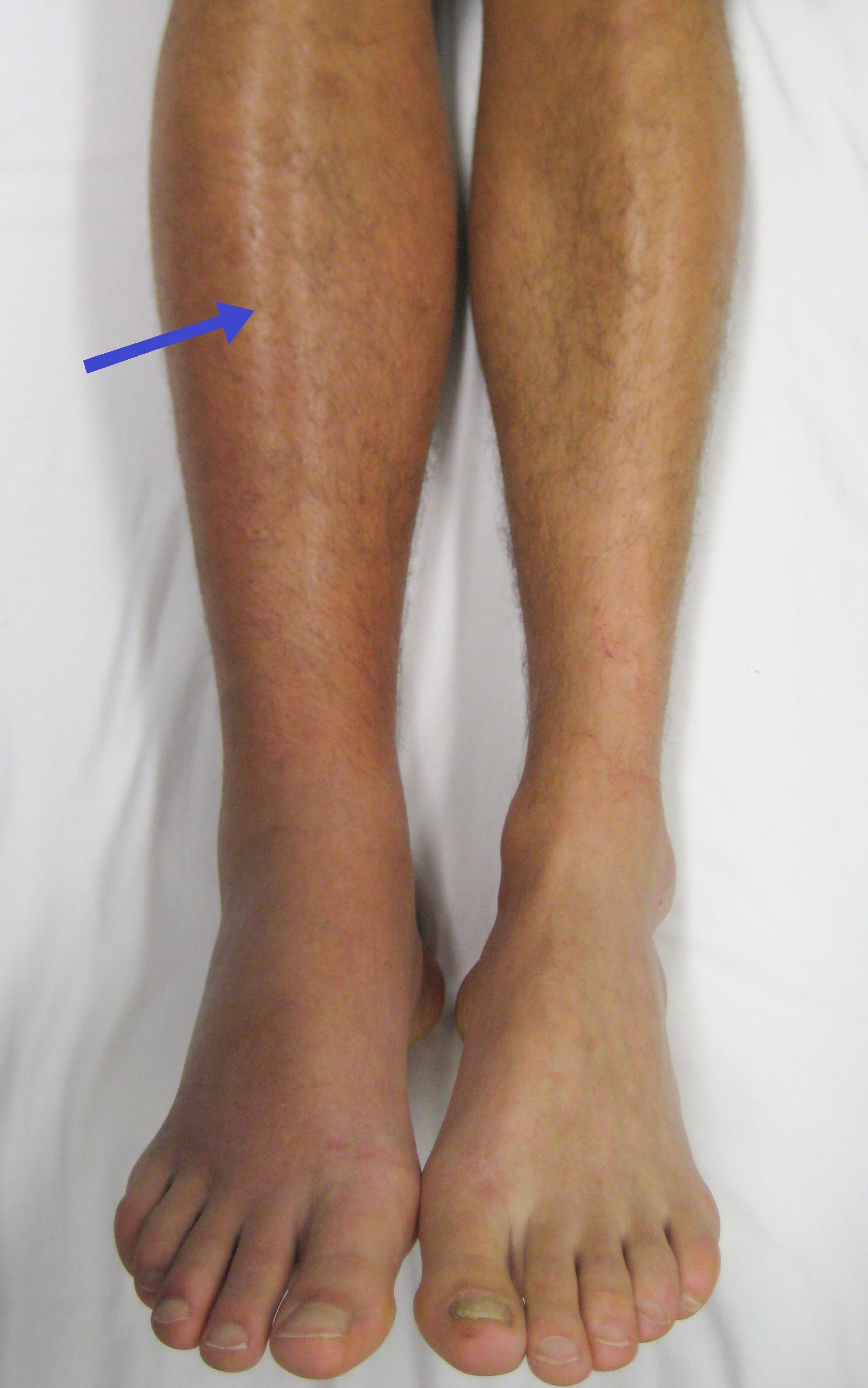
Pulmonary embolism (PE) is a blockage of an pulmonary artery, artery in the lungs by a substance that has moved from elsewhere in the body through the bloodstream (embolism). Symptoms of a PE may include dyspnea, shortness of breath, chest pain particularly upon breathing in, and coughing up blood. Symptoms of a deep vein thrombosis, blood clot in the leg may also be present, such as a erythema, red, warm, swollen, and painful leg. Signs of a PE include low blood oxygen saturation, oxygen levels, tachypnea, rapid breathing, tachycardia, rapid heart rate, and sometimes a mild fever. Severe cases can lead to Syncope (medicine), passing out, shock (circulatory), abnormally low blood pressure, obstructive shock, and cardiac arrest, sudden death. PE usually results from a blood clot in the leg that travels to the lung. The risk of blood clots is increased by advanced age, cancer, prolonged bed rest and immobilization, smoking, stroke, long-haul travel over 4 hours, certain genetics, ... [...More Info...] [...Related Items...] OR: [Wikipedia] [Google] [Baidu] |
Cholescintigraphy
Cholescintigraphy or hepatobiliary scintigraphy is scintigraphy of the hepatobiliary tract, including the gallbladder and bile ducts. The image produced by this type of medical imaging, called a cholescintigram, is also known by other names depending on which radiotracer is used, such as HIDA scan, PIPIDA scan, DISIDA scan, or BrIDA scan. Cholescintigraphic scanning is a nuclear medicine procedure to evaluate the health and function of the gallbladder and biliary system. A radioactive tracer is injected through any accessible vein and then allowed to circulate to the liver, where it is excreted into the bile ducts and stored by the gallbladder until released into the duodenum. Use of cholescintigraphic scans as a first-line form of imaging varies depending on indication. For example for cholecystitis, cheaper and less invasive ultrasound imaging may be preferred, while for bile reflux cholescintigraphy may be the first choice. Etymology and pronunciation The word ''cholescin ... [...More Info...] [...Related Items...] OR: [Wikipedia] [Google] [Baidu] |

