|
Pulmonary Venous System
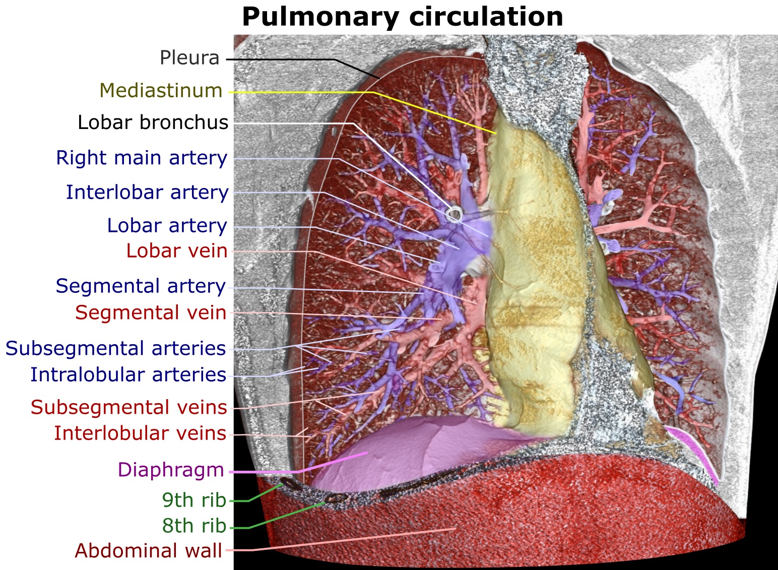
The pulmonary circulation is a division of the circulatory system in all vertebrates. The circuit begins with deoxygenated blood returned from the body to the right atrium of the heart where it is pumped out from the right ventricle to the lungs. In the lungs the blood is oxygenated and returned to the left atrium to complete the circuit. The other division of the circulatory system is the systemic circulation that begins with receiving the oxygenated blood from the pulmonary circulation into the left atrium. From the atrium the oxygenated blood enters the left ventricle where it is pumped out to the rest of the body, returning as deoxygenated blood back to the pulmonary circulation. The blood vessels of the pulmonary circulation are the pulmonary arteries and the pulmonary veins. A separate circulatory circuit known as the bronchial circulation supplies oxygenated blood to the tissue of the larger airways of the lung. Structure De-oxygenated blood leaves the heart, goe ... [...More Info...] [...Related Items...] OR: [Wikipedia] [Google] [Baidu] |
Circulatory System
The blood circulatory system is a system of organs that includes the heart, blood vessels, and blood which is circulated throughout the entire body of a human or other vertebrate. It includes the cardiovascular system, or vascular system, that consists of the heart and blood vessels (from Greek ''kardia'' meaning ''heart'', and from Latin ''vascula'' meaning ''vessels''). The circulatory system has two divisions, a systemic circulation or circuit, and a pulmonary circulation or circuit. Some sources use the terms ''cardiovascular system'' and ''vascular system'' interchangeably with the ''circulatory system''. The network of blood vessels are the great vessels of the heart including large elastic arteries, and large veins; other arteries, smaller arterioles, capillaries that join with venules (small veins), and other veins. The Closed circulatory system, circulatory system is closed in vertebrates, which means that the blood never leaves the network of blood vessels. Some in ... [...More Info...] [...Related Items...] OR: [Wikipedia] [Google] [Baidu] |
Right Ventricle
A ventricle is one of two large chambers toward the bottom of the heart that collect and expel blood towards the peripheral beds within the body and lungs. The blood pumped by a ventricle is supplied by an atrium, an adjacent chamber in the upper heart that is smaller than a ventricle. Interventricular means between the ventricles (for example the interventricular septum), while intraventricular means within one ventricle (for example an intraventricular block). In a four-chambered heart, such as that in humans, there are two ventricles that operate in a double circulatory system: the right ventricle pumps blood into the pulmonary circulation to the lungs, and the left ventricle pumps blood into the systemic circulation through the aorta. Structure Ventricles have thicker walls than atria and generate higher blood pressures. The physiological load on the ventricles requiring pumping of blood throughout the body and lungs is much greater than the pressure generated by the atria t ... [...More Info...] [...Related Items...] OR: [Wikipedia] [Google] [Baidu] |
Pulmonary Embolism
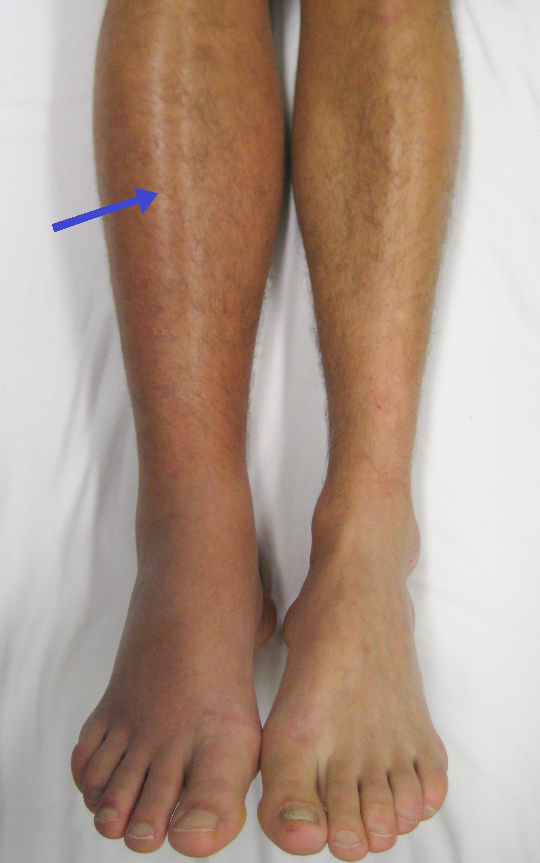
Pulmonary embolism (PE) is a blockage of an pulmonary artery, artery in the lungs by a substance that has moved from elsewhere in the body through the bloodstream (embolism). Symptoms of a PE may include dyspnea, shortness of breath, chest pain particularly upon breathing in, and coughing up blood. Symptoms of a deep vein thrombosis, blood clot in the leg may also be present, such as a erythema, red, warm, swollen, and painful leg. Signs of a PE include low blood oxygen saturation, oxygen levels, tachypnea, rapid breathing, tachycardia, rapid heart rate, and sometimes a mild fever. Severe cases can lead to Syncope (medicine), passing out, shock (circulatory), abnormally low blood pressure, obstructive shock, and cardiac arrest, sudden death. PE usually results from a blood clot in the leg that travels to the lung. The risk of blood clots is increased by advanced age, cancer, prolonged bed rest and immobilization, smoking, stroke, long-haul travel over 4 hours, certain genetics, g ... [...More Info...] [...Related Items...] OR: [Wikipedia] [Google] [Baidu] |
Fossa Ovalis (heart)
The ''fossa ovalis'' is a depression in the right atrium of the heart, at the level of the interatrial septum, the wall between right and left atrium. The ''fossa ovalis'' is the remnant of a thin fibrous sheet that covered the '' foramen ovale'' during fetal development. Function During fetal development, the foramen ovale allows blood to pass from the right atrium to the left atrium, bypassing the nonfunctional fetal lungs while the fetus obtains its oxygen from the placenta. A flap of tissue called the septum primum acts as a valve over the foramen ovale during that time. After birth, the introduction of air into the lungs causes the pressure in the pulmonary circulatory system to drop. This change in pressure pushes the septum primum against the atrial septum, closing the foramen. The septum primum and atrial septum eventually fuse together to form a complete seal, leaving a depression called the fossa ovalis. By age two, about 75% of people have a completely sealed fossa ova ... [...More Info...] [...Related Items...] OR: [Wikipedia] [Google] [Baidu] |
Ductus Arteriosus
The ''ductus arteriosus'', also called the ''ductus Botalli'', named after the Italian physiologist Leonardo Botallo, is a blood vessel in the developing fetus connecting the trunk of the pulmonary artery to the proximal descending aorta. It allows most of the blood from the right ventricle to bypass the fetus's fluid-filled non-functioning lungs. Upon closure at birth, it becomes the ''ligamentum arteriosum''. Development and structure The ''ductus arteriosus'' is formed from the left 6th aortic arch during embryonic development and attaches to the final part of the aortic arch (the isthmus of aorta) and the first part of the pulmonary artery. Disorder: Patent ductus arteriosus Consequences Failure of the ''ductus arteriosus'' to close after birth results in a condition called ''patent ductus arteriosus'', which results in the abnormal flow of blood from the aorta to the pulmonary artery: a left-to-right shunt. If left uncorrected, this usually leads to pulmonary hypertensi ... [...More Info...] [...Related Items...] OR: [Wikipedia] [Google] [Baidu] |
Foramen Ovale (heart)
In the fetal heart, the foramen ovale (), also foramen Botalli, or the ostium secundum of Born, allows blood to enter the left atrium from the right atrium. It is one of two fetal cardiac shunts, the other being the ductus arteriosus (which allows blood that still escapes to the right ventricle to bypass the pulmonary circulation). Another similar adaptation in the fetus is the ductus venosus. In most individuals, the foramen ovale closes at birth. It later forms the fossa ovalis. Development The foramen ovale () forms in the late fourth week of gestation, as a small passageway between the septum secundum and the ostium secundum. Initially the atria are separated from one another by the septum primum except for a small opening below the septum, the ostium primum. As the septum primum grows, the ostium primum narrows and eventually closes. Before it does so, bloodflow from the inferior vena cava wears down a portion of the septum primum, forming the ostium secundum. Some embryo ... [...More Info...] [...Related Items...] OR: [Wikipedia] [Google] [Baidu] |
Fetal Circulation
In humans, the circulatory system is different before and after birth. The fetal circulation is composed of the placenta, umbilical blood vessels encapsulated by the umbilical cord, heart and systemic blood vessels. A major difference between the fetal circulation and postnatal circulation is that the lungs are not used during the fetal stage resulting in the presence of shunts to move oxygenated blood and nutrients from the placenta to the fetal tissue. At birth, the start of breathing and the severance of the umbilical cord prompt various changes that quickly transforms fetal circulation into postnatal circulation. Oxygenation, nutrient, and waste exchange Placenta The placenta functions as the exchange site of nutrients and wastes between the maternal and fetal circulation. Water, glucose, amino acids, vitamins, and inorganic salts freely diffuse across the placenta along with oxygen. Two umbilical arteries carry deoxygenated blood and waste from the fetus to the placenta ... [...More Info...] [...Related Items...] OR: [Wikipedia] [Google] [Baidu] |
Aortic Valve
The aortic valve is a valve in the heart of humans and most other animals, located between the left ventricle and the aorta. It is one of the four valves of the heart and one of the two semilunar valves, the other being the pulmonary valve. The aortic valve normally has three cusps or leaflets, although in 1–2% of the population it is found to congenitally have two leaflets. The aortic valve is the last structure in the heart the blood travels through before stopping the flow through the systemic circulation. Structure The aortic valve normally has three cusps however there is some discrepancy in their naming. They may be called the left coronary, right coronary and non-coronary cusp. Some sources also advocate they be named as a left, right and posterior cusp. Anatomists have traditionally named them the left posterior (origin of left coronary), anterior (origin of the right coronary) and right posterior. The three cusps, when the valve is closed, contain a sinus called an a ... [...More Info...] [...Related Items...] OR: [Wikipedia] [Google] [Baidu] |
Left Ventricle
A ventricle is one of two large chambers toward the bottom of the heart that collect and expel blood towards the peripheral beds within the body and lungs. The blood pumped by a ventricle is supplied by an atrium, an adjacent chamber in the upper heart that is smaller than a ventricle. Interventricular means between the ventricles (for example the interventricular septum), while intraventricular means within one ventricle (for example an intraventricular block). In a four-chambered heart, such as that in humans, there are two ventricles that operate in a double circulatory system: the right ventricle pumps blood into the pulmonary circulation to the lungs, and the left ventricle pumps blood into the systemic circulation through the aorta. Structure Ventricles have thicker walls than atria and generate higher blood pressures. The physiological load on the ventricles requiring pumping of blood throughout the body and lungs is much greater than the pressure generated by the atria ... [...More Info...] [...Related Items...] OR: [Wikipedia] [Google] [Baidu] |
Mitral Valve
The mitral valve (), also known as the bicuspid valve or left atrioventricular valve, is one of the four heart valves. It has two cusps or flaps and lies between the left atrium and the left ventricle of the heart. The heart valves are all one-way valves allowing blood flow in just one direction. The mitral valve and the tricuspid valve are known as the atrioventricular valves because they lie between the atria and the ventricles. In normal conditions, blood flows through an open mitral valve during diastole with contraction of the left atrium, and the mitral valve closes during systole with contraction of the left ventricle. The valve opens and closes because of pressure differences, opening when there is greater pressure in the left atrium than ventricle and closing when there is greater pressure in the left ventricle than atrium. In abnormal conditions, blood may flow backward through the valve ( mitral regurgitation) or the mitral valve may be narrowed (mitral stenosis). Rh ... [...More Info...] [...Related Items...] OR: [Wikipedia] [Google] [Baidu] |
Capillaries
A capillary is a small blood vessel from 5 to 10 micrometres (μm) in diameter. Capillaries are composed of only the tunica intima, consisting of a thin wall of simple squamous endothelial cells. They are the smallest blood vessels in the body: they convey blood between the arterioles and venules. These microvessels are the site of exchange of many substances with the interstitial fluid surrounding them. Substances which cross capillaries include water, oxygen, carbon dioxide, urea, glucose, uric acid, lactic acid and creatinine. Lymph capillaries connect with larger lymph vessels to drain lymphatic fluid collected in the microcirculation. During early embryonic development, new capillaries are formed through vasculogenesis, the process of blood vessel formation that occurs through a '' de novo'' production of endothelial cells that then form vascular tubes. The term ''angiogenesis'' denotes the formation of new capillaries from pre-existing blood vessels and already present endo ... [...More Info...] [...Related Items...] OR: [Wikipedia] [Google] [Baidu] |


