|
Psilolechiaceae
''Psilolechia'' is a genus of four species of crustose lichens. It is the only member of Psilolechiaceae, a family that was created in 2014 to contain this genus. Taxonomy The genus ''Psilolechia'' was established by Abramo Bartolommeo Massalongo in 1860. Formerly classified in the family Pilocarpaceae, molecular phylogenetic analysis showed that ''Psilolechia'' represented a distinct lineage that deserved placement at the familial level, the Psilolechiaceae, which was formally circumscribed in 2014. This arrangement was accepted in later large-scale updates of fungal classification. Psilolechiaceae is in the order Lecanorales, in the suborder Sphaerophorineae, which also includes the families Pilocarpaceae, Psoraceae, and Ramalinaceae. Description Psilolechiaceae is a monogeneric family of crustose lichens with effuse, ecorticate (lacking a cortex), leprose thalli formed by goniocysts (aggregations of photobiont cells surrounded by short-celled hyphae) containing ''Trebouxia'' ... [...More Info...] [...Related Items...] OR: [Wikipedia] [Google] [Baidu] |
Psilolechia Clavulifera
''Psilolechia'' is a genus of four species of crustose lichens. It is the only member of Psilolechiaceae, a family that was created in 2014 to contain this genus. Taxonomy The genus ''Psilolechia'' was established by Abramo Bartolommeo Massalongo in 1860. Formerly classified in the family Pilocarpaceae, molecular phylogenetic analysis showed that ''Psilolechia'' represented a distinct lineage that deserved placement at the familial level, the Psilolechiaceae, which was formally circumscribed in 2014. This arrangement was accepted in later large-scale updates of fungal classification. Psilolechiaceae is in the order Lecanorales, in the suborder Sphaerophorineae, which also includes the families Pilocarpaceae, Psoraceae, and Ramalinaceae. Description Psilolechiaceae is a monogeneric family of crustose lichens with effuse, ecorticate (lacking a cortex), leprose thalli formed by goniocysts (aggregations of photobiont cells surrounded by short-celled hyphae) containing ''Trebouxia'' ... [...More Info...] [...Related Items...] OR: [Wikipedia] [Google] [Baidu] |
Psilolechia Leprosa
''Psilolechia'' is a genus of four species of crustose lichens. It is the only member of Psilolechiaceae, a family that was created in 2014 to contain this genus. Taxonomy The genus ''Psilolechia'' was established by Abramo Bartolommeo Massalongo in 1860. Formerly classified in the family Pilocarpaceae, molecular phylogenetic analysis showed that ''Psilolechia'' represented a distinct lineage that deserved placement at the familial level, the Psilolechiaceae, which was formally circumscribed in 2014. This arrangement was accepted in later large-scale updates of fungal classification. Psilolechiaceae is in the order Lecanorales, in the suborder Sphaerophorineae, which also includes the families Pilocarpaceae, Psoraceae, and Ramalinaceae. Description Psilolechiaceae is a monogeneric family of crustose lichens with effuse, ecorticate (lacking a cortex), leprose thalli formed by goniocysts (aggregations of photobiont cells surrounded by short-celled hyphae) containing ''Trebouxia'' ... [...More Info...] [...Related Items...] OR: [Wikipedia] [Google] [Baidu] |
Psilolechia Purpurascens
''Psilolechia'' is a genus of four species of crustose lichens. It is the only member of Psilolechiaceae, a family that was created in 2014 to contain this genus. Taxonomy The genus ''Psilolechia'' was established by Abramo Bartolommeo Massalongo in 1860. Formerly classified in the family Pilocarpaceae, molecular phylogenetic analysis showed that ''Psilolechia'' represented a distinct lineage that deserved placement at the familial level, the Psilolechiaceae, which was formally circumscribed in 2014. This arrangement was accepted in later large-scale updates of fungal classification. Psilolechiaceae is in the order Lecanorales, in the suborder Sphaerophorineae, which also includes the families Pilocarpaceae, Psoraceae, and Ramalinaceae. Description Psilolechiaceae is a monogeneric family of crustose lichens with effuse, ecorticate (lacking a cortex), leprose thalli formed by goniocysts (aggregations of photobiont cells surrounded by short-celled hyphae) containing ''Trebouxia'' ... [...More Info...] [...Related Items...] OR: [Wikipedia] [Google] [Baidu] |
Lecanorales
The Lecanorales are an order of mostly lichen-forming fungi belonging to the class Lecanoromycetes in the division Ascomycota. The order contains 26 families, 269 genera, and 5695 species. Families * Aphanopsidaceae * Biatorellaceae * Brigantiaeaceae * Bruceomycetaceae * Carbonicolaceae * Catillariaceae * Cladoniaceae * Crocyniaceae * Dactylosporaceae * Gypsoplacaceae * Haematommataceae * Lecanoraceae * Malmideaceae * Pachyascaceae * Parmeliaceae * Pilocarpaceae * Psilolechiaceae * Psoraceae * Ramalinaceae * Ramboldiaceae * Scoliciosporaceae * Sphaerophoraceae * Stereocaulaceae * Tephromelataceae * Vezdaeaceae Genera of uncertain placement There are several genera in the Lecanorales that have not been placed with certainty into any family. These are: *'' Coronoplectrum'' – 1 sp. *'' Ivanpisutia'' – 1 sp. *'' Joergensenia'' – 1 sp. *'' Myochroidea'' – 4 spp. *'' Neopsoromopsis'' – 1 sp. *''Psoromella ''Psoromella'' is a genus of lichenized fungi ... [...More Info...] [...Related Items...] OR: [Wikipedia] [Google] [Baidu] |
Psilolechia Lucida
''Psilolechia lucida'' is a species of saxicolous lichen in the family Psilolechiaceae. It is widely distributed through the world, where it grows on natural and artificial rocky substrates in the shade, often in sheltered underhangs. It forms a greenish crust on the surface of its substrate. Taxonomy It was originally described by lichenologist Erik Acharius in 1799. Maurice Choisy placed it in the genus ''Psilolechia'' in 1949. There are known to be two chemical races of ''P. lucida''. The first, which is known all over the world, contains rhizocarpic acid as a major secondary substance as well as some unknown substances. The second, reported only from Australia and New Zealand, has both rhizocarpic acid and zeorin. Description ''Psilolechia lucida'' forms a sulphur-yellow to yellowish green crust, although the colour is greener when the surface is wet. The crust comprises powdery soredia that can be thin or thick, and sometimes divided into irregular areoles. The ap ... [...More Info...] [...Related Items...] OR: [Wikipedia] [Google] [Baidu] |
Siliceous
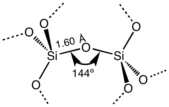
Silicon dioxide, also known as silica, is an oxide of silicon with the chemical formula , most commonly found in nature as quartz and in various living organisms. In many parts of the world, silica is the major constituent of sand. Silica is one of the most complex and most abundant families of materials, existing as a compound of several minerals and as a synthetic product. Notable examples include fused quartz, fumed silica, silica gel, opal and aerogels. It is used in structural materials, microelectronics (as an electrical insulator), and as components in the food and pharmaceutical industries. Structure In the majority of silicates, the silicon atom shows tetrahedral coordination, with four oxygen atoms surrounding a central Si atomsee 3-D Unit Cell. Thus, SiO2 forms 3-dimensional network solids in which each silicon atom is covalently bonded in a tetrahedral manner to 4 oxygen atoms. In contrast, CO2 is a linear molecule. The starkly different structures of the dioxide ... [...More Info...] [...Related Items...] OR: [Wikipedia] [Google] [Baidu] |
Tasmania
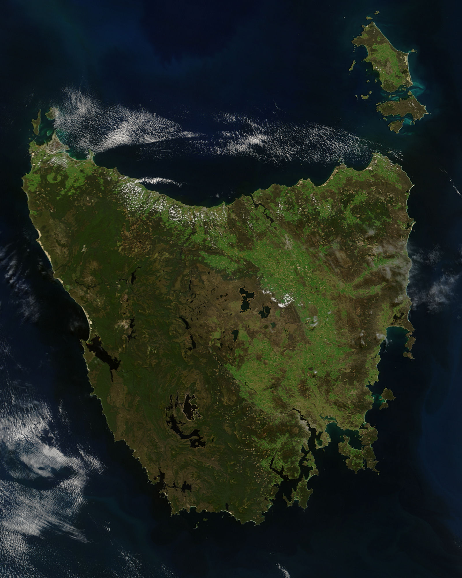
) , nickname = , image_map = Tasmania in Australia.svg , map_caption = Location of Tasmania in AustraliaCoordinates: , subdivision_type = Country , subdivision_name = Australia , established_title = Before federation , established_date = Colony of Tasmania , established_title2 = Federation , established_date2 = 1 January 1901 , named_for = Abel Tasman , demonym = , capital = Hobart , largest_city = capital , coordinates = , admin_center = 29 local government areas , admin_center_type = Administration , leader_title1 = Monarch , leader_name1 = Charles III , leader_title2 = Governor , leader_name2 ... [...More Info...] [...Related Items...] OR: [Wikipedia] [Google] [Baidu] |
Hyaline
A hyaline substance is one with a glassy appearance. The word is derived from el, ὑάλινος, translit=hyálinos, lit=transparent, and el, ὕαλος, translit=hýalos, lit=crystal, glass, label=none. Histopathology Hyaline cartilage is named after its glassy appearance on fresh gross pathology. On light microscopy of H&E stained slides, the extracellular matrix of hyaline cartilage looks homogeneously pink, and the term "hyaline" is used to describe similarly homogeneously pink material besides the cartilage. Hyaline material is usually acellular and proteinaceous. For example, arterial hyaline is seen in aging, high blood pressure, diabetes mellitus and in association with some drugs (e.g. calcineurin inhibitors). It is bright pink with PAS staining. Ichthyology and entomology In ichthyology and entomology, ''hyaline'' denotes a colorless, transparent substance, such as unpigmented fins of fishes or clear insect wings. Resh, Vincent H. and R. T. Cardé, Eds. Encyclo ... [...More Info...] [...Related Items...] OR: [Wikipedia] [Google] [Baidu] |
Hypha
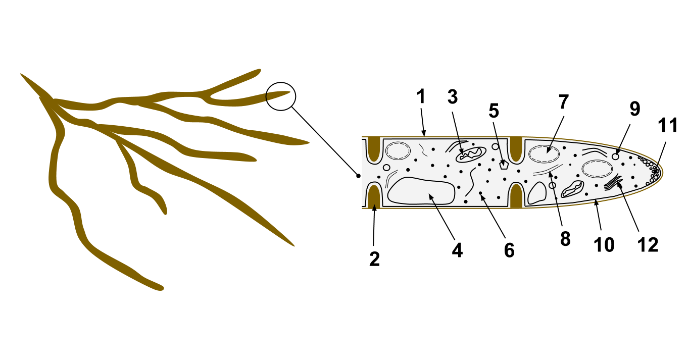
A hypha (; ) is a long, branching, filamentous structure of a fungus, oomycete, or actinobacterium. In most fungi, hyphae are the main mode of vegetative growth, and are collectively called a mycelium. Structure A hypha consists of one or more cells surrounded by a tubular cell wall. In most fungi, hyphae are divided into cells by internal cross-walls called "septa" (singular septum). Septa are usually perforated by pores large enough for ribosomes, mitochondria, and sometimes nuclei to flow between cells. The major structural polymer in fungal cell walls is typically chitin, in contrast to plants and oomycetes that have cellulosic cell walls. Some fungi have aseptate hyphae, meaning their hyphae are not partitioned by septa. Hyphae have an average diameter of 4–6 µm. Growth Hyphae grow at their tips. During tip growth, cell walls are extended by the external assembly and polymerization of cell wall components, and the internal production of new cell membrane. The S ... [...More Info...] [...Related Items...] OR: [Wikipedia] [Google] [Baidu] |
Staining
Staining is a technique used to enhance contrast in samples, generally at the microscopic level. Stains and dyes are frequently used in histology (microscopic study of biological tissues), in cytology (microscopic study of cells), and in the medical fields of histopathology, hematology, and cytopathology that focus on the study and diagnoses of diseases at the microscopic level. Stains may be used to define biological tissues (highlighting, for example, muscle fibers or connective tissue), cell populations (classifying different blood cells), or organelles within individual cells. In biochemistry, it involves adding a class-specific ( DNA, proteins, lipids, carbohydrates) dye to a substrate to qualify or quantify the presence of a specific compound. Staining and fluorescent tagging can serve similar purposes. Biological staining is also used to mark cells in flow cytometry, and to flag proteins or nucleic acids in gel electrophoresis. Light microscopes are used for viewin ... [...More Info...] [...Related Items...] OR: [Wikipedia] [Google] [Baidu] |
Amyloid (mycology)
In mycology a tissue or feature is said to be amyloid if it has a positive amyloid reaction when subjected to a crude chemical test using iodine as an ingredient of either Melzer's reagent or Lugol's solution, producing a blue to blue-black staining. The term "amyloid" is derived from the Latin ''amyloideus'' ("starch-like"). It refers to the fact that starch gives a similar reaction, also called an amyloid reaction. The test can be on microscopic features, such as spore walls or hyphal walls, or the apical apparatus or entire ascus wall of an ascus, or be a macroscopic reaction on tissue where a drop of the reagent is applied. Negative reactions, called inamyloid or nonamyloid, are for structures that remain pale yellow-brown or clear. A reaction producing a deep reddish to reddish-brown staining is either termed a dextrinoid reaction (pseudoamyloid is a synonym) or a hemiamyloid reaction. Melzer's reagent reactions Hemiamyloidity Hemiamyloidity in mycology refers to a special ... [...More Info...] [...Related Items...] OR: [Wikipedia] [Google] [Baidu] |
Ascus
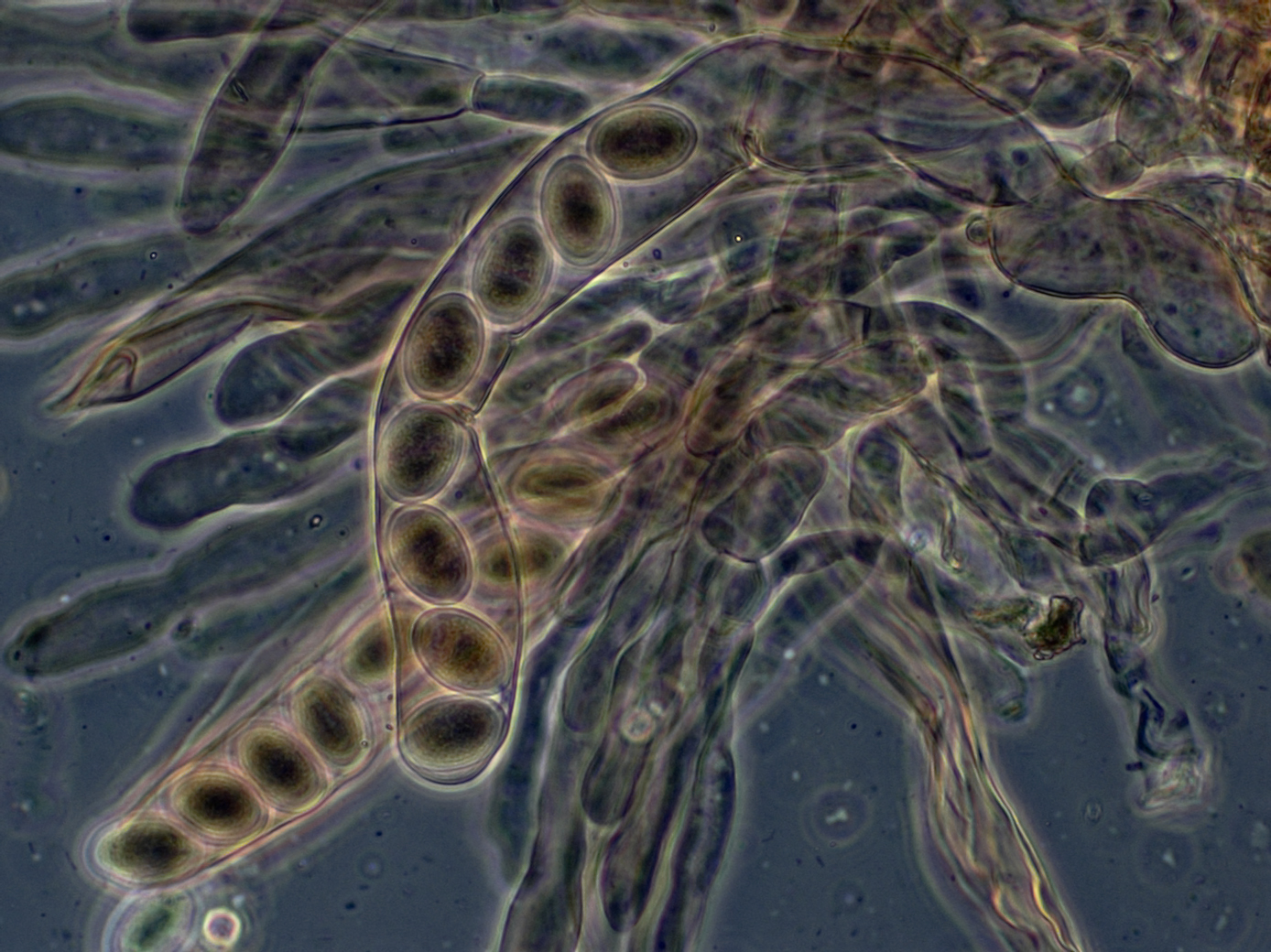
An ascus (; ) is the sexual spore-bearing cell produced in ascomycete fungi. Each ascus usually contains eight ascospores (or octad), produced by meiosis followed, in most species, by a mitotic cell division. However, asci in some genera or species can occur in numbers of one (e.g. ''Monosporascus cannonballus''), two, four, or multiples of four. In a few cases, the ascospores can bud off conidia that may fill the asci (e.g. ''Tympanis'') with hundreds of conidia, or the ascospores may fragment, e.g. some ''Cordyceps'', also filling the asci with smaller cells. Ascospores are nonmotile, usually single celled, but not infrequently may be coenocytic (lacking a septum), and in some cases coenocytic in multiple planes. Mitotic divisions within the developing spores populate each resulting cell in septate ascospores with nuclei. The term ocular chamber, or oculus, refers to the epiplasm (the portion of cytoplasm not used in ascospore formation) that is surrounded by the "bourrelet ... [...More Info...] [...Related Items...] OR: [Wikipedia] [Google] [Baidu] |


