|
Prostaglandin EP1 Receptor
Prostaglandin E2 receptor 1 (EP1) is a 42kDa prostaglandin receptor encoded by the PTGER1 gene. EP1 is one of four identified EP receptors, EP1, EP2, EP3, and EP4 which bind with and mediate cellular responses principally to prostaglandin E2) (PGE2) and also but generally with lesser affinity and responsiveness to certain other prostanoids (see Prostaglandin receptors). Animal model studies have implicated EP1 in various physiological and pathological responses. However, key differences in the distribution of EP1 between these test animals and humans as well as other complicating issues make it difficult to establish the function(s) of this receptor in human health and disease. Gene The ''PTGER1'' gene is located on human chromosome 19 at position p13.12 (i.e. 19p13.12), contains 2 introns and 3 exons, and codes for a G protein-coupled receptor (GPCR) of the rhodopsin-like receptor family, Subfamily A14 (see rhodopsin-like receptors#Subfamily A14). Expression Studies in mice, ... [...More Info...] [...Related Items...] OR: [Wikipedia] [Google] [Baidu] |
Prostaglandin Receptor
Prostaglandin receptors or prostanoid receptors represent a sub-class of cell surface membrane Receptor (biochemistry), receptors that are regarded as the primary receptors for one or more of the classical, naturally occurring prostanoids viz., prostaglandin D2, (i.e. PGD2), PGE2, PGF2alpha, prostacyclin (PGI2), thromboxane A2 (TXA2), and PGH2. They are named based on the prostanoid to which they preferentially bind and respond, e.g. the receptor responsive to PGI2 at lower concentrations than any other prostanoid is named the Prostacyclin receptor (IP). One exception to this rule is the receptor for thromboxane A2 (TP) which binds and responds to PGH2 and TXA2 equally well. All of the prostanoid receptors are G protein-coupled receptors belonging to the Rhodopsin-like receptors, Subfamily A14 of the rhodopsin-like receptor family except for the Prostaglandin DP2 receptor which is more closely related in amino acid sequence and functionality to chemotactic factor receptors such as th ... [...More Info...] [...Related Items...] OR: [Wikipedia] [Google] [Baidu] |
Binding Affinity
In biochemistry and pharmacology, a ligand is a substance that forms a complex with a biomolecule to serve a biological purpose. The etymology stems from ''ligare'', which means 'to bind'. In protein-ligand binding, the ligand is usually a molecule which produces a signal by binding to a site on a target protein. The binding typically results in a change of conformational isomerism (conformation) of the target protein. In DNA-ligand binding studies, the ligand can be a small molecule, ion, or protein which binds to the DNA double helix. The relationship between ligand and binding partner is a function of charge, hydrophobicity, and molecular structure. Binding occurs by intermolecular forces, such as ionic bonds, hydrogen bonds and Van der Waals forces. The association or docking is actually reversible through dissociation. Measurably irreversible covalent bonding between a ligand and target molecule is atypical in biological systems. In contrast to the definition of ligan ... [...More Info...] [...Related Items...] OR: [Wikipedia] [Google] [Baidu] |
P38 Mitogen-activated Protein Kinases
p38 mitogen-activated protein kinases are a class of mitogen-activated protein kinases (MAPKs) that are responsive to stress stimuli, such as cytokines, ultraviolet irradiation, heat shock, and osmotic shock, and are involved in cell differentiation, apoptosis and autophagy. Persistent activation of the p38 MAPK pathway in muscle satellite cells (muscle stem cells) due to ageing, impairs muscle regeneration. p38 MAP Kinase (MAPK), also called RK or CSBP (Cytokinin Specific Binding Protein), is the mammalian orthologue of the yeast Hog1p MAP kinase, which participates in a signaling cascade controlling cellular responses to cytokines and stress. Four p38 MAP kinases, p38-α (MAPK14), -β (MAPK11), -γ ( MAPK12 / ERK6), and -δ ( MAPK13 / SAPK4), have been identified. Similar to the SAPK/JNK pathway, p38 MAP kinase is activated by a variety of cellular stresses including osmotic shock, inflammatory cytokines, lipopolysaccharides (LPS), ultraviolet light, and growth facto ... [...More Info...] [...Related Items...] OR: [Wikipedia] [Google] [Baidu] |
Extracellular Signal-regulated Kinases
In molecular biology, extracellular signal-regulated kinases (ERKs) or classical MAP kinases are widely expressed protein kinase intracellular signalling molecules that are involved in functions including the regulation of meiosis, mitosis, and postmitotic functions in differentiated cells. Many different stimuli, including growth factors, cytokines, virus infection, ligands for heterotrimeric G protein-coupled receptors, transforming agents, and carcinogens, activate the ERK pathway. The term, "extracellular signal-regulated kinases", is sometimes used as a synonym for mitogen-activated protein kinase (MAPK), but has more recently been adopted for a specific subset of the mammalian MAPK family. In the MAPK/ERK pathway, Ras activates c-Raf, followed by mitogen-activated protein kinase kinase (abbreviated as MKK, MEK, or MAP2K) and then MAPK1/2 (below). Ras is typically activated by growth hormones through receptor tyrosine kinases and GRB2/ SOS, but may also receive other s ... [...More Info...] [...Related Items...] OR: [Wikipedia] [Google] [Baidu] |
Heterologous Desensitization
Heterologous desensitization (also known as cross-desensitization) is the term for the unresponsiveness of cells to one or more agonists to which they are normally responsive. Typically, desensitization is a receptor (biochemistry)-based phenomenon in which one receptor type, when bound to its ligand, becomes unable to further influence the signalling pathways by which it regulates cells and, in the case of cell surface membrane receptors, may thereafter be internalized. The desensitized receptor is degraded or freed of its activating ligand and re-cycled to a state where it is again able to respond to cognate ligands by activating its signalling pathways. This type of desensitization, termed homologous desensitization, leaves a cell transiently unresponsive to agents that activate the desensitized receptor but not to agents that activate other receptors. It commonly occurs with G protein-coupled receptors where it is mediated by the G protein-coupled receptor kinases (GRK) and are ... [...More Info...] [...Related Items...] OR: [Wikipedia] [Google] [Baidu] |
Homologous Desensitization
Homologous desensitization occurs when a receptor decreases its response to an agonist at high concentration. It is a process through which, after prolonged agonist exposure, the receptor is uncoupled from its signaling cascade and thus the cellular effect of receptor activation is attenuated. Homologous desensitization is distinguished from heterologous desensitization, a process in which repeated stimulation of a receptor by an agonist results in desensitization of the stimulated receptor as well as other, usually inactive, receptors on the same cell. They are sometimes denoted as agonist-dependent and agonist-independent desensitization respectively. While heterologous desensitization occurs rapidly at low agonist concentrations, homologous desensitization shows a dose dependent response and usually begins at significantly higher concentrations. Homologous desensitization serves as a mechanism for tachyphylaxis and helps organisms to maintain homeostasis. The process of homolo ... [...More Info...] [...Related Items...] OR: [Wikipedia] [Google] [Baidu] |
Protein Kinase C
In cell biology, Protein kinase C, commonly abbreviated to PKC (EC 2.7.11.13), is a family of protein kinase enzymes that are involved in controlling the function of other proteins through the phosphorylation of hydroxyl groups of serine and threonine amino acid residues on these proteins, or a member of this family. PKC enzymes in turn are activated by signals such as increases in the concentration of diacylglycerol (DAG) or calcium ions (Ca2+). Hence PKC enzymes play important roles in several signal transduction cascades. In biochemistry, the PKC family consists of fifteen isozymes in humans. They are divided into three subfamilies, based on their second messenger requirements: conventional (or classical), novel, and atypical. Conventional (c)PKCs contain the isoforms α, βI, βII, and γ. These require Ca2+, DAG, and a phospholipid such as phosphatidylserine for activation. Novel (n)PKCs include the δ, ε, η, and θ isoforms, and require DAG, but do not require Ca2+ for ... [...More Info...] [...Related Items...] OR: [Wikipedia] [Google] [Baidu] |
Phosphoinositide 3-kinase
Phosphoinositide 3-kinases (PI3Ks), also called phosphatidylinositol 3-kinases, are a family of enzymes involved in cellular functions such as cell growth, proliferation, differentiation, motility, survival and intracellular trafficking, which in turn are involved in cancer. PI3Ks are a family of related intracellular signal transducer enzymes capable of phosphorylating the 3 position hydroxyl group of the inositol ring of phosphatidylinositol (PtdIns). The pathway, with oncogene PIK3CA and tumor suppressor gene PTEN, is implicated in the sensitivity of cancer tumors to insulin and IGF1, and in calorie restriction. Discovery The discovery of PI3Ks by Lewis Cantley and colleagues began with their identification of a previously unknown phosphoinositide kinase associated with the polyoma middle T protein. They observed unique substrate specificity and chromatographic properties of the products of the lipid kinase, leading to the discovery that this phosphoinositide kinase ha ... [...More Info...] [...Related Items...] OR: [Wikipedia] [Google] [Baidu] |
G Beta-gamma Complex
The G beta-gamma complex (Gβγ) is a tightly bound dimeric protein complex, composed of one Gβ and one Gγ subunit, and is a component of heterotrimeric G proteins. Heterotrimeric G proteins, also called guanosine nucleotide-binding proteins, consist of three subunits, called alpha, beta, and gamma subunits, or Gα, Gβ, and Gγ. When a G protein-coupled receptor (GPCR) is activated, Gα dissociates from Gβγ, allowing both subunits to perform their respective downstream signaling effects. One of the major functions of Gβγ is the inhibition of the Gα subunit. History The individual subunits of the G protein complex were first identified in 1980 when the regulatory component of adenylate cyclase was successfully purified, yielding three polypeptides of different molecular weights. Initially, it was thought that Gα, the largest subunit, was the major effector regulatory subunit, and that Gβγ was largely responsible for inactivating the Gα subunit and enhancing membrane bi ... [...More Info...] [...Related Items...] OR: [Wikipedia] [Google] [Baidu] |
Gq Alpha Subunit
Gq protein alpha subunit is a family of heterotrimeric G protein alpha subunits. This family is also commonly called the Gq/11 (Gq/G11) family or Gq/11/14/15 family to include closely related family members. G alpha subunits may be referred to as Gq alpha, Gαq, or Gqα. Gq proteins couple to G protein-coupled receptors to activate beta-type phospholipase C (PLC-β) enzymes. PLC-β in turn hydrolyzes phosphatidylinositol 4,5-bisphosphate (PIP2) to diacyl glycerol (DAG) and inositol trisphosphate (IP3). IP3 acts as a second messenger to release stored calcium into the cytoplasm, while DAG acts as a second messenger that activates protein kinase C (PKC). Family members In humans, there are four distinct proteins in the Gq alpha subunit family: * Gαq is encoded by the gene GNAQ. * Gα11 is encoded by the gene GNA11. * Gα14 is encoded by the gene GNA14. * Gα15 is encoded by the gene GNA15. Function The general function of Gq is to activate intracellular signaling p ... [...More Info...] [...Related Items...] OR: [Wikipedia] [Google] [Baidu] |
G Proteins
G proteins, also known as guanine nucleotide-binding proteins, are a family of proteins that act as molecular switches inside cells, and are involved in transmitting signals from a variety of stimuli outside a cell to its interior. Their activity is regulated by factors that control their ability to bind to and hydrolyze guanosine triphosphate (GTP) to guanosine diphosphate (GDP). When they are bound to GTP, they are 'on', and, when they are bound to GDP, they are 'off'. G proteins belong to the larger group of enzymes called GTPases. There are two classes of G proteins. The first function as monomeric small GTPases (small G-proteins), while the second function as heterotrimeric G protein complexes. The latter class of complexes is made up of '' alpha'' (α), ''beta'' (β) and ''gamma'' (γ) subunits. In addition, the beta and gamma subunits can form a stable dimeric complex referred to as the beta-gamma complex . Heterotrimeric G proteins located within the cell are activ ... [...More Info...] [...Related Items...] OR: [Wikipedia] [Google] [Baidu] |
Competitive Antagonist
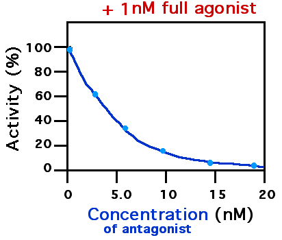
A receptor antagonist is a type of receptor ligand or drug that blocks or dampens a biological response by binding to and blocking a receptor rather than activating it like an agonist. Antagonist drugs interfere in the natural operation of receptor proteins.Pharmacology Guide: In vitro pharmacology: concentration-response curves " '' GlaxoWellcome.'' Retrieved on December 6, 2007. They are sometimes called blockers; examples include s, |


