|
Preputial Glands
Preputial glands are exocrine glands that are located in the folds of skin front of the genitals of some mammals. They occur in several species, including mice, ferrets, rhinoceroses, and even-toed ungulates and produce pheromones. The glands play a role in the urine-marking behavior of canids such as gray wolves and African wild dogs. The preputial glands of female animals are sometimes called clitoral glands. The preputial glands of male musk deer produce strong-smelling deer musk which is of economic importance, as it is used in perfumes. Human homologues There is debate about whether humans have functional homologues to preputial glands. Preputial glands were first noted by Edward Tyson and in 1694 fully described by William Cowper who named them Tyson's glands after Tyson. They are described as modified sebaceous glands located around the corona and inner surface of the prepuce of the human penis. They are believed to be most frequently found in the balanopreputial sulcu ... [...More Info...] [...Related Items...] OR: [Wikipedia] [Google] [Baidu] |
Exocrine Gland
Exocrine glands are glands that secrete substances on to an epithelial surface by way of a duct. Examples of exocrine glands include sweat, salivary, mammary, ceruminous, lacrimal, sebaceous, prostate and mucous. Exocrine glands are one of two types of glands in the human body, the other being endocrine glands, which secrete their products directly into the bloodstream. The liver and pancreas are both exocrine and endocrine glands; they are exocrine glands because they secrete products—bile and pancreatic juice—into the gastrointestinal tract through a series of ducts, and endocrine because they secrete other substances directly into the bloodstream. Exocrine sweat glands are part of the integumentary system; they have eccrine and apocrine types. Classification Structure Exocrine glands contain a glandular portion and a duct portion, the structures of which can be used to classify the gland. * The duct portion may be branched (called compound) or unbranched (called simple ... [...More Info...] [...Related Items...] OR: [Wikipedia] [Google] [Baidu] |
Edward Tyson
Edward Tyson (20 January 1651 – 1 August 1708) was an English scientist and physician. He is commonly regarded as the founder of modern comparative anatomy, which compares the anatomy between species. Biography Tyson was born the son of Edward Tyson at Clevedon, in Somerset. He became a BA from Oxford on 8 February 1670, an MA from Oxford on 4 November 1673, and an MD from Cambridge in 1678. He was admitted to the College of Physicians on 30 September 1680 and as a Fellow in April 1683. In 1684 he was appointed physician and governor to the Bethlem Hospital in London (the first mental hospital in Britain and the second in Europe). He is credited with changing the hospital from a zoo of sorts to a place intended to assist its inmates. He was elected a Fellow of the Royal Society in November 1679. He is buried at St Dionis Backchurch. Anatomical research In 1680, Tyson studied a porpoise and established that it is a mammal. He noted that the convoluted structures of the brains ... [...More Info...] [...Related Items...] OR: [Wikipedia] [Google] [Baidu] |
List Of Specialized Glands Within The Human Integumentary System ...
This article contains a list of glands of the human body List of endocrine and exocrine glands Skin There are several specialized glands within the human integumentary system that are derived from apocrine or sebaceous gland precursors. There are no specialized variants of eccrine glands. Endocrine glands See List of human endocrine organs and actions References {{reflist # Michael H. Ross & Wojciech Pawlina, Histology: A Text and Atlas Glands In animals, a gland is a group of cells in an animal's body that synthesizes substances (such as hormones) for release into the bloodstream (endocrine gland) or into cavities inside the body or its outer surface (exocrine gland). Structure De ... [...More Info...] [...Related Items...] OR: [Wikipedia] [Google] [Baidu] |
Pearly Penile Papules
Pearly penile papules (PPP) are benign small bumps on the human penis. They vary in size from 1–4 mm, are pearly or flesh-colored, smooth and dome-topped or filiform, and appear in one or several rows around the corona, the ridge of the head of the penis and sometimes on the penile shaft. They are painless, non-cancerous and not harmful. Symptoms and signs PPPs are small bumps on the human penis. They vary in size from 1–4 mm, are pearly or flesh-colored, smooth and dome-topped or filiform, and appear in one or several rows around the corona, the ridge of the glans and sometimes on the penile shaft. They are painless, non-cancerous and not harmful. Cause and mechanism PPPs are a type of angiofibroma. Their function is not well-understood. They are sometimes described as vestigial remnants of penile spines, sensitive features found in the same location in other primates. PPPs secrete oil that moistens the glans of the penis. They do not spread and often spont ... [...More Info...] [...Related Items...] OR: [Wikipedia] [Google] [Baidu] |
Corona Glandis
The corona of glans penis or penis crown refers to the rounded projecting border that forms at the base of the glans in human males. The corona overhangs a mucosal surface, known as the neck of the penis, which separates the shaft and the glans. The deep retro-glandular coronal sulcus forms between the corona and the neck of the penis. The corona and the neck are highly vascularized areas of the penis. The axial and dorsal penile arteries merge together at the neck before entering the glans. Small venous tributaries deriving from the corona drain the glans forming a venous retro-coronal plexus before merging with the dorsal veins. The circumference and the underside of the corona are densely innervated by several types of nerve terminals and are considered by males an erogenous area of the glans. In some males, small skin-colored bumps, known as pearly penile papules Pearly penile papules (PPP) are benign small bumps on the human penis. They vary in size from 1–4&nb ... [...More Info...] [...Related Items...] OR: [Wikipedia] [Google] [Baidu] |
The Journal Of Urology
''The Journal of Urology'' is a peer-reviewed medical journal covering urology published by Elsevier on behalf of the American Urological Association. It was established in 1917. A special centenary issue was released in 2017 to celebrate 100 years of the publication of the journal. Over the years, it absorbed the ''Transactions of the American Urological Association'' (1907–1920), as well as ''Investigative Urology'' (1963–1981) and ''Urological Survey'' (1951–1981). ''Urological Survey'' was known as ''Quarterly Review of Urology'' from 1946 to 1950. Editors The following persons have been editor-in-chief of the journal: Abstracting and indexing The journal is abstracted and indexed in BIOSIS, Current Contents/Clinical Medicine, EMBASE/Excerpta Medica, MEDLINE, and Scopus Scopus is Elsevier's abstract and citation database launched in 2004. Scopus covers nearly 36,377 titles (22,794 active titles and 13,583 inactive titles) from approximately 11,678 publishers, of whi ... [...More Info...] [...Related Items...] OR: [Wikipedia] [Google] [Baidu] |
Smegma
Smegma (Ancient Greek σμῆγμα : ''smēgma'') is a combination of shed skin cells, skin oils, and moisture. It occurs in both male and female mammalian genitalia. In females, it collects around the clitoris and in the folds of the labia minora; in males, smegma collects under the foreskin. Females The accumulation of sebum combined with dead skin cells forms smegma. ''Smegma clitoridis'' is defined as the secretion of the apocrine glands of the clitoris, in combination with desquamating epithelial cells. Glands that are located around the clitoris, the labia minora, and the labia majora secrete sebum. If smegma is not removed frequently it can lead to clitoral adhesion which can make clitoral stimulation (such as masturbation) painful (clitorodynia). Males In males, smegma helps keep the glans moist and facilitates sexual intercourse by acting as a lubricant. Smegma, itself, is completely benign, but uncircumcised "men with phimosis have an increased risk of penile ... [...More Info...] [...Related Items...] OR: [Wikipedia] [Google] [Baidu] |
Human Penis
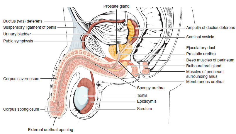
The human penis is an external male intromittent organ that additionally serves as the urinary duct. The main parts are the root (radix); the body (corpus); and the epithelium of the penis including the shaft skin and the foreskin (prepuce) covering the glans penis. The body of the penis is made up of three columns of tissue: two corpora cavernosa on the dorsal side and corpus spongiosum between them on the ventral side. The human male urethra passes through the prostate gland, where it is joined by the ejaculatory duct, and then through the penis. The urethra traverses the corpus spongiosum, and its opening, the meatus (), lies on the tip of the glans penis. It is a passage both for urination and ejaculation of semen (''see'' male reproductive system.) Most of the penis develops from the same embryonic tissue as the clitoris in females. The skin around the penis and the urethra share the same embryonic origin as the labia minora in females. An erection is the stiffening e ... [...More Info...] [...Related Items...] OR: [Wikipedia] [Google] [Baidu] |
Foreskin
In male human anatomy, the foreskin, also known as the prepuce, is the double-layered fold of skin, mucosal and muscular tissue at the distal end of the human penis that covers the glans and the urinary meatus. The foreskin is attached to the glans by an elastic band of tissue, known as the frenulum. The outer skin of the foreskin meets with the inner preputial mucosa at the area of the mucocutaneous junction. The foreskin is mobile, fairly stretchable and sustains the glans in a moist environment. Except for humans, a similar structure, known as penile sheath, appears in the male sexual organs of all primates and the vast majority of mammals. In humans, foreskin length varies widely and coverage of the glans in a flaccid and erect state can also vary. The foreskin is fused to the glans at birth and is generally not retractable in infancy and early childhood. Inability to retract the foreskin in childhood should not be considered a problem unless there are other symptoms. Retr ... [...More Info...] [...Related Items...] OR: [Wikipedia] [Google] [Baidu] |
Sebaceous Gland
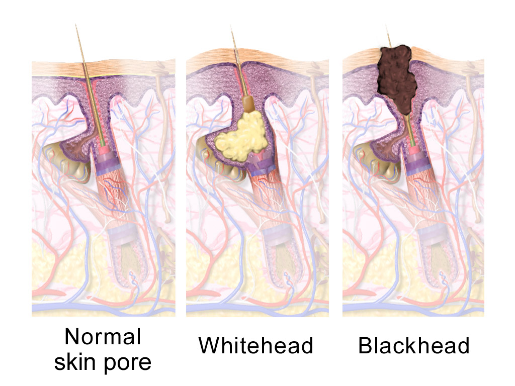
A sebaceous gland is a microscopic exocrine gland in the skin that opens into a hair follicle to secrete an oily or waxy matter, called sebum, which lubricates the hair and skin of mammals. In humans, sebaceous glands occur in the greatest number on the face and scalp, but also on all parts of the skin except the palms of the hands and soles of the feet. In the eyelids, meibomian glands, also called tarsal glands, are a type of sebaceous gland that secrete a special type of sebum into tears. Surrounding the female nipple, areolar glands are specialized sebaceous glands for lubricating the nipple. Fordyce spots are benign, visible, sebaceous glands found usually on the lips, gums and inner cheeks, and genitals. Structure Location Sebaceous glands are found throughout all areas of the skin, except the palms of the hands and soles of the feet. There are two types of sebaceous glands, those connected to hair follicles and those that exist independently. Sebaceous glands are found ... [...More Info...] [...Related Items...] OR: [Wikipedia] [Google] [Baidu] |
William Cowper (anatomist)
William Cowper ( ; c. 1666 – 8 March 1709) was an English surgery, surgeon and anatomist, famous for his early description of what is now known as Cowper's gland. Cowper was born in Petersfield, Hampshire, and he was apprenticed to a London surgeon, William Bignall, in March 1682. He was admitted to the Company of Barber-Surgeons in 1691 and began practising in London the same year. In 1694, he published his noted work, ''Myotomia Reformata, or a New Administration of the Muscles'', and he was elected a member of the Royal Society in 1696. In 1698, he published ''The Anatomy of the Humane Bodies'', which gained him great fame and notoriety, and over the next eleven years he published a number of tracts on topics ranging from surgery and pathology to physiology and anatomy. He died on 8 March 1709, and was buried in St Peter's Church, Petersfield. Some have called Cowper's ''Anatomy of the Humane Bodies'' one of the greatest acts of plagiarism in all of medical publishing, t ... [...More Info...] [...Related Items...] OR: [Wikipedia] [Google] [Baidu] |
Homologous Structures
In biology, homology is similarity due to shared ancestry between a pair of structures or genes in different taxa. A common example of homologous structures is the forelimbs of vertebrates, where the wings of bats and birds, the arms of primates, the front flippers of whales and the forelegs of four-legged vertebrates like dogs and crocodiles are all derived from the same ancestral tetrapod structure. Evolutionary biology explains homologous structures adapted to different purposes as the result of descent with modification from a common ancestor. The term was first applied to biology in a non-evolutionary context by the anatomist Richard Owen in 1843. Homology was later explained by Charles Darwin's theory of evolution in 1859, but had been observed before this, from Aristotle onwards, and it was explicitly analysed by Pierre Belon in 1555. In developmental biology, organs that developed in the embryo in the same manner and from similar origins, such as from matching primord ... [...More Info...] [...Related Items...] OR: [Wikipedia] [Google] [Baidu] |


.jpeg)
