|
Pilon Fracture
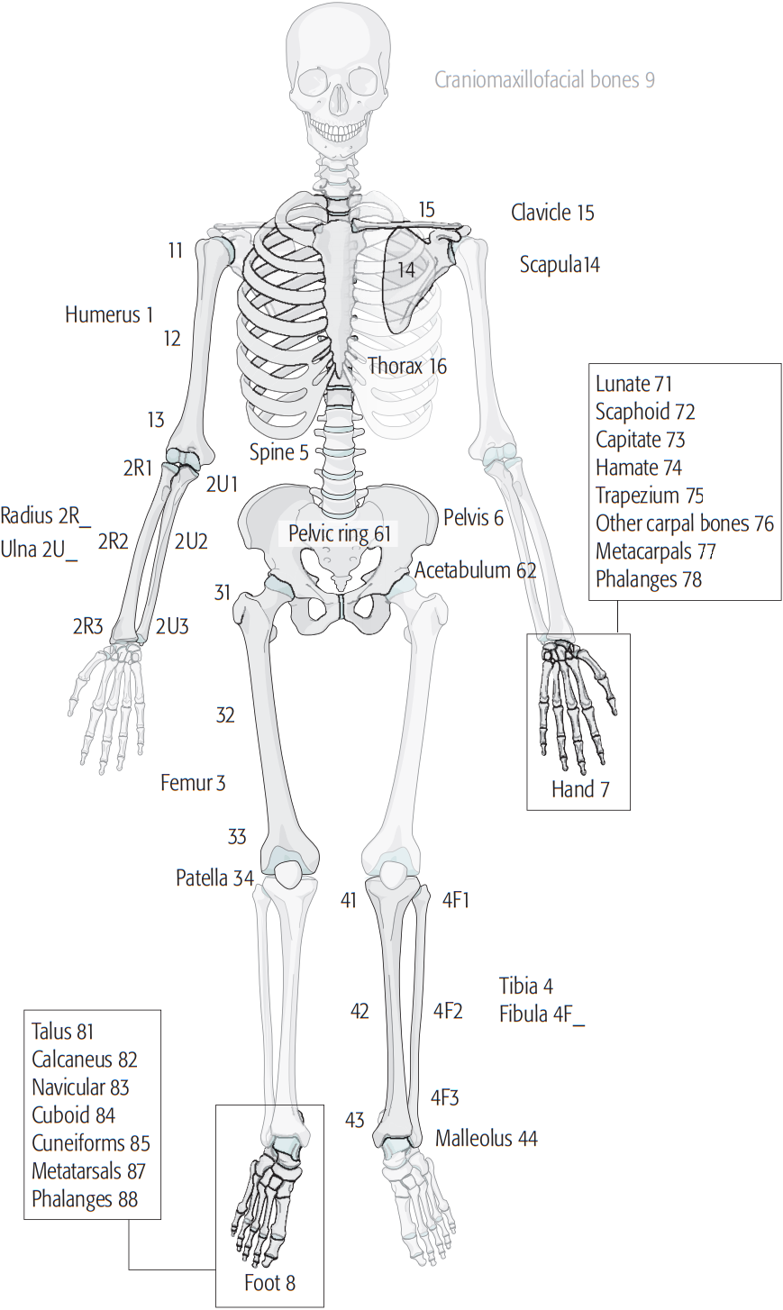
A pilon fracture, is a fracture of the distal part of the tibia, involving its articular surface at the ankle joint. Pilon fractures are caused by rotational or axial forces, mostly as a result of falls from a height or motor vehicle accidents. Pilon fractures are rare, comprising 3 to 10 percent of all fractures of the tibia and 1 percent of all lower extremity fractures, but they involve a large part of the weight-bearing surface of the tibia in the ankle joint. Because of this, they may be difficult to fixate and are historically associated with high rates of complications and poor outcome. ''Pilon'' is the French word for "pestle" and was introduced into orthopedic literature in 1911 by pioneer French radiologist Étienne Destot. Classification Pilon fractures are categorized by two main X-ray schemes, Ruedi-Allgower classification system. and Müller AO Classification of fractures. Treatment The treatment of pilon fractures depends on the extent of the injury. This i ... [...More Info...] [...Related Items...] OR: [Wikipedia] [Google] [Baidu] |
X-ray
An X-ray, or, much less commonly, X-radiation, is a penetrating form of high-energy electromagnetic radiation. Most X-rays have a wavelength ranging from 10 picometers to 10 nanometers, corresponding to frequencies in the range 30 petahertz to 30 exahertz ( to ) and energies in the range 145 eV to 124 keV. X-ray wavelengths are shorter than those of UV rays and typically longer than those of gamma rays. In many languages, X-radiation is referred to as Röntgen radiation, after the German scientist Wilhelm Conrad Röntgen, who discovered it on November 8, 1895. He named it ''X-radiation'' to signify an unknown type of radiation.Novelline, Robert (1997). ''Squire's Fundamentals of Radiology''. Harvard University Press. 5th edition. . Spellings of ''X-ray(s)'' in English include the variants ''x-ray(s)'', ''xray(s)'', and ''X ray(s)''. The most familiar use of X-rays is checking for fractures (broken bones), but X-rays are also used in other ways. ... [...More Info...] [...Related Items...] OR: [Wikipedia] [Google] [Baidu] |
Müller AO Classification Of Fractures
The Müller AO Classification of fractures is a system for classifying bone fractures initially published in 1987 by the AO Foundation The AO Foundation is a nonprofit organization dedicated to improving the care of patients with musculoskeletal injuries or pathologies and their sequelae through research, development, and education of surgeons and operating room personnel. The AO ... as a method of categorizing injuries according to therognosis of the patient's anatomical and functional outcome. "AO" is an initialism for the German "Arbeitsgemeinschaft für Osteosynthesefragen", the predecessor of the AO Foundation. It is one of the few complete fracture classification systems to remain in use today after validation. Comprehensive classification of the long bones The English language version of the system allows consistent in detail description of a fracture in defined terminology by creating a 5-element alphanumeric code: Localisation First, each fracture is given 2 numbers ... [...More Info...] [...Related Items...] OR: [Wikipedia] [Google] [Baidu] |
Ankle Fracture
An ankle fracture is a break of one or more of the bones that make up the ankle joint. Symptoms may include pain, swelling, bruising, and an inability to walk on the injured leg. Complications may include an associated high ankle sprain, compartment syndrome, stiffness, malunion, and post-traumatic arthritis. Ankle fractures may result from excessive stress on the joint such as from rolling an ankle or from blunt trauma. Types of ankle fractures include lateral malleolus, medial malleolus, posterior malleolus, bimalleolar, and trimalleolar fractures. The Ottawa ankle rule can help determine the need for X-rays. Special X-ray views called stress views help determine whether an ankle fracture is unstable. Treatment depends on the fracture type. Ankle stability largely dictates non-operative vs. operative treatment. Non-operative treatment includes splinting or casting while operative treatment includes fixing the fracture with metal implants through an open reduction internal fi ... [...More Info...] [...Related Items...] OR: [Wikipedia] [Google] [Baidu] |
Vacuum Assisted Closure Wound Therapy
Negative-pressure wound therapy (NPWT), also known as a vacuum assisted closure (VAC), is a therapeutic technique using a suction pump, tubing and a dressing to remove excess exudate and promote healing in acute or chronic wounds and second- and third-degree burns. The therapy involves the controlled application of subatmospheric pressure to the local wound environment using a sealed wound dressing connected to a vacuum pump. The use of this technique in wound management started in the 1990s and this technique is often recommended for treatment of a range of wounds including dehisced surgical wounds, closed surgical wounds, open abdominal wounds, open fractures, pressure injuries or pressure ulcers, diabetic foot ulcers, venous insufficiency ulcers, some types of skin grafts, burns, sternal wounds. It may also be considered after a clean surgery in a person who is obese. NPWT is performed by applying a vacuum through a special sealed dressing. The continued vacuum draws ou ... [...More Info...] [...Related Items...] OR: [Wikipedia] [Google] [Baidu] |
Open Fracture
An open fracture, also called a compound fracture, is a type of bone fracture in orthopedics that is frequently caused by high energy trauma. It is a bone fracture, also known as a broken bone, associated with a break in the skin continuity which can cause complications such as infection, malunion, and nonunion. Gustilo open fracture classification is the most commonly used method to classify open fractures, to guide treatment and to predict clinical outcomes. Advanced trauma life support is the first line of action in dealing with open fractures and to rule out other life-threatening condition in cases of trauma. Cephalosporins are generally the first line of antibiotics. The antibiotics are continued for 24 hours to minimize the risk of infections. Therapeutic irrigation, wound debridement, early wound closure and bone fixation are the main management of open fractures. All these actions aimed to reduce the risk of infections. Classification There are a number of classificat ... [...More Info...] [...Related Items...] OR: [Wikipedia] [Google] [Baidu] |
External Fixation
External fixation is a surgical treatment wherein rods are screwed into bone and exit the body to be attached to a stabilizing structure on the outside of the body. It is an alternative to internal fixation, where the components used to provide stability are positioned entirely within the patient's body. It is used to stabilize bone and soft tissues at a distance from the operative or injury focus. They provide unobstructed access to the relevant skeletal and soft tissue structures for their initial assessment and also for secondary interventions needed to restore bony continuity and a functional soft tissue cover. Indications # Stabilization of severe open fractures # Stabilization of infected nonunions # Correction of extremity malalignments and length discrepancies # Initial stabilization of soft tissue and bony disruption in poly trauma patients (damage control orthopaedics) # Closed fracture with associated severe soft tissue injuries # Severely comminuted diaphyseal and per ... [...More Info...] [...Related Items...] OR: [Wikipedia] [Google] [Baidu] |
Soft Tissue
Soft tissue is all the tissue in the body that is not hardened by the processes of ossification or calcification such as bones and teeth. Soft tissue connects, surrounds or supports internal organs and bones, and includes muscle, tendons, ligaments, fat, fibrous tissue, lymph and blood vessels, fasciae, and synovial membranes. with :q=a_E_E_ \qquad Q=b_E_E_ quadratic forms of Green-Lagrange strains E_ and a_, b_ and c material constants. W is the strain energy function per volume unit, which is the mechanical strain energy for a given temperature. Isotropic simplification The Fung-model, simplified with isotropic hypothesis (same mechanical properties in all directions). This written in respect of the principal stretches (\lambda_i): :W = \frac\left (\lambda_1^2 + \lambda_2^2 + \lambda_3^2 - 3) + b\left( e^ -1 \right) \right/math> , where a, b and c are constants. Simplification for small and big stretches For small strains, the exponential term is very small, thus neg ... [...More Info...] [...Related Items...] OR: [Wikipedia] [Google] [Baidu] |
Talus Bone
The talus (; Latin for ankle or ankle bone), talus bone, astragalus (), or ankle bone is one of the group of foot bones known as the tarsus. The tarsus forms the lower part of the ankle joint. It transmits the entire weight of the body from the lower legs to the foot.Platzer (2004), p 216 The talus has joints with the two bones of the lower leg, the tibia and thinner fibula. These leg bones have two prominences (the lateral and medial malleoli) that articulate with the talus. At the foot end, within the tarsus, the talus articulates with the calcaneus (heel bone) below, and with the curved navicular bone in front; together, these foot articulations form the ball-and-socket-shaped talocalcaneonavicular joint. The talus is the second largest of the tarsal bones; it is also one of the bones in the human body with the highest percentage of its surface area covered by articular cartilage. It is also unusual in that it has a retrograde blood supply, i.e. arterial blood enters the ... [...More Info...] [...Related Items...] OR: [Wikipedia] [Google] [Baidu] |
Fibula
The fibula or calf bone is a leg bone on the lateral side of the tibia, to which it is connected above and below. It is the smaller of the two bones and, in proportion to its length, the most slender of all the long bones. Its upper extremity is small, placed toward the back of the head of the tibia, below the knee joint and excluded from the formation of this joint. Its lower extremity inclines a little forward, so as to be on a plane anterior to that of the upper end; it projects below the tibia and forms the lateral part of the ankle joint. Structure The bone has the following components: * Lateral malleolus * Interosseous membrane connecting the fibula to the tibia, forming a syndesmosis joint * The superior tibiofibular articulation is an arthrodial joint between the lateral condyle of the tibia and the head of the fibula. * The inferior tibiofibular articulation (tibiofibular syndesmosis) is formed by the rough, convex surface of the medial side of the lower end of the f ... [...More Info...] [...Related Items...] OR: [Wikipedia] [Google] [Baidu] |
Fracture (bone)
A bone fracture (abbreviated FRX or Fx, Fx, or #) is a medical condition in which there is a partial or complete break in the continuity of any bone in the body. In more severe cases, the bone may be broken into several fragments, known as a ''comminuted fracture''. A bone fracture may be the result of high force impact or stress, or a minimal trauma injury as a result of certain medical conditions that weaken the bones, such as osteoporosis, osteopenia, bone cancer, or osteogenesis imperfecta, where the fracture is then properly termed a pathologic fracture. Signs and symptoms Although bone tissue contains no pain receptors, a bone fracture is painful for several reasons: * Breaking in the continuity of the periosteum, with or without similar discontinuity in endosteum, as both contain multiple pain receptors. * Edema and hematoma of nearby soft tissues caused by ruptured bone marrow evokes pressure pain. * Involuntary muscle spasms trying to hold bone fragments in place. D ... [...More Info...] [...Related Items...] OR: [Wikipedia] [Google] [Baidu] |
Étienne Destot
Étienne Destot (1 March 1864 – 3 December 1918) was a French radiologist and anatomist who was a native of Dijon. He studied medicine in Lyon, and later worked in the hospitals of Hôtel Dieu, Croix-Rousse and Charité in Lyon. In addition to his work in medicine, he was an accomplished sculptor. Destot was a pioneer in the field of radiology. In February 1896, less than two months after Wilhelm Conrad Röntgen (1845–1923) announced his discovery of the X-ray, Destot was making radiographs of patients at the Hôtel-Dieu de Lyon. He made thousands of radiographies, many of which were of patients supplied by surgeon Louis Léopold Ollier (1830–1900). In 1913, due to severe radiation damage to his hands, he was forced to relinquish his position as radiologist. Destot also made contributions in the field of orthopedics, and in 1911 is credited as being the first physician to use the term ''pilon'' in orthopedic literature. [...More Info...] [...Related Items...] OR: [Wikipedia] [Google] [Baidu] |


