|
Photoactivatable Fluorescent Protein
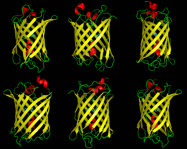
Photoactivatable fluorescent proteins (PAFPs) is a type of fluorescent protein that exhibit fluorescence that can be modified by a light-induced chemical reaction. History The first PAFP, Kaede (protein), was isolated from '' Trachyphyllia geoffroyi'' in a cDNA library screen designed to identify new fluorescent proteins. A fluorescent green protein derived from this screen was serendipitously discovered to have sensitivity to ultraviolet light-- We happened to leave one of the protein aliquots on the laboratory bench overnight. The next day, we found that the protein sample on the bench had turned red, whereas the others that were kept in a paper box remained green. Although the sky had been partly cloudy, the red sample had been exposed to sunlight through the south-facing windows. Properties Many PAFPs have been engineered from existing fluorescent proteins or identified from large-scale screens in the wake of Kaede's discovery. Many of these undergo green-to-red ph ... [...More Info...] [...Related Items...] OR: [Wikipedia] [Google] [Baidu] |
Fluorescent Protein
Fluorescent proteins include: * Green fluorescent protein (GFP) * Yellow fluorescent protein Yellow fluorescent protein (YFP) is a genetic mutant of green fluorescent protein (GFP) originally derived from the jellyfish '' Aequorea victoria''. Its excitation peak is 513 nm and its emission peak is 527 nm. Like the parent GFP, YFP ... (YFP) * Red fluorescent protein (RFP) {{Short pages monitor ... [...More Info...] [...Related Items...] OR: [Wikipedia] [Google] [Baidu] |
Photochemistry
Photochemistry is the branch of chemistry concerned with the chemical effects of light. Generally, this term is used to describe a chemical reaction caused by absorption of ultraviolet (wavelength from 100 to 400 nm), visible light (400–750 nm) or infrared radiation (750–2500 nm). In nature, photochemistry is of immense importance as it is the basis of photosynthesis, vision, and the formation of vitamin D with sunlight. Photochemical reactions proceed differently than temperature-driven reactions. Photochemical paths access high energy intermediates that cannot be generated thermally, thereby overcoming large activation barriers in a short period of time, and allowing reactions otherwise inaccessible by thermal processes. Photochemistry can also be destructive, as illustrated by the photodegradation of plastics. Concept Grotthuss–Draper law and Stark-Einstein law Photoexcitation is the first step in a photochemical process where the reactant is elevated ... [...More Info...] [...Related Items...] OR: [Wikipedia] [Google] [Baidu] |
Kaede (protein)
Kaede is a photoactivatable fluorescent protein naturally originated from a stony coral, '' Trachyphyllia geoffroyi''. Its name means "maple" in Japanese. With the irradiation of ultraviolet light (350–400 nm), Kaede undergoes irreversible photoconversion from green fluorescence to red fluorescence. Kaede is a homotetrameric protein with the size of 116 kDa. The tetrameric structure was deduced as its primary structure is only 28 kDa. This tetramerization possibly makes Kaede have a low tendency to form aggregates when fused to other proteins. Discovery The property of photoconverted fluorescence Kaede protein was serendipitously discovered and first reported by Ando et al. in Proceedings of the United States National Academy of Sciences. An aliquot of Kaede protein was discovered to emit red fluorescence after being left on the bench and exposed to sunlight. Subsequent verification revealed that Kaede, which is originally green fluorescent, after exposure to UV light i ... [...More Info...] [...Related Items...] OR: [Wikipedia] [Google] [Baidu] |
Trachyphyllia Geoffroyi
The open brain coral (''Trachyphyllia geoffroyi'') is a brightly colored free-living coral species in the family Merulinidae. It is the only species in the monotypic genus ''Trachyphyllia'' and can be found throughout the Indo-Pacific. Description Open brain corals can be solitary or colonial. They are small corals, rarely reaching over 20 cm in diameter. They are free-living and exhibit a flabello-meandroid growth form, meaning they have distinct valley regions separated by walls. In colonial forms, the valley regions can contain multiple individual polyps. Complexity of valley regions can range; some are hourglass shaped while other cans be highly lobed. They typically have bilateral symmetry. During the day when the polyp is closed, the coral is covered by a mantle that extends beyond the skeleton, but can retract when disturbed. Polyps and mantle are very fleshy. Colonies can be blue, green, yellow, brown, and are often vibrantly colored. The open brain coral is know ... [...More Info...] [...Related Items...] OR: [Wikipedia] [Google] [Baidu] |
High-throughput Screening
High-throughput screening (HTS) is a method for scientific experimentation especially used in drug discovery and relevant to the fields of biology, materials science and chemistry. Using robotics, data processing/control software, liquid handling devices, and sensitive detectors, high-throughput screening allows a researcher to quickly conduct millions of chemical, genetic, or pharmacological tests. Through this process one can quickly recognize active compounds, antibodies, or genes that modulate a particular biomolecular pathway. The results of these experiments provide starting points for drug design and for understanding the noninteraction or role of a particular location. Assay plate preparation The key labware or testing vessel of HTS is the microtiter plate, which is a small container, usually disposable and made of plastic, that features a grid of small, open divots called ''wells''. In general, microplates for HTS have either 96, 192, 384, 1536, 3456 or 6144 wells. The ... [...More Info...] [...Related Items...] OR: [Wikipedia] [Google] [Baidu] |
Eos (protein)
EosFP is a photoactivatable green to red fluorescent protein. Its green fluorescence (516 nm) switches to red (581 nm) upon UV irradiation of ~390 nm (violet/blue light) due to a photo-induced modification resulting from a break in the peptide backbone near the chromophore. Eos was first discovered as a tetrameric protein in the stony coral '' Lobophyllia hemprichii''''.'' Like other fluorescent proteins, Eos allows for applications such as the tracking of fusion proteins, multicolour labelling and tracking of cell movement. Several variants of Eos have been engineered for use in specific study systems including mEos2, mEos4 and CaMPARI. History EosFP was first discovered in 2005 during a large scale screen for PAFPs (photoactivatable fluorescent proteins) within the stony coral '' Lobophyllia hemprichii.'' It has since been successfully cloned in ''Escherichia coli'' and fusion constructs have been developed for use in human cells. Eos was named after the Gr ... [...More Info...] [...Related Items...] OR: [Wikipedia] [Google] [Baidu] |
Dronpa
Dronpa is a reversibly switchable photoactivatable fluorescent protein that is 2.5 times as bright as EGFP. Dronpa gets switched off by strong illumination with 488 nm (blue) light and this can be reversed by weak 405 nm UV light. A single dronpa molecule can be switched on and off over 100 times. It has an excitation peak at 503 nm and an emission peak at 518 nm. History A tetrameric, reversibly switchable fluorescent protein was discovered in a cDNA screen of a stony coral (Pectiniidae Pectiniidae was a family of stony corals, commonly known as chalice corals, but the name is no longer considered valid. Taxonomy The "robust" stony coral families of Faviidae, Merulinidae, Mussidae and Pectiniidae, have traditionally been recog ...). A monomeric variant of this protein was named "Dronpa" after "''Dron''" a ninja term for vanishing and ''pa'' for photoactivation. Structure and mechanism of photoswitching Dronpa is 257 amino acids long and is a 28.8 k ... [...More Info...] [...Related Items...] OR: [Wikipedia] [Google] [Baidu] |
Bioluminescence
Bioluminescence is the production and emission of light by living organisms. It is a form of chemiluminescence. Bioluminescence occurs widely in marine vertebrates and invertebrates, as well as in some fungi, microorganisms including some bioluminescent bacteria, and terrestrial arthropods such as fireflies. In some animals, the light is bacteriogenic, produced by symbiotic bacteria such as those from the genus ''Vibrio''; in others, it is autogenic, produced by the animals themselves. In a general sense, the principal chemical reaction in bioluminescence involves a light-emitting molecule and an enzyme, generally called luciferin and luciferase, respectively. Because these are generic names, luciferins and luciferases are often distinguished by the species or group, e.g. firefly luciferin. In all characterized cases, the enzyme catalyzes the oxidation of the luciferin. In some species, the luciferase requires other cofactors, such as calcium or magnesium ions, and somet ... [...More Info...] [...Related Items...] OR: [Wikipedia] [Google] [Baidu] |


