|
Petrous Ganglion
The inferior ganglion of the glossopharyngeal nerve (petrosal ganglion) is a sensory ganglion. It is larger than and inferior to the superior ganglion of the glossopharyngeal nerve. It is located within the jugular foramen. The pseudounipolar neurons of the inferior ganglion of the glossopharyngeal nerve provide sensory innervation to areas around the tongue and pharynx. More specifically: # innervation of taste buds on the posterior 1/3 of tongue # general sensory innervation of posterior 1/3 of tongue, soft palate, palatine tonsils, upper pharynx and Eustachian tubes # innervation of baroreceptor cells in the carotid sinus # innervation of glomus type I chemoreceptor cells in the carotid body The central processes of the neurons which provide taste sensation synapse in the rostral portion of the solitary nucleus (also called the gustatory nucleus). The central processes of the neurons which provide general sensory information synapse in the spinal trigeminal nucleus. Final ... [...More Info...] [...Related Items...] OR: [Wikipedia] [Google] [Baidu] |
Glossopharyngeal
The glossopharyngeal nerve (), also known as the ninth cranial nerve, cranial nerve IX, or simply CN IX, is a cranial nerve that exits the brainstem from the sides of the upper Medulla oblongata, medulla, just anterior (closer to the nose) to the vagus nerve. Being a mixed nerve (sensorimotor), it carries afferent sensory and efferent motor information. The motor division of the glossopharyngeal nerve is derived from the Basal plate (neural tube), basal plate of the embryonic medulla oblongata, whereas the sensory division originates from the cranial neural crest. Structure From the anterior portion of the medulla oblongata, the glossopharyngeal nerve passes laterally across or below the Flocculus (cerebellar), flocculus, and leaves the skull through the central part of the jugular foramen. From the superior and inferior ganglia in jugular foramen, it has its own sheath of dura mater. The inferior ganglion on the inferior surface of petrous part of temporal is related with a tri ... [...More Info...] [...Related Items...] OR: [Wikipedia] [Google] [Baidu] |
Carotid Sinus
In human anatomy, the carotid sinus is a dilated area at the base of the internal carotid artery just superior to the bifurcation of the internal carotid and external carotid at the level of the superior border of thyroid cartilage. The carotid sinus extends from the bifurcation to the "true" internal carotid artery. The carotid sinus is sensitive to pressure changes in the arterial blood at this level. It is the major baroreception site in humans and most mammals. Structure The carotid sinus is the reflex area of the carotid artery, consisting of baroreceptors which monitor blood pressure. Function The carotid sinus contains numerous baroreceptors which function as a "sampling area" for many homeostatic mechanisms for maintaining blood pressure. The carotid sinus baroreceptors are innervated by the carotid sinus nerve, which is a branch of the glossopharyngeal nerve (CN IX). The neurons which innervate the carotid sinus centrally project to the solitary nucleus in th ... [...More Info...] [...Related Items...] OR: [Wikipedia] [Google] [Baidu] |
Inferior Salivatory Nucleus
The salivatory nuclei are the superior salivatory nucleus, and the inferior salivatory nucleus that innervate the salivary glands. They are located in the pontine tegmentum in the brainstem. They both are examples of cranial nerve nuclei. The superior salivatory nucleus innervates the submandibular gland and the sublingual gland and is part of the facial nerve The inferior salivatory nucleus innervates the parotid gland by way of the otic ganglion and forms the parasympathetic component of the glossopharyngeal nerve. Superior salivatory nucleus The superior salivatory nucleus (or nucleus salivatorius superior) of the facial nerve is a visceromotor cranial nerve nucleus located in the pontine tegmentum. It is one of the salivatory nuclei. Parasympathetic efferent fibers of the facial nerve (preganglionic fibers) arise according to some authors from the small cells of the facial nucleus, or according to others from a special nucleus of cells scattered in the reticular ... [...More Info...] [...Related Items...] OR: [Wikipedia] [Google] [Baidu] |
Tympanic Nerve
The tympanic nerve (Jacobson’s nerve) is a branch of the glossopharyngeal nerve found near the ear. It gives sensation to the middle ear, the Eustachian tube, the parotid gland, and mastoid air cells. It gives parasympathetic to supply to the parotid gland via the otic ganglion and the auriculotemporal nerve. Structure The tympanic nerve arises from the inferior ganglion of the glossopharyngeal nerve. It ascends to the tympanic cavity through a small canal, the inferior tympanic canaliculus, on the under surface of the petrous portion of the temporal bone on the ridge which separates the carotid canal from the jugular fossa. In the tympanic cavity, it divides into branches which form the tympanic plexus and are contained in grooves upon the surface of the promontory. The tympanic nerve contains sensory axons to the middle ear, including the internal surface of the tympanic membrane. Their cell bodies are found in the superior ganglion of the glossopharyngeal nerve. It al ... [...More Info...] [...Related Items...] OR: [Wikipedia] [Google] [Baidu] |
Spinal Trigeminal Nucleus
The spinal trigeminal nucleus is a nucleus in the medulla that receives information about deep/crude touch, pain, and temperature from the ipsilateral face. In addition to the trigeminal nerve (CN V), the facial (CN VII), glossopharyngeal (CN IX), and vagus nerves (CN X) also convey pain information from their areas to the spinal trigeminal nucleus. Thus the spinal trigeminal nucleus receives input from V, , [...More Info...] [...Related Items...] OR: [Wikipedia] [Google] [Baidu] |
Gustatory Nucleus
The gustatory nucleus is the rostral part of the solitary nucleus located in the medulla. The gustatory nucleus is associated with the sense of taste and has two sections, the rostral and lateral regions. A close association between the gustatory nucleus and visceral information exists for this function in the gustatory system, assisting in homeostasis - via the identification of food that might be possibly poisonous or harmful for the body. There are many gustatory nuclei in the brain stem. Each of these nuclei corresponds to three cranial nerves, the facial nerve (VII), the glossopharyngeal nerve (IX), and the vagus nerve (X) and GABA is the primary inhibitory neurotransmitter involved in its functionality. All visceral afferents in the vagus and glossopharyngeal nerves first arrive in the nucleus of the solitary tract and information from the gustatory system can then be relayed to the thalamus and cortex. The central axons on primary sensory neurons in the taste system in th ... [...More Info...] [...Related Items...] OR: [Wikipedia] [Google] [Baidu] |
Solitary Nucleus
In the human brainstem, the solitary nucleus, also called nucleus of the solitary tract, nucleus solitarius, and nucleus tractus solitarii, (SN or NTS) is a series of purely sensory nuclei (clusters of nerve cell bodies) forming a vertical column of grey matter embedded in the medulla oblongata. Through the center of the SN runs the solitary tract, a white bundle of nerve fibers, including fibers from the facial, glossopharyngeal and vagus nerves, that innervate the SN. The SN projects to, among other regions, the reticular formation, parasympathetic preganglionic neurons, hypothalamus and thalamus, forming circuits that contribute to autonomic regulation. Cells along the length of the SN are arranged roughly in accordance with function; for instance, cells involved in taste are located in the rostral part, while those receiving information from cardio-respiratory and gastrointestinal processes are found in the caudal part. Inputs * Taste information from the facial nerve ... [...More Info...] [...Related Items...] OR: [Wikipedia] [Google] [Baidu] |
Carotid Body
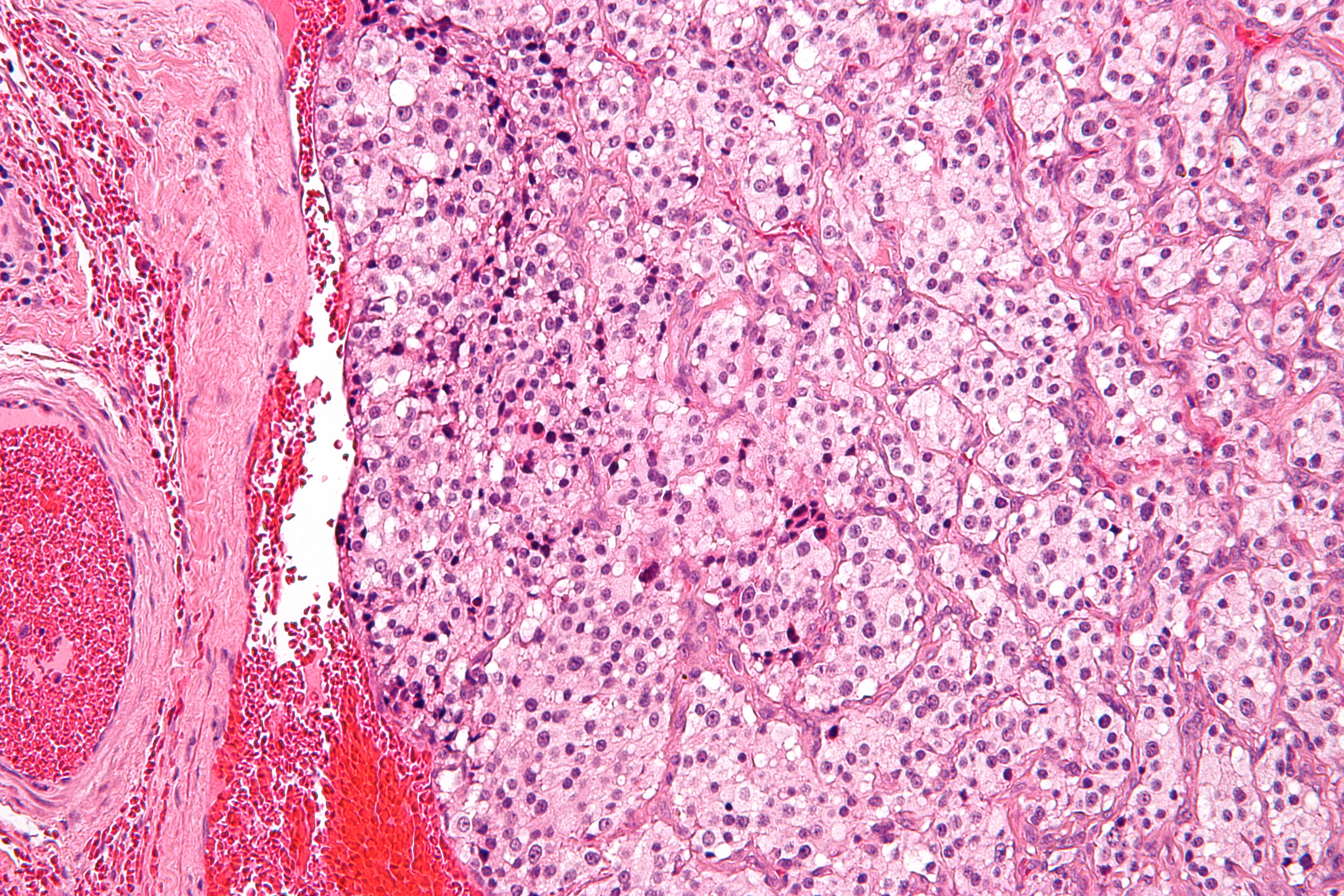
The carotid body is a small cluster of chemoreceptor cells, and supporting sustentacular cells. The carotid body is located in the adventitia, in the bifurcation (fork) of the common carotid artery, which runs along both sides of the neck. The carotid body detects changes in the composition of arterial blood flowing through it, mainly the partial pressure of arterial oxygen, but also of carbon dioxide. It is also sensitive to changes in blood pH, and temperature. Structure The carotid body is made up of two types of cells, called glomus cells: glomus type I cells are peripheral chemoreceptors, and glomus type II cells are sustentacular supportive cells. * Glomus type I cells are derived from the neural crest. They release a variety of neurotransmitters, including acetylcholine, ATP, and dopamine that trigger EPSPs in synapsed neurons leading to the respiratory center. They are innervated by axons of the glossopharyngeal nerve which collectively are called the carotid sinus ne ... [...More Info...] [...Related Items...] OR: [Wikipedia] [Google] [Baidu] |
Glomus Type I Cell
Glomus cells are the cell type mainly located in the carotid bodies and aortic bodies. Glomus type I cells are peripheral chemoreceptors which sense the oxygen, carbon dioxide and pH levels of the blood. When there is a decrease in the blood's pH, a decrease in oxygen (pO2), or an increase in carbon dioxide ( pCO2), the carotid bodies and the aortic bodies signal the dorsal respiratory group in the medulla oblongata to increase the volume and rate of breathing. The glomus cells have a high metabolic rate and good blood perfusion and thus are sensitive to changes in arterial blood gas tension. Glomus type II cells are sustentacular cells having a similar supportive function to glial cells. Structure The signalling within the chemoreceptors is thought to be mediated by the release of neurotransmitters by the glomus cells, including dopamine, noradrenaline, acetylcholine, substance P, vasoactive intestinal peptide and enkephalins. Vasopressin has been found to inhibit t ... [...More Info...] [...Related Items...] OR: [Wikipedia] [Google] [Baidu] |
Eustachian Tube
In anatomy, the Eustachian tube, also known as the auditory tube or pharyngotympanic tube, is a tube that links the nasopharynx to the middle ear, of which it is also a part. In adult humans, the Eustachian tube is approximately long and in diameter. It is named after the sixteenth-century Italian anatomist Bartolomeo Eustachi. In humans and other tetrapods, both the middle ear and the ear canal are normally filled with air. Unlike the air of the ear canal, however, the air of the middle ear is not in direct contact with the atmosphere outside the body; thus, a pressure difference can develop between the atmospheric pressure of the ear canal and the middle ear. Normally, the Eustachian tube is collapsed, but it gapes open with swallowing and with positive pressure, allowing the middle ear's pressure to adjust to the atmospheric pressure. When taking off in an aircraft, the ambient air pressure goes from higher (on the ground) to lower (in the sky). The air in the middle ear ... [...More Info...] [...Related Items...] OR: [Wikipedia] [Google] [Baidu] |
Vagus
The vagus nerve, also known as the tenth cranial nerve, cranial nerve X, or simply CN X, is a cranial nerve that interfaces with the parasympathetic control of the heart, lungs, and digestive tract. It comprises two nerves—the left and right vagus nerves—but they are typically referred to collectively as a single subsystem. The vagus is the longest nerve of the autonomic nervous system in the human body and comprises both sensory and motor fibers. The sensory fibers originate from neurons of the nodose ganglion, whereas the motor fibers come from neurons of the dorsal motor nucleus of the vagus and the nucleus ambiguus. The vagus was also historically called the pneumogastric nerve. Structure Upon leaving the medulla oblongata between the olive and the inferior cerebellar peduncle, the vagus nerve extends through the jugular foramen, then passes into the carotid sheath between the internal carotid artery and the internal jugular vein down to the neck, chest, and abdomen, wh ... [...More Info...] [...Related Items...] OR: [Wikipedia] [Google] [Baidu] |
Palatine Tonsil
Palatine tonsils, commonly called the tonsils and occasionally called the faucial tonsils, are tonsils located on the left and right sides at the back of the throat, which can often be seen as flesh-colored, pinkish lumps. Tonsils only present as "white lumps" if they are inflamed or infected with symptoms of exudates (pus drainage) and severe swelling. Tonsillitis is an inflammation of the tonsils and will often, but not necessarily, cause a sore throat and fever. In Chronic (medicine), chronic cases tonsillectomy may be indicated. Structure The palatine tonsils are located in the isthmus of the fauces, between the palatoglossal arch and the palatopharyngeal arch of the soft palate. The palatine tonsil is one of the mucosa-associated lymphoid tissues (MALT), located at the entrance to the upper respiratory and gastrointestinal tracts to protect the body from the entry of exogenous material through mucosal sites. In consequence it is a site of, and potential focus for, infection ... [...More Info...] [...Related Items...] OR: [Wikipedia] [Google] [Baidu] |


.jpg)