|
Petrolacosaurus Kansensis
''Petrolacosaurus'' ("rock lake lizard") is an extinct genus of diapsid reptile from the late Carboniferous period. It was a small, long reptile, and the earliest known reptile with two temporal fenestrae (holes at the rear part of the skull). This means that it was at the base of Diapsida, the largest and most successful radiation of reptiles that would eventually include all modern reptile groups, as well as dinosaurs (which survive to the modern day as birds) and other famous extinct reptiles such as plesiosaurs, ichthyosaurs, and pterosaurs. However, ''Petrolacosaurus'' itself was part of Araeoscelida, a short-lived early branch of the diapsid family tree which went extinct in the mid-Permian. Discovery The first ''Petrolacosaurus'' fossil was found in 1932 in Garnett, Kansas, by a field expedition from the University of Kansas Natural History Museum. The party consisted of Henry H. Lane, Claude Hibbard, David Dunkle, Wallace Lane, Louis Coghill, and Curtis Hesse. Unfortun ... [...More Info...] [...Related Items...] OR: [Wikipedia] [Google] [Baidu] |
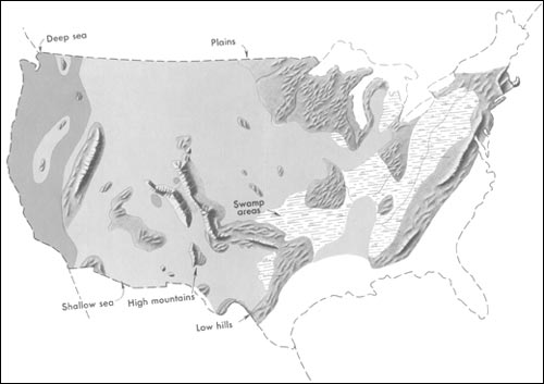
Pennsylvanian (geology)
The Pennsylvanian ( , also known as Upper Carboniferous or Late Carboniferous) is, in the International Commission on Stratigraphy, ICS geologic timescale, the younger of two period (geology), subperiods (or upper of two system (stratigraphy), subsystems) of the Carboniferous Period. It lasted from roughly . As with most other geochronology, geochronologic units, the stratum, rock beds that define the Pennsylvanian are well identified, but the exact date of the start and end are uncertain by a few hundred thousand years. The Pennsylvanian is named after the U.S. state of Pennsylvania, where the coal-productive beds of this age are widespread. The division between Pennsylvanian and Mississippian (geology), Mississippian comes from North American stratigraphy. In North America, where the early Carboniferous beds are primarily marine limestones, the Pennsylvanian was in the past treated as a full-fledged geologic period between the Mississippian and the Permian. In parts of Europe, ... [...More Info...] [...Related Items...] OR: [Wikipedia] [Google] [Baidu] |
Petrolacosaurus Skull Diagram
''Petrolacosaurus'' ("rock lake lizard") is an extinct genus of diapsid reptile from the late Carboniferous period. It was a small, long reptile, and the earliest known reptile with two temporal fenestrae (holes at the rear part of the skull). This means that it was at the base of Diapsida, the largest and most successful radiation of reptiles that would eventually include all modern reptile groups, as well as dinosaurs (which survive to the modern day as birds) and other famous extinct reptiles such as plesiosaurs, ichthyosaurs, and pterosaurs. However, ''Petrolacosaurus'' itself was part of Araeoscelida, a short-lived early branch of the diapsid family tree which went extinct in the mid-Permian. Discovery The first ''Petrolacosaurus'' fossil was found in 1932 in Garnett, Kansas, by a field expedition from the University of Kansas Natural History Museum. The party consisted of Henry H. Lane, Claude Hibbard, David Dunkle, Wallace Lane, Louis Coghill, and Curtis Hesse. Unfortun ... [...More Info...] [...Related Items...] OR: [Wikipedia] [Google] [Baidu] |
Vertebra
The spinal column, a defining synapomorphy shared by nearly all vertebrates,Hagfish are believed to have secondarily lost their spinal column is a moderately flexible series of vertebrae (singular vertebra), each constituting a characteristic irregular bone whose complex structure is composed primarily of bone, and secondarily of hyaline cartilage. They show variation in the proportion contributed by these two tissue types; such variations correlate on one hand with the cerebral/caudal rank (i.e., location within the backbone), and on the other with phylogenetic differences among the vertebrate taxa. The basic configuration of a vertebra varies, but the bone is its ''body'', with the central part of the body constituting the ''centrum''. The upper (closer to) and lower (further from), respectively, the cranium and its central nervous system surfaces of the vertebra body support attachment to the intervertebral discs. The posterior part of a vertebra forms a vertebral arch ... [...More Info...] [...Related Items...] OR: [Wikipedia] [Google] [Baidu] |
Sacrum
The sacrum (plural: ''sacra'' or ''sacrums''), in human anatomy, is a large, triangular bone at the base of the spine that forms by the fusing of the sacral vertebrae (S1S5) between ages 18 and 30. The sacrum situates at the upper, back part of the pelvic cavity, between the two wings of the pelvis. It forms joints with four other bones. The two projections at the sides of the sacrum are called the alae (wings), and articulate with the ilium at the L-shaped sacroiliac joints. The upper part of the sacrum connects with the last lumbar vertebra (L5), and its lower part with the coccyx (tailbone) via the sacral and coccygeal cornua. The sacrum has three different surfaces which are shaped to accommodate surrounding pelvic structures. Overall it is concave (curved upon itself). The base of the sacrum, the broadest and uppermost part, is tilted forward as the sacral promontory internally. The central part is curved outward toward the posterior, allowing greater room for the pel ... [...More Info...] [...Related Items...] OR: [Wikipedia] [Google] [Baidu] |
Cervical Vertebrae
In tetrapods, cervical vertebrae (singular: vertebra) are the vertebrae of the neck, immediately below the skull. Truncal vertebrae (divided into thoracic and lumbar vertebrae in mammals) lie caudal (toward the tail) of cervical vertebrae. In sauropsid species, the cervical vertebrae bear cervical ribs. In lizards and saurischian dinosaurs, the cervical ribs are large; in birds, they are small and completely fused to the vertebrae. The vertebral transverse processes of mammals are homologous to the cervical ribs of other amniotes. Most mammals have seven cervical vertebrae, with the only three known exceptions being the manatee with six, the two-toed sloth with five or six, and the three-toed sloth with nine. In humans, cervical vertebrae are the smallest of the true vertebrae and can be readily distinguished from those of the thoracic or lumbar regions by the presence of a foramen (hole) in each transverse process, through which the vertebral artery, vertebral veins, an ... [...More Info...] [...Related Items...] OR: [Wikipedia] [Google] [Baidu] |
Youngina
''Youngina'' is an extinct genus of diapsid reptile from the Late Permian Beaufort Group (''Tropidostoma''-''Dicynodon'' zones) of the Karoo Red Beds of South Africa. This, and a few related forms, make up the family Younginidae, within the Order Eosuchia (proposed by Broom in 1914). Eosuchia, having become a wastebasket taxon for many probably distantly-related primitive diapsid reptiles ranging from the Late Carboniferous to the Eocene, Romer proposed that it be replaced by Younginiformes (that included Younginidae and the Tangasauridae, ranging from the Permian to the Triassic). ''Youngina'' is known from several specimens. Many of these were attributed to as separate genera and species (such as ''Youngoides'' and ''Youngopsis''), but it was later realized that they were not distinct from ''Y. capensis''. The holotype specimen of ''Youngina'' was described briefly in 1914. The "''Youngoides romeri''" specimen was first attributed to ''Youngina'', but later given its eponymous ... [...More Info...] [...Related Items...] OR: [Wikipedia] [Google] [Baidu] |
Mandible
In anatomy, the mandible, lower jaw or jawbone is the largest, strongest and lowest bone in the human facial skeleton. It forms the lower jaw and holds the lower tooth, teeth in place. The mandible sits beneath the maxilla. It is the only movable bone of the skull (discounting the ossicles of the middle ear). It is connected to the temporal bones by the temporomandibular joints. The bone is formed prenatal development, in the fetus from a fusion of the left and right mandibular prominences, and the point where these sides join, the mandibular symphysis, is still visible as a faint ridge in the midline. Like other symphyses in the body, this is a midline articulation where the bones are joined by fibrocartilage, but this articulation fuses together in early childhood.Illustrated Anatomy of the Head and Neck, Fehrenbach and Herring, Elsevier, 2012, p. 59 The word "mandible" derives from the Latin word ''mandibula'', "jawbone" (literally "one used for chewing"), from ''wikt:mandere ... [...More Info...] [...Related Items...] OR: [Wikipedia] [Google] [Baidu] |
Synapsid
Synapsids + (, 'arch') > () "having a fused arch"; synonymous with ''theropsids'' (Greek, "beast-face") are one of the two major groups of animals that evolved from basal amniotes, the other being the sauropsids, the group that includes reptiles and birds. The group includes mammals and every animal more closely related to mammals than to sauropsids. Unlike other amniotes, synapsids have a single temporal fenestra, an opening low in the skull roof behind each eye orbit, leaving a bony arch beneath each; this accounts for their name. The distinctive temporal fenestra developed about 318 million years ago during the Late Carboniferous period, when synapsids and sauropsids diverged, but was subsequently merged with the orbit in early mammals. Traditionally, non-mammalian synapsids were believed to have evolved from reptiles, and therefore described as mammal-like reptiles in classical systematics, and primitive synapsids were also referred to as pelycosaurs, or pelycosaur-gr ... [...More Info...] [...Related Items...] OR: [Wikipedia] [Google] [Baidu] |
Sauropsida
Sauropsida ("lizard faces") is a clade of amniotes, broadly equivalent to the class Reptilia. Sauropsida is the sister taxon to Synapsida, the other clade of amniotes which includes mammals as its only modern representatives. Although early synapsids have historically been referred to as "mammal-like reptiles", all synapsids are more closely related to mammals than to any modern reptile. Sauropsids, on the other hand, include all amniotes more closely related to modern reptiles than to mammals. This includes Aves (birds), which are now recognized as a subgroup of archosaurian reptiles despite originally being named as a separate class in Linnaean taxonomy. The base of Sauropsida forks into two main groups of "reptiles": Eureptilia ("true reptiles") and Parareptilia ("next to reptiles"). Eureptilia encompasses all living reptiles (including birds), as well as various extinct groups. Parareptilia is typically considered to be an entirely extinct group, though a few hypotheses for ... [...More Info...] [...Related Items...] OR: [Wikipedia] [Google] [Baidu] |
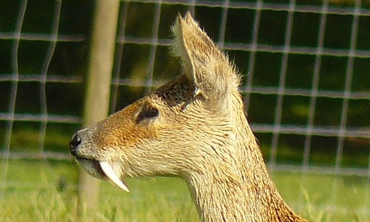
Canine Tooth
In mammalian oral anatomy, the canine teeth, also called cuspids, dog teeth, or (in the context of the upper jaw) fangs, eye teeth, vampire teeth, or vampire fangs, are the relatively long, pointed teeth. They can appear more flattened however, causing them to resemble incisors and leading them to be called ''incisiform''. They developed and are used primarily for firmly holding food in order to tear it apart, and occasionally as weapons. They are often the largest teeth in a mammal's mouth. Individuals of most species that develop them normally have four, two in the upper jaw and two in the lower, separated within each jaw by incisors; humans and dogs are examples. In most species, canines are the anterior-most teeth in the maxillary bone. The four canines in humans are the two maxillary canines and the two mandibular canines. Details There are generally four canine teeth: two in the upper (maxillary) and two in the lower (mandibular) arch. A canine is placed laterally to ... [...More Info...] [...Related Items...] OR: [Wikipedia] [Google] [Baidu] |
Maxilla
The maxilla (plural: ''maxillae'' ) in vertebrates is the upper fixed (not fixed in Neopterygii) bone of the jaw formed from the fusion of two maxillary bones. In humans, the upper jaw includes the hard palate in the front of the mouth. The two maxillary bones are fused at the intermaxillary suture, forming the anterior nasal spine. This is similar to the mandible (lower jaw), which is also a fusion of two mandibular bones at the mandibular symphysis. The mandible is the movable part of the jaw. Structure In humans, the maxilla consists of: * The body of the maxilla * Four processes ** the zygomatic process ** the frontal process of maxilla ** the alveolar process ** the palatine process * three surfaces – anterior, posterior, medial * the Infraorbital foramen * the maxillary sinus * the incisive foramen Articulations Each maxilla articulates with nine bones: * two of the cranium: the frontal and ethmoid * seven of the face: the nasal, zygomatic, lacrimal, inferior n ... [...More Info...] [...Related Items...] OR: [Wikipedia] [Google] [Baidu] |
Premaxilla
The premaxilla (or praemaxilla) is one of a pair of small cranial bones at the very tip of the upper jaw of many animals, usually, but not always, bearing teeth. In humans, they are fused with the maxilla. The "premaxilla" of therian mammal has been usually termed as the incisive bone. Other terms used for this structure include premaxillary bone or ''os premaxillare'', intermaxillary bone or ''os intermaxillare'', and Goethe's bone. Human anatomy In human anatomy, the premaxilla is referred to as the incisive bone (') and is the part of the maxilla which bears the incisor teeth, and encompasses the anterior nasal spine and alar region. In the nasal cavity, the premaxillary element projects higher than the maxillary element behind. The palatal portion of the premaxilla is a bony plate with a generally transverse orientation. The incisive foramen is bound anteriorly and laterally by the premaxilla and posteriorly by the palatine process of the maxilla. It is formed from the ... [...More Info...] [...Related Items...] OR: [Wikipedia] [Google] [Baidu] |


.jpg)

