|
Periodontists
Periodontology or periodontics (from Ancient Greek , – 'around'; and , – 'tooth', genitive , ) is the specialty of dentistry that studies supporting structures of teeth, as well as diseases and conditions that affect them. The supporting tissues are known as the periodontium, which includes the gingiva (gums), alveolar bone, cementum, and the periodontal ligament. A periodontist is a dentist that specializes in the prevention, diagnosis and treatment of periodontal disease and in the placement of dental implants. The periodontium The term ''periodontium'' is used to describe the group of structures that directly surround, support and protect the teeth. The periodontium is composed largely of the gingival tissue and the supporting bone. Gingivae Normal gingiva may range in color from light coral pink to heavily pigmented. The soft tissues and connective fibres that cover and protect the underlying cementum, periodontal ligament and alveolar bone are known as the gingiva ... [...More Info...] [...Related Items...] OR: [Wikipedia] [Google] [Baidu] |
Specialty (dentistry)
In the United States and Canada, there are twelve recognized dental specialties in which some dentists choose to train and practice, in addition to or instead of general dentistry. In the United Kingdom and Australia, there are thirteen. To become a specialist requires training in a residency or advanced graduate training program. Once a residency is completed, the doctor is granted a certificate of specialty training. Many specialty programs have optional or required advanced degrees such as a master's degree, such as the Master of Science (MS or MSc), Master of Dental Surgery/Science (MDS/MDSc), Master of Dentistry (MDent), Master of Clinical Dentistry (MClinDent), Master of Philosophy (MPhil), Master of Medical Science (MMS or (MMSc); doctorate such as Doctor of Clinical Dentistry (DClinDent), Doctor of Medical Science/Sciences (DMSc), or PhD;or medical degree: Doctor of Medicine/ Bachelor of Medicine, Bachelor of Surgery (MD/MBBS) specific to maxillofacial surgery and so ... [...More Info...] [...Related Items...] OR: [Wikipedia] [Google] [Baidu] |
Gingival Sulcus
The gingival sulcus is an area of potential space between a tooth and the surrounding gingival tissue and is lined by sulcular epithelium. The depth of the sulcus (Latin for ''groove'') is bounded by two entities: apically by the gingival fibers of the connective tissue attachment and coronally by the free gingival margin. A healthy sulcular depth is three millimeters or less, which is readily self-cleansable with a properly used toothbrush or the supplemental use of other oral hygiene aids. Anatomy The Dentogingival tissues consist of many constituents, such as the enamel or cementum of the tooth and the connective tissue supporting epithelia like the junctional epithelium, the gingival epithelium and the sulcular epithelium. The junctional epithelium is developed during the eruption of teeth when the reduced enamel epithelium merges with the oral epithelium The reduced enamel epithelium forms the first junctional epithelium and is firmly attached to the enamel. In ... [...More Info...] [...Related Items...] OR: [Wikipedia] [Google] [Baidu] |
Gingivitis
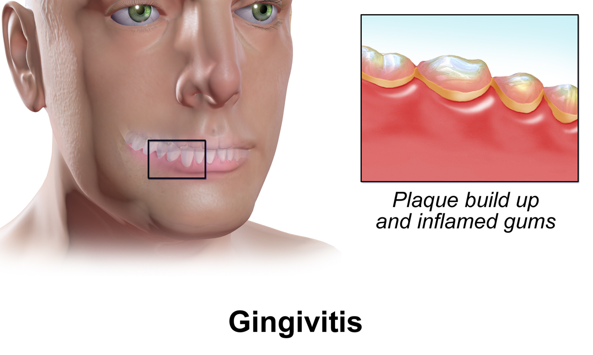
Gingivitis is a non-destructive disease that causes inflammation of the gums. The most common form of gingivitis, and the most common form of periodontal disease overall, is in response to bacterial biofilms (also called plaque) that is attached to tooth surfaces, termed ''plaque-induced gingivitis''. Most forms of gingivitis are plaque-induced. While some cases of gingivitis never progress to periodontitis, periodontitis is always preceded by gingivitis. Gingivitis is reversible with good oral hygiene; however, without treatment, gingivitis can progress to periodontitis, in which the inflammation of the gums results in tissue destruction and bone resorption around the teeth. Periodontitis can ultimately lead to tooth loss. Signs and symptoms The symptoms of gingivitis are somewhat non-specific and manifest in the gum tissue as the classic signs of inflammation: *Swollen gums *Bright red gums *Gums that are tender or painful to the touch *Bleeding gums or bleeding after br ... [...More Info...] [...Related Items...] OR: [Wikipedia] [Google] [Baidu] |
Dental Canaliculi
Bone canaliculi are microscopic canals between the lacunae of ossified bone. The radiating processes of the osteocytes (called filopodia) project into these canals. These cytoplasmic processes are joined together by gap junctions. Osteocytes do not entirely fill up the canaliculi. The remaining space is known as the periosteocytic space, which is filled with periosteocytic fluid. This fluid contains substances too large to be transported through the gap junctions that connect the osteocytes. In cartilage, the lacunae and hence, the chondrocytes, are isolated from each other. Materials picked up by osteocytes adjacent to blood vessels are distributed throughout the bone matrix via the canaliculi. Dental canaliculi The dental canaliculi (sometimes called dentinal tubules) are the blood supply of a tooth. Odontoblast process run in the canaliculi that transverse the dentin layer and are referred as dentinal tubules. The number and size of the canaliculi decrease as the tubule ... [...More Info...] [...Related Items...] OR: [Wikipedia] [Google] [Baidu] |
Dentin
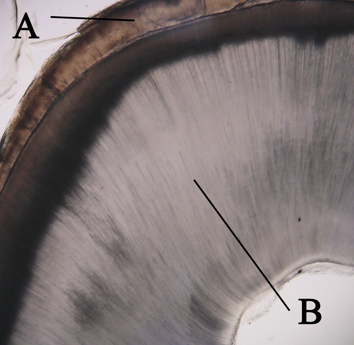
Dentin () (American English) or dentine ( or ) (British English) ( la, substantia eburnea) is a calcified tissue of the body and, along with enamel, cementum, and pulp, is one of the four major components of teeth. It is usually covered by enamel on the crown and cementum on the root and surrounds the entire pulp. By volume, 45% of dentin consists of the mineral hydroxyapatite, 33% is organic material, and 22% is water. Yellow in appearance, it greatly affects the color of a tooth due to the translucency of enamel. Dentin, which is less mineralized and less brittle than enamel, is necessary for the support of enamel. Dentin rates approximately 3 on the Mohs scale of mineral hardness. There are two main characteristics which distinguish dentin from enamel: firstly, dentin forms throughout life; secondly, dentin is sensitive and can become hypersensitive to changes in temperature due to the sensory function of odontoblasts, especially when enamel recedes and dentin channels b ... [...More Info...] [...Related Items...] OR: [Wikipedia] [Google] [Baidu] |
Cementum
Cementum is a specialized calcified substance covering the root of a tooth. The cementum is the part of the periodontium that attaches the teeth to the alveolar bone by anchoring the periodontal ligament.Illustrated Dental Embryology, Histology, and Anatomy, Bath-Balogh and Fehrenbach, Elsevier, 2011, page 170. Structure The cells of cementum are the entrapped cementoblasts, the cementocytes. Each cementocyte lies in its lacuna, similar to the pattern noted in bone. These lacunae also have canaliculi or canals. Unlike those in bone, however, these canals in cementum do not contain nerves, nor do they radiate outward. Instead, the canals are oriented toward the periodontal ligament and contain cementocytic processes that exist to diffuse nutrients from the ligament because it is vascularized. After the apposition of cementum in layers, the cementoblasts that do not become entrapped in cementum line up along the cemental surface along the length of the outer covering of the per ... [...More Info...] [...Related Items...] OR: [Wikipedia] [Google] [Baidu] |
Alveolar Process
The alveolar process () or alveolar bone is the thickened ridge of bone that contains the tooth sockets on the jaw bones (in humans, the maxilla and the mandible). The structures are covered by gums as part of the oral cavity. The synonymous terms ''alveolar ridge'' and ''alveolar margin'' are also sometimes used more specifically to refer to the ridges on the inside of the mouth which can be felt with the tongue, either on roof of the mouth between the upper teeth and the hard palate or on the bottom of the mouth behind the lower teeth. Terminology The term ''alveolar'' () ('hollow') refers to the cavities of the tooth sockets, known as dental alveoli. The alveolar process is also called the ''alveolar bone'' or ''alveolar ridge''. The curved portion is referred to as the alveolar arch. The alveolar bone proper, also called bundle bone, directly surrounds the teeth. The term alveolar crest describes the extreme rim of the bone nearest to the crowns of the teeth. The portio ... [...More Info...] [...Related Items...] OR: [Wikipedia] [Google] [Baidu] |
Sharpey's Fibres
Sharpey's fibres (bone fibres, or perforating fibres) are a matrix of connective tissue consisting of bundles of strong predominantly type I collagen fibres connecting periosteum to bone. They are part of the outer fibrous layer of periosteum, entering into the outer circumferential and interstitial lamellae of bone tissue. Sharpey's fibres are also used to attach muscle to the periosteum of bone by merging with the fibrous periosteum and underlying bone as well. A good example is the attachment of the rotator cuff muscles to the blade of the scapula. In the teeth, Sharpey's fibres are the terminal ends of principal fibres (of the periodontal ligament) that insert into the cementum and into the periosteum of the alveolar bone. A study on rats suggests that the three-dimensional structure of Sharpey's fibres intensifies the continuity between the periodontal ligament fibre and the alveolar bone (tooth socket), and acts as a buffer medium against stress. Sharpey's fibres in the ... [...More Info...] [...Related Items...] OR: [Wikipedia] [Google] [Baidu] |
Periodontal Fiber
The periodontal ligament, commonly abbreviated as the PDL, is a group of specialized connective tissue fibers that essentially attach a tooth to the alveolar bone within which it sits. It inserts into root cementum one side and onto alveolar bone on the other. Structure The PDL consists of principal fibres, loose connective tissue, blast and clast cells, oxytalan fibres and Cell Rest of Malassez. Alveolodental ligament The main principal fiber group is the alveolodental ligament, which consists of five fiber subgroups: alveolar crest, horizontal, oblique, apical, and interradicular on multirooted teeth. Principal fibers other than the alveolodental ligament are the transseptal fibers. All these fibers help the tooth withstand the naturally substantial compressive forces that occur during chewing and remain embedded in the bone. The ends of the principal fibers that are within either cementum or alveolar bone proper are considered Sharpey fibers. * Alveolar crest fibers ('' ... [...More Info...] [...Related Items...] OR: [Wikipedia] [Google] [Baidu] |
Interdental Gingiva
The interdental papilla, also known as the interdental gingiva, is the part of the gums (gingiva) that exists coronal to the free gingival margin on the buccal and lingual surfaces of the teeth A tooth ( : teeth) is a hard, calcified structure found in the jaws (or mouths) of many vertebrates and used to break down food. Some animals, particularly carnivores and omnivores, also use teeth to help with capturing or wounding prey, te .... The interdental papillae fill in the area between the teeth apical to their contact areas to prevent food impaction; they assume a conical shape for the anterior teeth and a blunted shape buccolingually for the posterior teeth. A missing papilla is often visible as a small triangular gap between adjacent teeth. The relationship of interdental bone to the interproximal contact point between adjacent teeth is a determining factor in whether the interdental papilla will be present. If greater than 8mm exist between the interdental bone and t ... [...More Info...] [...Related Items...] OR: [Wikipedia] [Google] [Baidu] |
Chewing
Chewing or mastication is the process by which food is crushed and ground by teeth. It is the first step of digestion, and it increases the surface area of foods to allow a more efficient break down by enzymes. During the mastication process, the food is positioned by the cheek and tongue between the teeth for grinding. The muscles of mastication move the jaws to bring the teeth into intermittent contact, repeatedly occluding and opening. As chewing continues, the food is made softer and warmer, and the enzymes in saliva begin to break down carbohydrates in the food. After chewing, the food (now called a bolus) is swallowed. It enters the esophagus and via peristalsis continues on to the stomach, where the next step of digestion occurs. Increasing the number of chews per bite increases relevant gut hormones. Studies suggest that chewing may decrease self-reported hunger and food intake. Chewing gum has been around for many centuries; there is evidence that northern Europ ... [...More Info...] [...Related Items...] OR: [Wikipedia] [Google] [Baidu] |
Mucogingival Junction
A mucogingival junction is an anatomical feature found on the intraoral mucosa. The mucosa of the cheeks and floor of the mouth are freely moveable and fragile, whereas the mucosa around the teeth and on the palate are firm and keratinized. Where the two tissue types meet is known as a mucogingival junction. There are three mucogingival junctions: on the facial of the maxilla and on both the facial and lingual of the mandible. The palatal gingiva of the maxilla is continuous with the tissue of the palate, which is bound down to the palatal bones. Because the palate is devoid of freely moveable alveolar mucosa, there is no mucogingival junction.Carranza's Clinical Periodontology, W.B. Saunders 2002, page 17. Clinical importance The clinical importance of the mucogingival junction is in measuring the width of attached gingiva. Attached gingiva is important because it is bound very tightly to the underlying alveolar bone and provides protection to the mucosa during functional use ... [...More Info...] [...Related Items...] OR: [Wikipedia] [Google] [Baidu] |


.jpg)
