|
Periclimenes Pholeter
''Periclimenes pholeter'', is a species of shrimp belonging to the family Palaemonidae. The species is closest to '' Periclimenes indicus'', '' P. obscurus'' and '' P. toloensis'', resembling these species in the presence of an epigastric tooth on the carapace, the shape of the abdomen, the spinulation of the carapace, and the unarmed fingers of the first chelipeds. ''P. pholeter'' most resembles ''P. indicus'' by the elongate carpus and long fingers of the second pereiopods, differing in these features from ''P. toloensis'', which has the fingers slightly less than half as long as the palm. In ''P. obscurus'' the fingers are shorter than the palm, but the carpus is about as long as the palm. From ''P. indicus'', this species differs: by the greater size; by the much higher rostrum and the greater number of ventral rostral teeth; by the shorter eye; by the less slender antennular peduncle; by the more deeply cleft upper antennular flagellum ... [...More Info...] [...Related Items...] OR: [Wikipedia] [Google] [Baidu] |
Lipke Holthuis
Lipke Bijdeley Holthuis (21 April 1921 – 7 March 2008) was a Dutch crustacean, carcinologist, considered one of the "undisputed greats" of carcinology, and "the greatest carcinologist of our time". Holthuis was born in Probolinggo, East Java and obtained his doctorate from Leiden University on 23 January 1946. He was appointed the assistant curator of the ''Rijksmuseum van Natuurlijke Historie'' (now ''Naturalis'') in Leiden in 1941. He was the most prolific carcinologist of the 20th century, publishing 620 papers (108 of which were in the Leiden Museum Journals) totalling 12,795 pages which is an average of 185 pages per year and an average of approximately 21 pages per paper. These were published on many groups of crustaceans, their natural history and nomenclature, and the history of carcinology. This steady stream of publications resulted in the description of 428 new taxa: 2 new families, 5 subfamilies, 83 genera and 338 species. 67 taxa were named after him between 1953 ( ... [...More Info...] [...Related Items...] OR: [Wikipedia] [Google] [Baidu] |
Ischium
The ischium () forms the lower and back region of the (''os coxae''). Situated below the ilium and behind the pubis, it is one of three regions whose fusion creates the . The superior portion of this region forms approximately one-third of the acetabulum. |
Setae
In biology, setae (singular seta ; from the Latin word for "bristle") are any of a number of different bristle- or hair-like structures on living organisms. Animal setae Protostomes Annelid setae are stiff bristles present on the body. They help, for example, earthworms to attach to the surface and prevent backsliding during peristaltic motion. These hairs make it difficult to pull a worm straight from the ground. Setae in oligochaetes (a group including earthworms) are largely composed of chitin. They are classified according to the limb to which they are attached; for instance, notosetae are attached to notopodia; neurosetae to neuropodia. Crustaceans have mechano- and chemosensory setae. Setae are especially present on the mouthparts of crustaceans and can also be found on grooming limbs. In some cases, setae are modified into scale like structures. Setae on the legs of krill and other small crustaceans help them to gather phytoplankton. It captures them and allows ... [...More Info...] [...Related Items...] OR: [Wikipedia] [Google] [Baidu] |
Spinule
Spinules are small spines or thorns (vertebral columns) that are part of biological and manmade structures. The word originates from the Latin word and is often used in botany and zoology. The presence or absence of spinules, and their shape, can differentiate species and is used to describe and distinguish anatomical features. The development of spinules in the eye may be affected by dopamine, circadian rhythms, and exposure to light or dark environments, according to a studies of controlling mechanisms. liquid-crystal displays (LCDs) can employ an anisotropic Anisotropy () is the property of a material which allows it to change or assume different properties in different directions, as opposed to isotropy. It can be defined as a difference, when measured along different axes, in a material's physic ... conducting film (ACF) that "consists of an epoxy resin and nickel particles with spinules". [...More Info...] [...Related Items...] OR: [Wikipedia] [Google] [Baidu] |
Dactylus
The dactylus is the tip region of the tentacular club of cephalopods and of the leg of some crustaceans (see arthropod leg). In cephalopods, the dactylus is narrow and often characterized by the asymmetrical placement of suckers (i.e., the ventral expansion of the club) and the absence of a dorsal protective membrane. In crustaceans, the dactylus is the seventh and terminal segment of their thoracic appendages. In certain instances the dactylus, together with the propodus, form the claw. The term ''dactylus'' means "finger" in Greek Greek may refer to: Greece Anything of, from, or related to Greece, a country in Southern Europe: *Greeks, an ethnic group. *Greek language, a branch of the Indo-European language family. **Proto-Greek language, the assumed last common ancestor .... References Cephalopod zootomy Crustacean anatomy {{Animal anatomy-stub ... [...More Info...] [...Related Items...] OR: [Wikipedia] [Google] [Baidu] |
Chela (organ)
A chela ()also called a claw, nipper, or pinceris a pincer (biology), pincer-like organ at the end of certain limbs of some arthropods. The name comes from Ancient Greek , through New Latin '. The plural form is chelae. Legs bearing a chela are called chelipeds. Another name is ''claw'' because most chelae are curved and have a sharp point like a claw. Chelae can be present at the tips of arthropod legs as well as their pedipalps. Chelae are distinct from spider chelicerae in that they do not contain venomous glands and cannot distribute venom. See also * Pincer (biology) * Pincer (tool) References Arthropod anatomy {{Arthropod-anatomy-stub ... [...More Info...] [...Related Items...] OR: [Wikipedia] [Google] [Baidu] |
Maxilliped
An appendage (or outgrowth) is an external body part, or natural prolongation, that protrudes from an organism's body. In arthropods, an appendage refers to any of the homologous body parts that may extend from a body segment, including antennae, mouthparts (including mandibles, maxillae and maxillipeds), gills, locomotor legs ( pereiopods for walking, and pleopods for swimming), sexual organs (gonopods), and parts of the tail (uropods). Typically, each body segment carries one pair of appendages. An appendage which is modified to assist in feeding is known as a maxilliped or gnathopod. In vertebrates, an appendage can refer to a locomotor part such as a tail, fins on a fish, limbs (legs, flippers or wings) on a tetrapod; exposed sex organ; defensive parts such as horns and antlers; or sensory organs such as auricles, proboscis ( trunk and snout) and barbels. Appendages may become ''uniramous'', as in insects and centipedes, where each appendage comprises a single ... [...More Info...] [...Related Items...] OR: [Wikipedia] [Google] [Baidu] |
Endite
The arthropod leg is a form of jointed appendage of arthropods, usually used for walking. Many of the terms used for arthropod leg segments (called podomeres) are of Latin origin, and may be confused with terms for bones: ''coxa'' (meaning hip, plural ''coxae''), ''trochanter'', ''femur'' (plural ''femora''), ''tibia'' (plural ''tibiae''), ''tarsus'' (plural ''tarsi''), ''ischium'' (plural ''ischia''), ''metatarsus'', ''carpus'', ''dactylus'' (meaning finger), ''patella'' (plural ''patellae''). Homologies of leg segments between groups are difficult to prove and are the source of much argument. Some authors posit up to eleven segments per leg for the most recent common ancestor of extant arthropods but modern arthropods have eight or fewer. It has been argued that the ancestral leg need not have been so complex, and that other events, such as successive loss of function of a ''Hox''-gene, could result in parallel gains of leg segments. In arthropods, each of the leg segments artic ... [...More Info...] [...Related Items...] OR: [Wikipedia] [Google] [Baidu] |
Maxilla
The maxilla (plural: ''maxillae'' ) in vertebrates is the upper fixed (not fixed in Neopterygii) bone of the jaw formed from the fusion of two maxillary bones. In humans, the upper jaw includes the hard palate in the front of the mouth. The two maxillary bones are fused at the intermaxillary suture, forming the anterior nasal spine. This is similar to the mandible (lower jaw), which is also a fusion of two mandibular bones at the mandibular symphysis. The mandible is the movable part of the jaw. Structure In humans, the maxilla consists of: * The body of the maxilla * Four processes ** the zygomatic process ** the frontal process of maxilla ** the alveolar process ** the palatine process * three surfaces – anterior, posterior, medial * the Infraorbital foramen * the maxillary sinus * the incisive foramen Articulations Each maxilla articulates with nine bones: * two of the cranium: the frontal and ethmoid * seven of the face: the nasal, zygomatic, lacrimal, inferior n ... [...More Info...] [...Related Items...] OR: [Wikipedia] [Google] [Baidu] |
Molar (tooth)
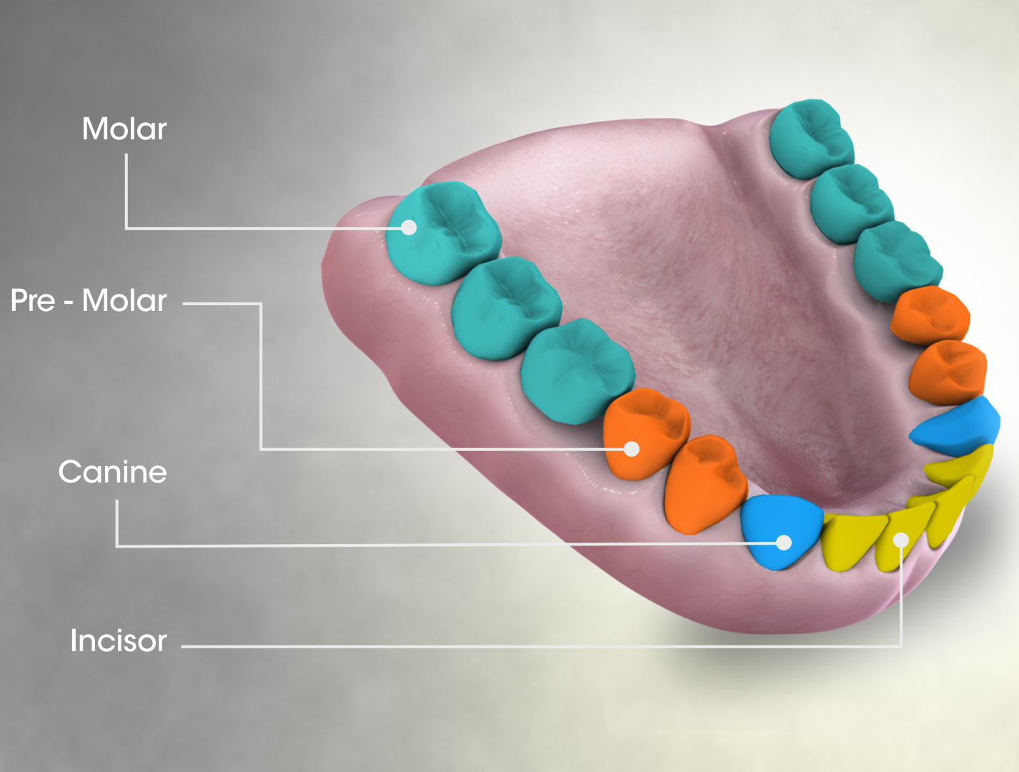
The molars or molar teeth are large, flat teeth at the back of the mouth. They are more developed in mammals. They are used primarily to grind food during chewing. The name ''molar'' derives from Latin, ''molaris dens'', meaning "millstone tooth", from ''mola'', millstone and ''dens'', tooth. Molars show a great deal of diversity in size and shape across mammal groups. The third molar of humans is sometimes vestigial. Human anatomy In humans, the molar teeth have either four or five cusps. Adult humans have 12 molars, in four groups of three at the back of the mouth. The third, rearmost molar in each group is called a wisdom tooth. It is the last tooth to appear, breaking through the front of the gum at about the age of 20, although this varies from individual to individual. Race can also affect the age at which this occurs, with statistical variations between groups. In some cases, it may not even erupt at all. The human mouth contains upper (maxillary) and lower (mandib ... [...More Info...] [...Related Items...] OR: [Wikipedia] [Google] [Baidu] |
Incisor
Incisors (from Latin ''incidere'', "to cut") are the front teeth present in most mammals. They are located in the premaxilla above and on the mandible below. Humans have a total of eight (two on each side, top and bottom). Opossums have 18, whereas armadillos have none. Structure Adult humans normally have eight incisors, two of each type. The types of incisor are: * maxillary central incisor (upper jaw, closest to the center of the lips) * maxillary lateral incisor (upper jaw, beside the maxillary central incisor) * mandibular central incisor (lower jaw, closest to the center of the lips) * mandibular lateral incisor (lower jaw, beside the mandibular central incisor) Children with a full set of deciduous teeth (primary teeth) also have eight incisors, named the same way as in permanent teeth. Young children may have from zero to eight incisors depending on the stage of their tooth eruption and tooth development. Typically, the mandibular central incisors erupt first, followed ... [...More Info...] [...Related Items...] OR: [Wikipedia] [Google] [Baidu] |


