|
Parsonage–Turner Syndrome
Parsonage–Turner syndrome, also known as acute brachial neuropathy, neuralgic amyotrophy and abbreviated PTS, is a syndrome of unknown cause; although many specific risk factors have been identified (such as; post-operative, post-infectious, post-traumatic or post-vaccination), the cause is still unknown. The condition manifests as a rare set of symptoms most likely resulting from autoimmune inflammation of unknown cause of the brachial plexus. Parsonage–Turner syndrome occurs in about 1.6 out of 100,000 people every year. Signs and symptoms This syndrome can begin with severe shoulder or arm pain followed by weakness and numbness. Those with Parsonage–Turner experience acute, sudden-onset pain radiating from the shoulder to the upper arm. Affected muscles become weak and atrophied, and in advanced cases, paralyzed. Occasionally, there will be no pain and just paralysis, and sometimes just pain, not ending in paralysis. MRI may assist in diagnosis. Scapular winging is com ... [...More Info...] [...Related Items...] OR: [Wikipedia] [Google] [Baidu] |
Brachial Plexus
The brachial plexus is a network () of nerves formed by the anterior rami of the lower four cervical nerves and first thoracic nerve ( C5, C6, C7, C8, and T1). This plexus extends from the spinal cord, through the cervicoaxillary canal in the neck, over the first rib, and into the armpit, it supplies afferent and efferent nerve fibers the to chest, shoulder, arm, forearm, and hand. Structure The brachial plexus is divided into five ''roots'', three ''trunks'', six ''divisions'' (three anterior and three posterior), three ''cords'', and five ''branches''. There are five "terminal" branches and numerous other "pre-terminal" or "collateral" branches, such as the subscapular nerve, the thoracodorsal nerve, and the long thoracic nerve, that leave the plexus at various points along its length. A common structure used to identify part of the brachial plexus in cadaver dissections is the M or W shape made by the musculocutaneous nerve, lateral cord, median nerve, medial cord, and ... [...More Info...] [...Related Items...] OR: [Wikipedia] [Google] [Baidu] |
Atrophy
Atrophy is the partial or complete wasting away of a part of the body. Causes of atrophy include mutations (which can destroy the gene to build up the organ), poor nourishment, poor circulation, loss of hormonal support, loss of nerve supply to the target organ, excessive amount of apoptosis of cells, and disuse or lack of exercise or disease intrinsic to the tissue itself. In medical practice, hormonal and nerve inputs that maintain an organ or body part are said to have ''trophic'' effects. A diminished muscular trophic condition is designated as ''atrophy''. Atrophy is reduction in size of cell, organ or tissue, after attaining its normal mature growth. In contrast, hypoplasia is the reduction in the cellular numbers of an organ, or tissue that has not attained normal maturity. Atrophy is the general physiological process of reabsorption and breakdown of tissues, involving apoptosis. When it occurs as a result of disease or loss of trophic support because of other diseases ... [...More Info...] [...Related Items...] OR: [Wikipedia] [Google] [Baidu] |
Magnetic Resonance Imaging
Magnetic resonance imaging (MRI) is a medical imaging technique used in radiology to form pictures of the anatomy and the physiological processes of the body. MRI scanners use strong magnetic fields, magnetic field gradients, and radio waves to generate images of the organs in the body. MRI does not involve X-rays or the use of ionizing radiation, which distinguishes it from CT and PET scans. MRI is a medical application of nuclear magnetic resonance (NMR) which can also be used for imaging in other NMR applications, such as NMR spectroscopy. MRI is widely used in hospitals and clinics for medical diagnosis, staging and follow-up of disease. Compared to CT, MRI provides better contrast in images of soft-tissues, e.g. in the brain or abdomen. However, it may be perceived as less comfortable by patients, due to the usually longer and louder measurements with the subject in a long, confining tube, though "Open" MRI designs mostly relieve this. Additionally, implants and oth ... [...More Info...] [...Related Items...] OR: [Wikipedia] [Google] [Baidu] |
Winged Scapula
A winged scapula (scapula alata) is a skeletal medical condition in which the shoulder blade protrudes from a person's back in an abnormal position. In rare conditions it has the potential to lead to limited functional activity in the upper extremity to which it is adjacent. It can affect a person's ability to lift, pull, and push weighty objects. In some serious cases, the ability to perform activities of daily living such as changing one's clothes and washing one's hair may be hindered. The name of this condition comes from its appearance, a wing-like resemblance, due to the medial border of the scapula sticking straight out from the back. Scapular winging has been observed to disrupt scapulohumeral rhythm, contributing to decreased flexion and abduction of the upper extremity, as well as a loss in power and the source of considerable pain. A winged scapula is considered normal posture in young children, but not older children and adults. Signs and symptoms The severity ... [...More Info...] [...Related Items...] OR: [Wikipedia] [Google] [Baidu] |
Supraspinatus
The supraspinatus (plural ''supraspinati'') is a relatively small muscle of the upper back that runs from the supraspinous fossa superior portion of the scapula (shoulder blade) to the greater tubercle of the humerus. It is one of the four rotator cuff muscles and also abducts the arm at the shoulder. The spine of the scapula separates the supraspinatus muscle from the infraspinatus muscle, which originates below the spine. Structure The supraspinatus muscle arises from the supraspinous fossa, a shallow depression in the body of the scapula above its spine. The supraspinatus muscle tendon passes laterally beneath the cover of the acromion. Research in 1996 showed that the postero-lateral origin was more lateral than classically described. The supraspinatus tendon is inserted into the superior facet of the greater tubercle of the humerus. The distal attachments of the three rotator cuff muscles that insert into the greater tubercle of the humerus can be abbreviated as SIT when vie ... [...More Info...] [...Related Items...] OR: [Wikipedia] [Google] [Baidu] |
Infraspinatus
In human anatomy, the infraspinatus muscle is a thick triangular muscle, which occupies the chief part of the infraspinatous fossa.''Gray's Anatomy'', see infobox. As one of the four muscles of the rotator cuff, the main function of the infraspinatus is to externally rotate the humerus and stabilize the shoulder joint. Structure It attaches medially to the infraspinous fossa of the scapula and laterally to the middle facet of the greater tubercle of the humerus. The muscle arises by fleshy fibers from the medial two-thirds of the infraspinatous fossa, and by tendinous fibers from the ridges on its surface; it also arises from the infraspinatous fascia which covers it, and separates it from the teres major and teres minor. The fibers converge to a tendon, which glides over the lateral border of the spine of the scapula and passing across the posterior part of the capsule of the shoulder-joint, is inserted into the middle impression on the greater tubercle of the humerus. The trape ... [...More Info...] [...Related Items...] OR: [Wikipedia] [Google] [Baidu] |
Deltoid Muscle
The deltoid muscle is the muscle forming the rounded contour of the human shoulder. It is also known as the 'common shoulder muscle', particularly in other animals such as the domestic cat. Anatomically, the deltoid muscle appears to be made up of three distinct sets of muscle fibers, namely the # anterior or clavicular part (pars clavicularis) # posterior or scapular part (pars scapularis) # intermediate or acromial part (pars acromialis) However, electromyography suggests that it consists of at least seven groups that can be independently coordinated by the nervous system. It was previously called the deltoideus (plural ''deltoidei'') and the name is still used by some anatomists. It is called so because it is in the shape of the Greek capital letter delta (Δ). Deltoid is also further shortened in slang as "delt". A study of 30 shoulders revealed an average mass of in humans, ranging from to . Structure Previous studies showed that the insertions of the tendons of the delto ... [...More Info...] [...Related Items...] OR: [Wikipedia] [Google] [Baidu] |
Axillary Nerve
The axillary nerve or the circumflex nerve is a nerve of the human body, that originates from the brachial plexus (upper trunk, posterior division, posterior cord) at the level of the axilla (armpit) and carries nerve fibers from C5 and C6. The axillary nerve travels through the quadrangular space with the posterior circumflex humeral artery and vein to innervate the deltoid and teres minor. Structure The nerve lies at first behind the axillary artery, and in front of the subscapularis, and passes downward to the lower border of that muscle. It then winds from anterior to posterior around the neck of the humerus, in company with the posterior humeral circumflex artery, through the quadrangular space (bounded above by the teres minor, below by the teres major, medially by the long head of the triceps brachii, and laterally by the surgical neck of the humerus), and divides into an anterior, a posterior, and a collateral branch to the long head of the triceps brachii branch. * The ... [...More Info...] [...Related Items...] OR: [Wikipedia] [Google] [Baidu] |
Quadrilateral Space Syndrome
Quadrilateral space syndrome is a rotator cuff denervation syndrome in which the axillary nerve is compressed at the quadrilateral space of the rotator cuff. Cause Diagnosis Diagnosis is usually suspected by clinical history and confirmed by MRI, in which edema of the teres minor is seen, with variable involvement of the deltoid. The circumflex humeral artery may also be compressed. Before the advent of MRI, compression of this vessel on angiography used to be the mechanism of diagnosis, although this is no longer done as it is an invasive procedure. Atrophy can occur in cases of chronic nerve impingement. It can be associated with a glenoid labral cyst, with the cyst also reflecting injury of the glenoid labrum.{{Cite journal, title = Quadrilateral space syndrome caused by glenoid labral cyst, journal = AJR. American Journal of Roentgenology, date = 2000-10-01, issn = 0361-803X, pmid = 11000173, pages = 1103–1105, volume = 175, issue = 4, doi = 10.2214/ajr.175.4.1751103, ... [...More Info...] [...Related Items...] OR: [Wikipedia] [Google] [Baidu] |
Teres Minor
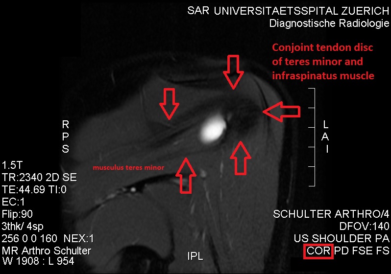
The teres minor (Latin ''teres'' meaning 'rounded') is a narrow, elongated muscle of the rotator cuff. The muscle originates from the lateral border and adjacent posterior surface of the corresponding right or left scapula and inserts at both the greater tubercle of the humerus and the posterior surface of the joint capsule. The primary function of the teres minor is to modulate the action of the deltoid, preventing the humeral head from sliding upward as the arm is abducted. It also functions to rotate the humerus laterally. The teres minor is innervated by the axillary nerve. Structure It arises from the dorsal surface of the axillary border of the scapula for the upper two-thirds of its extent, and from two aponeurotic laminae, one of which separates it from the infraspinatus muscle, the other from the teres major muscle. Its fibers run obliquely upwards and laterally; the upper ones end in a tendon which is inserted into the lowest of the three impressions on the greater tub ... [...More Info...] [...Related Items...] OR: [Wikipedia] [Google] [Baidu] |
Suprascapular Nerve
The suprascapular nerve is a nerve that branches from the upper trunk of the brachial plexus. It is responsible for the innervation of two of the muscles that originate from the scapula, namely the supraspinatus muscle, supraspinatus and infraspinatus muscles. Structure The suprascapular nerve arises from the upper trunk of the brachial plexus which is formed by the union of the Anterior ramus of spinal nerve, ventral rami of the fifth and sixth cervical nerves. After branching from the upper trunk, the nerve passes across the posterior triangle of the neck parallel to the inferior belly of the omohyoid muscle and deep to the trapezius muscle. It then runs along the Scapula#Borders, superior border of the scapula through the suprascapular canal, in which it enters via the suprascapular notch inferior to the superior transverse scapular ligament and enters the supraspinous fossa. It then passes beneath the supraspinatus, and curves around the lateral border of the spine of the scapul ... [...More Info...] [...Related Items...] OR: [Wikipedia] [Google] [Baidu] |
Spinoglenoid Notch
The great scapular notch (or ''spinoglenoid notch'') is a notch which serves to connect the supraspinous fossa and infraspinous fossa. It lies immediately medial to the attachment of the acromion to the lateral angle of the scapular spine. The Suprascapular artery and suprascapular nerve pass around the great scapular notch anteroposteriorly. Supraspinatus and infraspinatus are both supplied by the suprascapular nerve, which originates from the superior trunk of the brachial plexus (roots C5-C6). Additional images File:Great scapular notch - animation01.gif, Left scapula. Great scapular notch shown in red. File:Great scapular notch - animation04.gif, Animation. Great scapular notch shown in red. See also * Suprascapular notch The suprascapular notch (or ''scapular notch'') is a notch in the superior border of the scapula, just medial to the base of the coracoid process. It forms the entrance site into the suprascapular canal. Structure This notch is converted into ... ... [...More Info...] [...Related Items...] OR: [Wikipedia] [Google] [Baidu] |


