|
Paraventricular Nucleus Of Hypothalamus
The paraventricular nucleus (PVN, PVA, or PVH) is a nucleus in the hypothalamus. Anatomically, it is adjacent to the third ventricle and many of its neurons project to the posterior pituitary. These projecting neurons secrete oxytocin and a smaller amount of vasopressin, otherwise the nucleus also secretes corticotropin-releasing hormone (CRH) and thyrotropin-releasing hormone (TRH). CRH and TRH are secreted into the hypophyseal portal system and act on different targets neurons in the anterior pituitary. PVN is thought to mediate many diverse functions through these different hormones, including osmoregulation, appetite, and the response of the body to stress. Location The paraventricular nucleus lies adjacent to the third ventricle. It lies within the periventricular zone and is not to be confused with the periventricular nucleus, which occupies a more medial position, beneath the third ventricle. The PVN is highly vascularised and is protected by the blood–brain barrier, althou ... [...More Info...] [...Related Items...] OR: [Wikipedia] [Google] [Baidu] |
Vasopressin
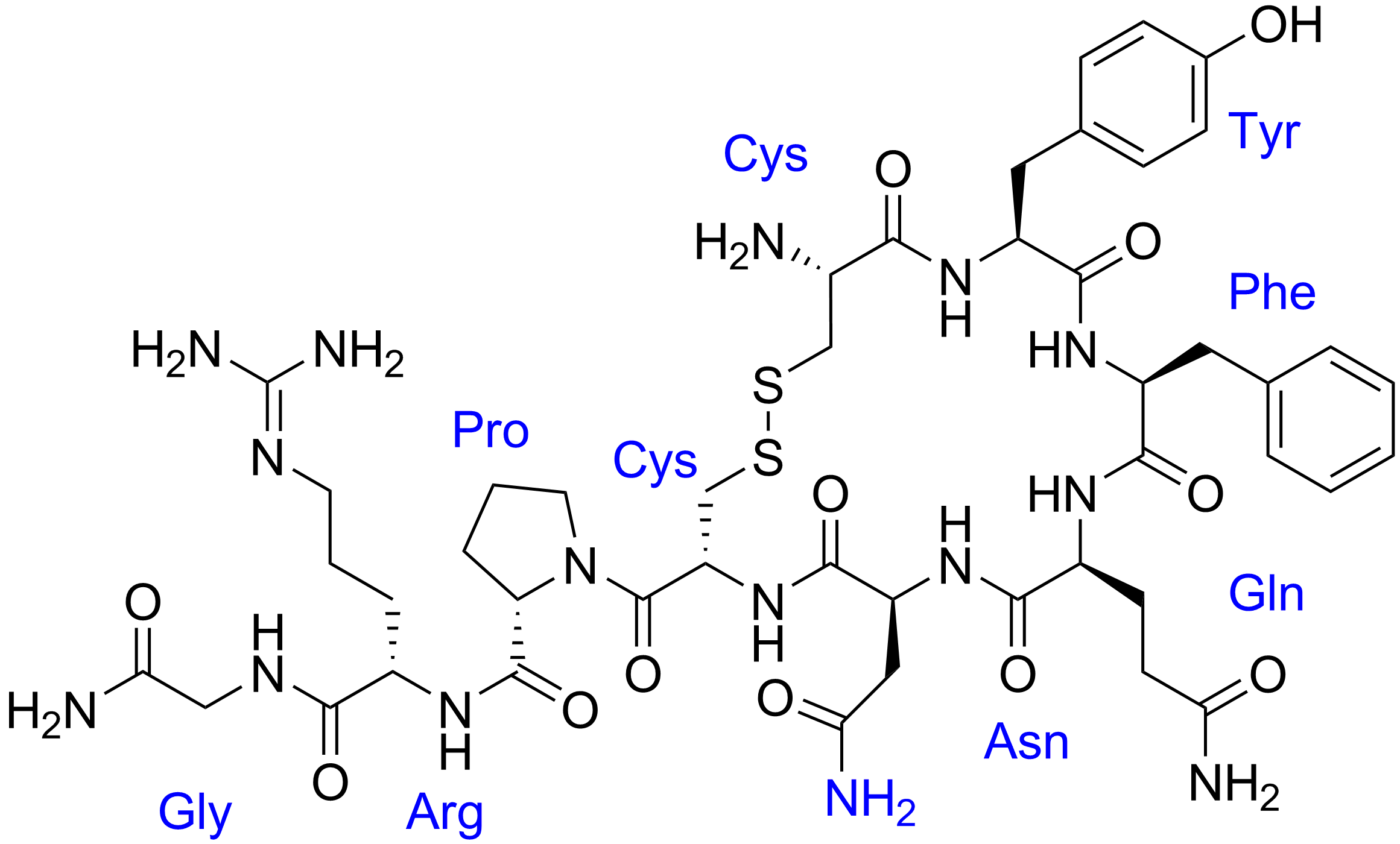
Human vasopressin, also called antidiuretic hormone (ADH), arginine vasopressin (AVP) or argipressin, is a hormone synthesized from the AVP gene as a peptide prohormone in neurons in the hypothalamus, and is converted to AVP. It then travels down the axon terminating in the posterior pituitary, and is released from vesicles into the circulation in response to extracellular fluid hypertonicity (hyperosmolality). AVP has two primary functions. First, it increases the amount of solute-free water reabsorbed back into the circulation from the filtrate in the kidney tubules of the nephrons. Second, AVP constricts arterioles, which increases peripheral vascular resistance and raises arterial blood pressure. A third function is possible. Some AVP may be released directly into the brain from the hypothalamus, and may play an important role in social behavior, sexual motivation and pair bonding, and maternal responses to stress. Vasopressin induces differentiation of stem cells in ... [...More Info...] [...Related Items...] OR: [Wikipedia] [Google] [Baidu] |
Neuroendocrine Cells
Neuroendocrine cells are cells that receive neuronal input (through neurotransmitters released by nerve cells or neurosecretory cells) and, as a consequence of this input, release messenger molecules (hormones) into the blood. In this way they bring about an integration between the nervous system and the endocrine system, a process known as neuroendocrine integration. An example of a neuroendocrine cell is a cell of the adrenal medulla (innermost part of the adrenal gland), which releases epinephrine, adrenaline to the blood. The adrenal medullary cells are controlled by the sympathetic nervous system, sympathetic division of the autonomic nervous system. These cells are modified postganglionic neurons. Autonomic nerve fibers lead directly to them from the central nervous system. The adrenal medullary hormones are kept in vesicles much in the same way neurotransmitters are kept in neuronal vesicles. Hormonal effects can last up to ten times longer than those of neurotransmitters. Sy ... [...More Info...] [...Related Items...] OR: [Wikipedia] [Google] [Baidu] |
Vasopressin
Human vasopressin, also called antidiuretic hormone (ADH), arginine vasopressin (AVP) or argipressin, is a hormone synthesized from the AVP gene as a peptide prohormone in neurons in the hypothalamus, and is converted to AVP. It then travels down the axon terminating in the posterior pituitary, and is released from vesicles into the circulation in response to extracellular fluid hypertonicity (hyperosmolality). AVP has two primary functions. First, it increases the amount of solute-free water reabsorbed back into the circulation from the filtrate in the kidney tubules of the nephrons. Second, AVP constricts arterioles, which increases peripheral vascular resistance and raises arterial blood pressure. A third function is possible. Some AVP may be released directly into the brain from the hypothalamus, and may play an important role in social behavior, sexual motivation and pair bonding, and maternal responses to stress. Vasopressin induces differentiation of stem cells in ... [...More Info...] [...Related Items...] OR: [Wikipedia] [Google] [Baidu] |
Anterior Pituitary Gland
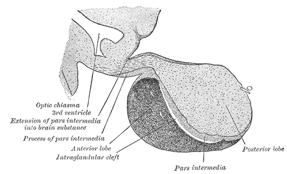
A major organ of the endocrine system, the anterior pituitary (also called the adenohypophysis or pars anterior) is the glandular, anterior lobe that together with the posterior lobe (posterior pituitary, or the neurohypophysis) makes up the pituitary gland (hypophysis). The anterior pituitary regulates several physiological processes, including stress, growth, reproduction, and lactation. Proper functioning of the anterior pituitary and of the organs it regulates can often be ascertained via blood tests that measure hormone levels. Structure The pituitary gland sits in a protective bony enclosure called the sella turcica (''Turkish chair/saddle''). It is composed of three lobes: the anterior, intermediate, and posterior lobes. In many animals, these lobes are distinct. However, in humans, the intermediate lobe is but a few cell layers thick and indistinct; as a result, it is often considered part of the anterior pituitary. In all animals, the fleshy, glandular anterior pituita ... [...More Info...] [...Related Items...] OR: [Wikipedia] [Google] [Baidu] |
ACTH
Adrenocorticotropic hormone (ACTH; also adrenocorticotropin, corticotropin) is a polypeptide tropic hormone produced by and secreted by the anterior pituitary gland. It is also used as a medication and diagnostic agent. ACTH is an important component of the hypothalamic-pituitary-adrenal axis and is often produced in response to biological stress (along with its precursor corticotropin-releasing hormone from the hypothalamus). Its principal effects are increased production and release of cortisol by the cortex of the adrenal gland. ACTH is also related to the circadian rhythm in many organisms. Deficiency of ACTH is an indicator of secondary adrenal insufficiency (suppressed production of ACTH due to an impairment of the pituitary gland or hypothalamus, cf. hypopituitarism) or tertiary adrenal insufficiency (disease of the hypothalamus, with a decrease in the release of corticotropin releasing hormone (CRH)). Conversely, chronically elevated ACTH levels occur in primary adren ... [...More Info...] [...Related Items...] OR: [Wikipedia] [Google] [Baidu] |
Corticotropin-releasing Hormone
Corticotropin-releasing hormone (CRH) (also known as corticotropin-releasing factor (CRF) or corticoliberin; corticotropin may also be spelled corticotrophin) is a peptide hormone involved in stress (biology), stress responses. It is a releasing hormone that belongs to corticotropin-releasing factor family. In humans, it is encoded by the ''CRH'' gene. Its main function is the stimulation of the pituitary synthesis of adrenocorticotropic hormone (ACTH), as part of the hypothalamic–pituitary–adrenal axis (HPA axis). Corticotropin-releasing hormone (CRH) is a 41-amino acid peptide derived from a 196-amino acid preprohormone. CRH is secreted by the paraventricular nucleus (PVN) of the hypothalamus in response to stress (biological), stress. Increased CRH production has been observed to be associated with Alzheimer's disease and major depression, and autosomal recessive hypothalamic corticotropin deficiency has multiple and potentially fatal metabolic consequences including hypo ... [...More Info...] [...Related Items...] OR: [Wikipedia] [Google] [Baidu] |
Myelin
Myelin is a lipid-rich material that surrounds nerve cell axons (the nervous system's "wires") to insulate them and increase the rate at which electrical impulses (called action potentials) are passed along the axon. The myelinated axon can be likened to an electrical wire (the axon) with insulating material (myelin) around it. However, unlike the plastic covering on an electrical wire, myelin does not form a single long sheath over the entire length of the axon. Rather, myelin sheaths the nerve in segments: in general, each axon is encased with multiple long myelinated sections with short gaps in between called nodes of Ranvier. Myelin is formed in the central nervous system (CNS; brain, spinal cord and optic nerve) by glial cells called oligodendrocytes and in the peripheral nervous system (PNS) by glial cells called Schwann cells. In the CNS, axons carry electrical signals from one nerve cell body to another. In the PNS, axons carry signals to muscles and glands or from senso ... [...More Info...] [...Related Items...] OR: [Wikipedia] [Google] [Baidu] |
Peptide Hormones
Peptide hormones or protein hormones are hormones whose molecules are peptide, or proteins, respectively. The latter have longer amino acid chain lengths than the former. These hormones have an effect on the endocrine system of animals, including humans. Most hormones can be classified as either amino acid–based hormones (amine, peptide, or protein) or steroid hormones. The former are water-soluble and act on the surface of target cells via second messengers; the latter, being lipid-soluble, move through the plasma membranes of target cells (both cytoplasmic and nuclear) to act within their nuclei. Like all peptides and proteins, peptide hormones and protein hormones are synthesized in cells from amino acids according to mRNA transcripts, which are synthesized from DNA templates inside the cell nucleus. Preprohormones, peptide hormone precursors, are then processed in several stages, typically in the endoplasmic reticulum, including removal of the N-terminal signal sequence an ... [...More Info...] [...Related Items...] OR: [Wikipedia] [Google] [Baidu] |
Spinal Cord
The spinal cord is a long, thin, tubular structure made up of nervous tissue, which extends from the medulla oblongata in the brainstem to the lumbar region of the vertebral column (backbone). The backbone encloses the central canal of the spinal cord, which contains cerebrospinal fluid. The brain and spinal cord together make up the central nervous system (CNS). In humans, the spinal cord begins at the occipital bone, passing through the foramen magnum and then enters the spinal canal at the beginning of the cervical vertebrae. The spinal cord extends down to between the first and second lumbar vertebrae, where it ends. The enclosing bony vertebral column protects the relatively shorter spinal cord. It is around long in adult men and around long in adult women. The diameter of the spinal cord ranges from in the cervical and lumbar regions to in the thoracic area. The spinal cord functions primarily in the transmission of nerve signals from the motor cortex to the body, ... [...More Info...] [...Related Items...] OR: [Wikipedia] [Google] [Baidu] |
Brainstem
The brainstem (or brain stem) is the posterior stalk-like part of the brain that connects the cerebrum with the spinal cord. In the human brain the brainstem is composed of the midbrain, the pons, and the medulla oblongata. The midbrain is continuous with the thalamus of the diencephalon through the tentorial notch, and sometimes the diencephalon is included in the brainstem. The brainstem is very small, making up around only 2.6 percent of the brain's total weight. It has the critical roles of regulating cardiac, and respiratory function, helping to control heart rate and breathing rate. It also provides the main motor and sensory nerve supply to the face and neck via the cranial nerves. Ten pairs of cranial nerves come from the brainstem. Other roles include the regulation of the central nervous system and the body's sleep cycle. It is also of prime importance in the conveyance of motor and sensory pathways from the rest of the brain to the body, and from the body back to t ... [...More Info...] [...Related Items...] OR: [Wikipedia] [Google] [Baidu] |
Parvocellular Neurosecretory Cell
Parvocellular neurosecretory cells are small neurons within paraventricular nucleus (PVN) of the hypothalamus. The axons of the parvocellular neurosecretory cells of the PVN project to the median eminence, at the base of the brain, where their neurosecretory nerve terminals release peptides into blood vessels in the hypothalamo-pituitary portal system. The blood vessels carry the peptides to the anterior pituitary gland, where they regulate the secretion of hormones into the systemic circulation. __TOC__ Types The parvocellular neurosecretory cells include those that make: * Thyrotropin-releasing hormone (TRH), which acts as the primary regulator of TSH and a regulator of prolactin * Corticotropin-releasing hormone (CRH), which acts as the primary regulator of ACTH * * * Neurotensin, which acts as a regulator of luteinizing hormone and prolactin See also * Magnocellular neurosecretory cell Magnocellular neurosecretory cells are large neuroendocrine cells within the supraoptic ... [...More Info...] [...Related Items...] OR: [Wikipedia] [Google] [Baidu] |


.jpg)


