|
Paraganglioma
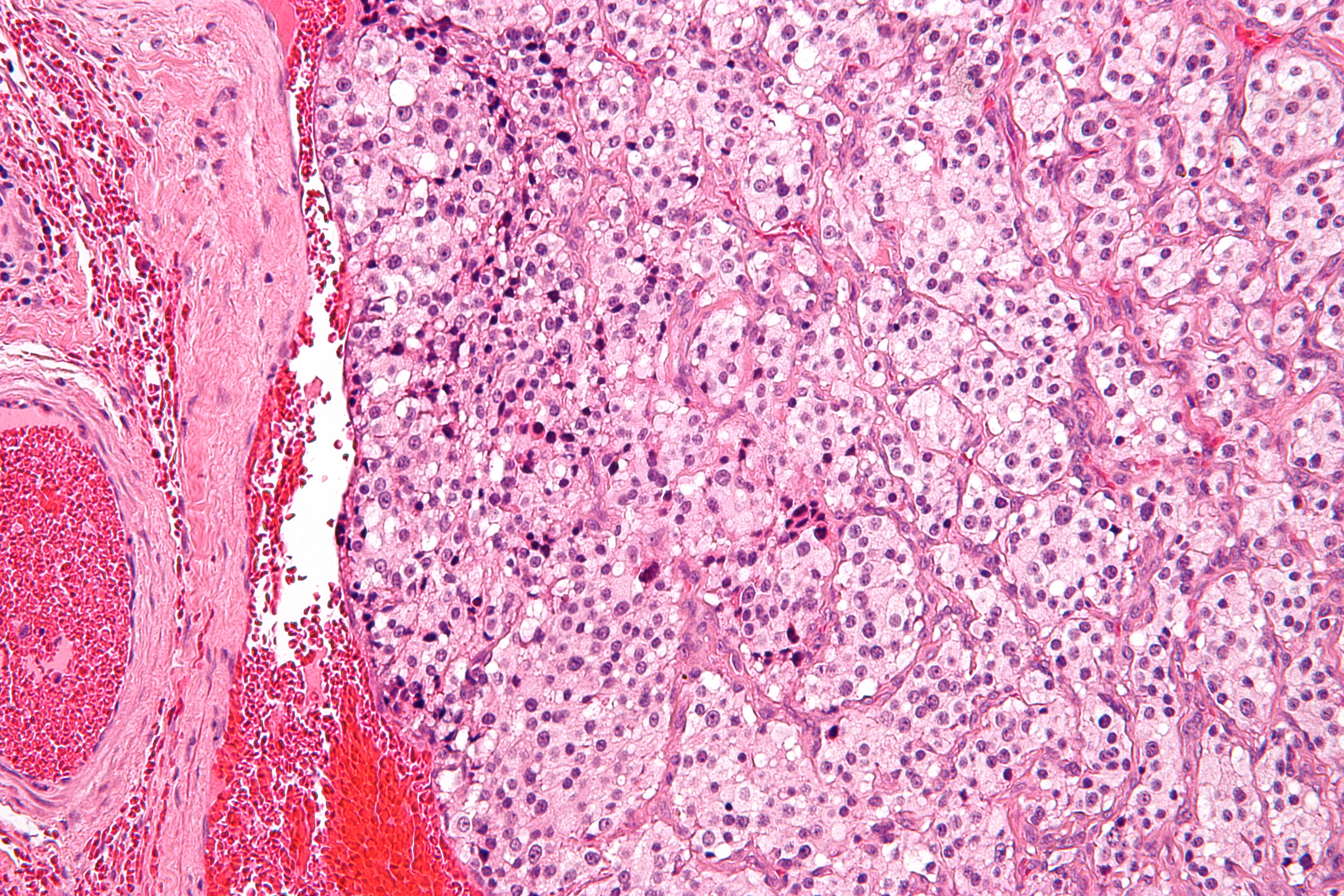
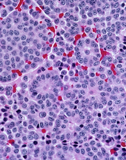
A paraganglioma is a rare neuroendocrine neoplasm that may develop at various body sites (including the head, neck, thorax and abdomen). When the same type of tumor is found in the adrenal gland, they are referred to as a pheochromocytoma. They are rare tumors, with an overall estimated incidence of 1/300,000. There is no test that determines benign from malignant tumors; long-term follow-up is therefore recommended for all individuals with paraganglioma. Signs and symptoms Most paragangliomas are asymptomatic, present as a painless mass, or create symptoms such as hypertension, tachycardia, headache, and palpitations. While all contain neurosecretory granules, only in 1–3% of cases is secretion of hormones such as catecholamines abundant enough to be clinically significant; in that case manifestations often resemble those of pheochromocytomas (intra-medullary paraganglioma). Genetics About 75% of paragangliomas are sporadic; the remaining 25% are hereditary (and have an increas ... [...More Info...] [...Related Items...] OR: [Wikipedia] [Google] [Baidu] |
Pheochromocytoma
Pheochromocytoma (PHEO or PCC) is a rare tumor of the adrenal medulla composed of chromaffin cells, also known as pheochromocytes. When a tumor composed of the same cells as a pheochromocytoma develops outside the adrenal gland, it is referred to as a paraganglioma. These neuroendocrine tumors are capable of producing and releasing massive amounts of catecholamines, metanephrines, or methoxytyramine, which result in the most common symptoms, including hypertension (high blood pressure), tachycardia (fast heart rate), and diaphoresis (sweating). However, not all of these tumors will secrete catecholamines. Those that do not are referred to as biochemically silent, and are predominantly located in the head and neck. While patients with biochemically silent disease will not develop the typical disease manifestations described above, the tumors grow and compress the surrounding structures of the head and neck, and can result in pulsatile tinnitus (ringing of the ear), hearing loss, au ... [...More Info...] [...Related Items...] OR: [Wikipedia] [Google] [Baidu] |
SDHB
Succinate dehydrogenase biquinoneiron-sulfur subunit, mitochondrial (SDHB) also known as iron-sulfur subunit of complex II (Ip) is a protein that in humans is encoded by the ''SDHB'' gene. The succinate dehydrogenase (also called SDH or Complex II) protein complex catalyzes the oxidation of succinate (succinate + ubiquinone => fumarate + ubiquinol). SDHB is one of four protein subunits forming succinate dehydrogenase, the other three being SDHA, SDHC and SDHD. The SDHB subunit is connected to the SDHA subunit on the hydrophilic, catalytic end of the SDH complex. It is also connected to the SDHC/SDHD subunits on the hydrophobic end of the complex anchored in the mitochondrial membrane. The subunit is an iron-sulfur protein with three iron-sulfur clusters. It weighs 30 kDa. Structure The gene that codes for the SDHB protein is nuclear, not mitochondrial DNA. However, the expressed protein is located in the inner membrane of the mitochondria. The location of the gene in humans ... [...More Info...] [...Related Items...] OR: [Wikipedia] [Google] [Baidu] |
Carotid Body
The carotid body is a small cluster of chemoreceptor cells, and supporting sustentacular cells. The carotid body is located in the adventitia, in the bifurcation (fork) of the common carotid artery, which runs along both sides of the neck. The carotid body detects changes in the composition of arterial blood flowing through it, mainly the partial pressure of arterial oxygen, but also of carbon dioxide. It is also sensitive to changes in blood pH, and temperature. Structure The carotid body is made up of two types of cells, called glomus cells: glomus type I cells are peripheral chemoreceptors, and glomus type II cells are sustentacular supportive cells. * Glomus type I cells are derived from the neural crest. They release a variety of neurotransmitters, including acetylcholine, ATP, and dopamine that trigger EPSPs in synapsed neurons leading to the respiratory center. They are innervated by axons of the glossopharyngeal nerve which collectively are called the carotid sinus ne ... [...More Info...] [...Related Items...] OR: [Wikipedia] [Google] [Baidu] |
Sustentacular Cell
A sustentacular cell is a type of cell primarily associated with structural support, they can be found in various tissues. * Sustentacular cells of the olfactory epithelium (also called supporting cells) have been shown to be involved in the phagocytosis of dead neurons, odorant transformation and xenobiotic metabolism. * One type of sustentacular cell is the Sertoli cell, in the testicle. It is located in the walls of the seminiferous tubules and supplies nutrients to sperm. They are responsible for the differentiation of spermatids, the maintenance of the blood-testis barrier, and the secretion of inhibin, androgen-binding protein and Mullerian-inhibiting factor. * The organ of Corti in the inner ear and taste buds also contain sustentacular cells. * Another type of sustentacular cell is found with glomus cells of the carotid and aortic bodies. * About 40% of carcinoid A carcinoid (also carcinoid tumor) is a slow-growing type of neuroendocrine tumor originating in the ce ... [...More Info...] [...Related Items...] OR: [Wikipedia] [Google] [Baidu] |
Neuroendocrine Tumour
Neuroendocrine tumors (NETs) are neoplasm A neoplasm () is a type of abnormal and excessive growth of tissue. The process that occurs to form or produce a neoplasm is called neoplasia. The growth of a neoplasm is uncoordinated with that of the normal surrounding tissue, and persists ...s that arise from cells of the endocrine (hormonal) and nervous systems. They most commonly occur in the intestine, where they are often called carcinoid tumors, but they are also found in the pancreas, lung, and the rest of the body. Although there are many kinds of NETs, they are treated as a group of tissue because the cells of these neoplasms share common features, such as looking similar, having special secretory granules, and often producing biogenic amines and polypeptide hormones. Classification WHO The World Health Organization (WHO) classification scheme places neuroendocrine tumors into three main categories, which emphasize the Grading (tumors), tumor grade rather than the anato ... [...More Info...] [...Related Items...] OR: [Wikipedia] [Google] [Baidu] |
Neuroendocrine Carcinoma
Neuroendocrine tumors (NETs) are neoplasms that arise from cells of the endocrine (hormonal) and nervous systems. They most commonly occur in the intestine, where they are often called carcinoid tumors, but they are also found in the pancreas, lung, and the rest of the body. Although there are many kinds of NETs, they are treated as a group of tissue because the cells of these neoplasms share common features, such as looking similar, having special secretory granules, and often producing biogenic amines and polypeptide hormones. Classification WHO The World Health Organization (WHO) classification scheme places neuroendocrine tumors into three main categories, which emphasize the tumor grade rather than the anatomical origin: * well-differentiated neuroendocrine tumours, further subdivided into tumors with benign and those with uncertain behavior * well-differentiated (low grade) neuroendocrine carcinomas with low-grade malignant behavior * poorly differentiated (high grade) ne ... [...More Info...] [...Related Items...] OR: [Wikipedia] [Google] [Baidu] |
SDHD
Succinate dehydrogenase biquinonecytochrome b small subunit, mitochondrial (CybS), also known as succinate dehydrogenase complex subunit D (SDHD), is a protein that in humans is encoded by the ''SDHD'' gene. Names previously used for SDHD were PGL and PGL1. Succinate dehydrogenase is an important enzyme in both the citric acid cycle and the electron transport chain. Structure The SDHD gene is located on chromosome 11 at locus 11q23 and it spans 8,978 base pairs. There are pseudogenes for this gene on chromosomes 1, 2, 3, 7, and 18. The SDHD gene produces a 17 kDa protein composed of 159 amino acids. The SDHD protein is one of the two integral transmembrane subunits anchoring the four-subunit succinate dehydrogenase (Complex II) protein complex to the matrix side of the mitochondrial inner membrane. The other transmembrane subunit is SDHC. The SDHC/SDHD dimer is connected to the SDHB electron transport subunit which, in turn, is connected to the SDHA subunit. Functi ... [...More Info...] [...Related Items...] OR: [Wikipedia] [Google] [Baidu] |
SDHAF2
Succinate dehydrogenase complex assembly factor 2, formerly known as SDH5 and also known as SDH assembly factor 2 or SDHAF2 is a protein that in humans is encoded by the SDHAF2 gene. This gene encodes a mitochondrial protein needed for the flavination of a succinate dehydrogenase complex subunit required for activity of the complex. Mutations in this gene are associated with pheochromocytoma and paraganglioma. Structure ''SDHAF2'' is located on the q arm of Chromosome 11 in position 12.2 and spans 16,642 base pairs. The ''SDHAF2'' gene produces a 6.7 kDa protein composed of 65 amino acids. This highly conserved protein is a cofactor of flavin adenine dinucleotide (FAD). The structure represents a five-helix bundle with a region of well-defined conserved surface residues. This conserved region includes a negatively charged periphery and a positively charged surface, and a patch that is hydrophobic. The region is located in α-helices I, II, and the connecting band. Function ... [...More Info...] [...Related Items...] OR: [Wikipedia] [Google] [Baidu] |
SDHC (gene)
Succinate dehydrogenase complex subunit C, also known as succinate dehydrogenase cytochrome b560 subunit, mitochondrial, is a protein that in humans is encoded by the ''SDHC'' gene. This gene encodes one of four nuclear-encoded subunits that comprise succinate dehydrogenase, also known as mitochondrial complex II, a key enzyme complex of the tricarboxylic acid cycle and aerobic respiratory chains of mitochondria. The encoded protein is one of two integral membrane proteins that anchor other subunits of the complex, which form the catalytic core, to the inner mitochondrial membrane. There are several related pseudogenes for this gene on different chromosomes. Mutations in this gene have been associated with pheochromocytomas and paragangliomas. Alternatively spliced transcript variants have been described. Structure The gene that codes for the SDHC protein is nuclear, even though the protein is located in the inner membrane of the mitochondria. The location of the gene in humans ... [...More Info...] [...Related Items...] OR: [Wikipedia] [Google] [Baidu] |
Carcinoid Tumor
A carcinoid (also carcinoid tumor) is a slow-growing type of neuroendocrine tumor originating in the cells of the neuroendocrine system. In some cases, metastasis may occur. Carcinoid tumors of the midgut (jejunum, ileum, appendix, and cecum) are associated with carcinoid syndrome. Carcinoid tumors are the most common malignant tumor of the appendix, but they are most commonly associated with the small intestine, and they can also be found in the rectum and stomach. They are known to grow in the liver, but this finding is usually a manifestation of metastatic disease from a primary carcinoid occurring elsewhere in the body. They have a very slow growth rate compared to most malignant tumors. The median age at diagnosis for all patients with neuroendocrine tumors is 63 years. Signs and symptoms While most carcinoids are asymptomatic through the natural life and are discovered only upon surgery for unrelated reasons (so-called ''coincidental carcinoids''), all carcinoids are co ... [...More Info...] [...Related Items...] OR: [Wikipedia] [Google] [Baidu] |
Medullary Carcinoma Of The Thyroid
Medullary thyroid cancer is a form of Thyroid cancer, thyroid carcinoma which originates from the parafollicular cells (C cells), which produce the hormone calcitonin.Hu MI, Vassilopoulou-Sellin R, Lustig R, Lamont JP"Thyroid and Parathyroid Cancers"in Pazdur R, Wagman LD, Camphausen KA, Hoskins WJ (EdsCancer Management: A Multidisciplinary Approach 11 ed. 2008. Medullary tumors are the third most common of all thyroid cancers and together make up about 3% of all thyroid cancer cases. MTC was first characterized in 1959. Approximately 25% of medullary thyroid cancer cases are Genetic disorder, genetic in nature, caused by a mutation in the RET proto-oncogene. When MTC occurs by itself it is termed sporadic medullary thyroid cancer. Medullary thyroid cancer is seen in people with Multiple endocrine neoplasia type 2, multiple endocrine neoplasia type 2A and Multiple endocrine neoplasia type 2B, 2B. When medullary thyroid cancer due to a hereditary genetic disorder occurs without othe ... [...More Info...] [...Related Items...] OR: [Wikipedia] [Google] [Baidu] |
Micrograph
A micrograph or photomicrograph is a photograph or digital image taken through a microscope or similar device to show a magnified image of an object. This is opposed to a macrograph or photomacrograph, an image which is also taken on a microscope but is only slightly magnified, usually less than 10 times. Micrography is the practice or art of using microscopes to make photographs. A micrograph contains extensive details of microstructure. A wealth of information can be obtained from a simple micrograph like behavior of the material under different conditions, the phases found in the system, failure analysis, grain size estimation, elemental analysis and so on. Micrographs are widely used in all fields of microscopy. Types Photomicrograph A light micrograph or photomicrograph is a micrograph prepared using an optical microscope, a process referred to as ''photomicroscopy''. At a basic level, photomicroscopy may be performed simply by connecting a camera to a microscope, th ... [...More Info...] [...Related Items...] OR: [Wikipedia] [Google] [Baidu] |


