|
Pancoast Tumor
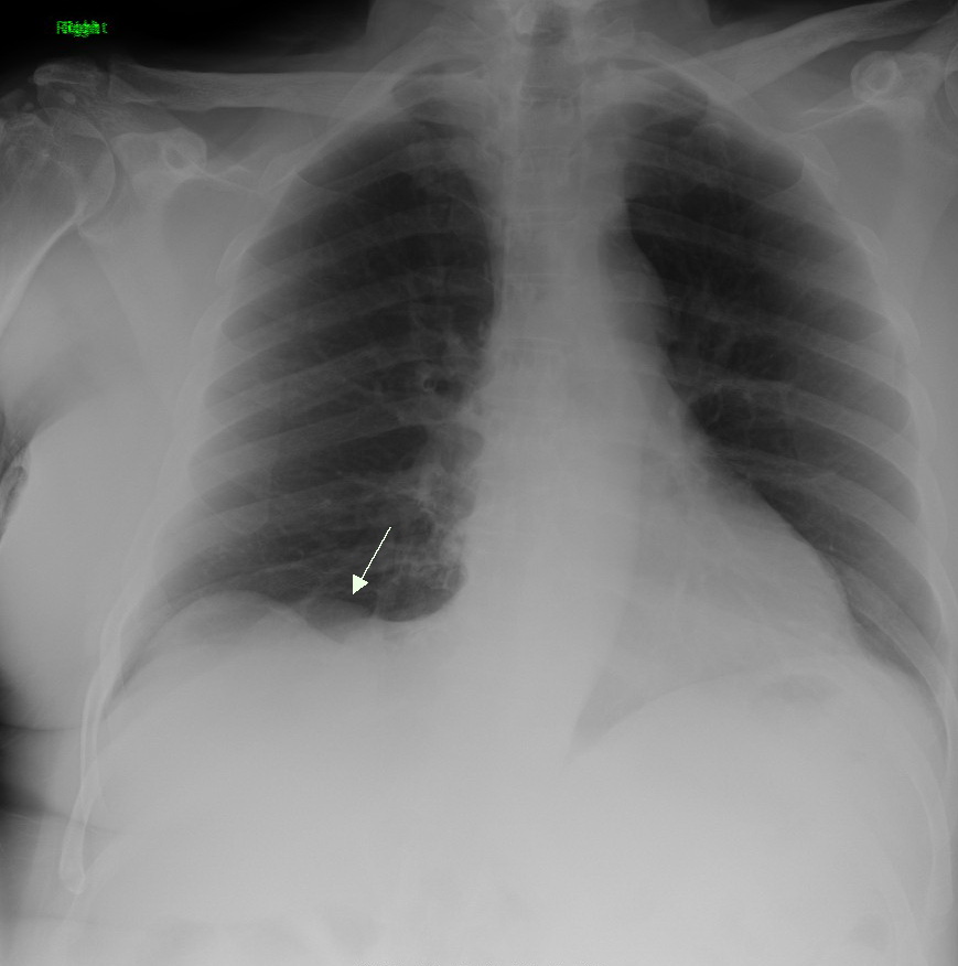
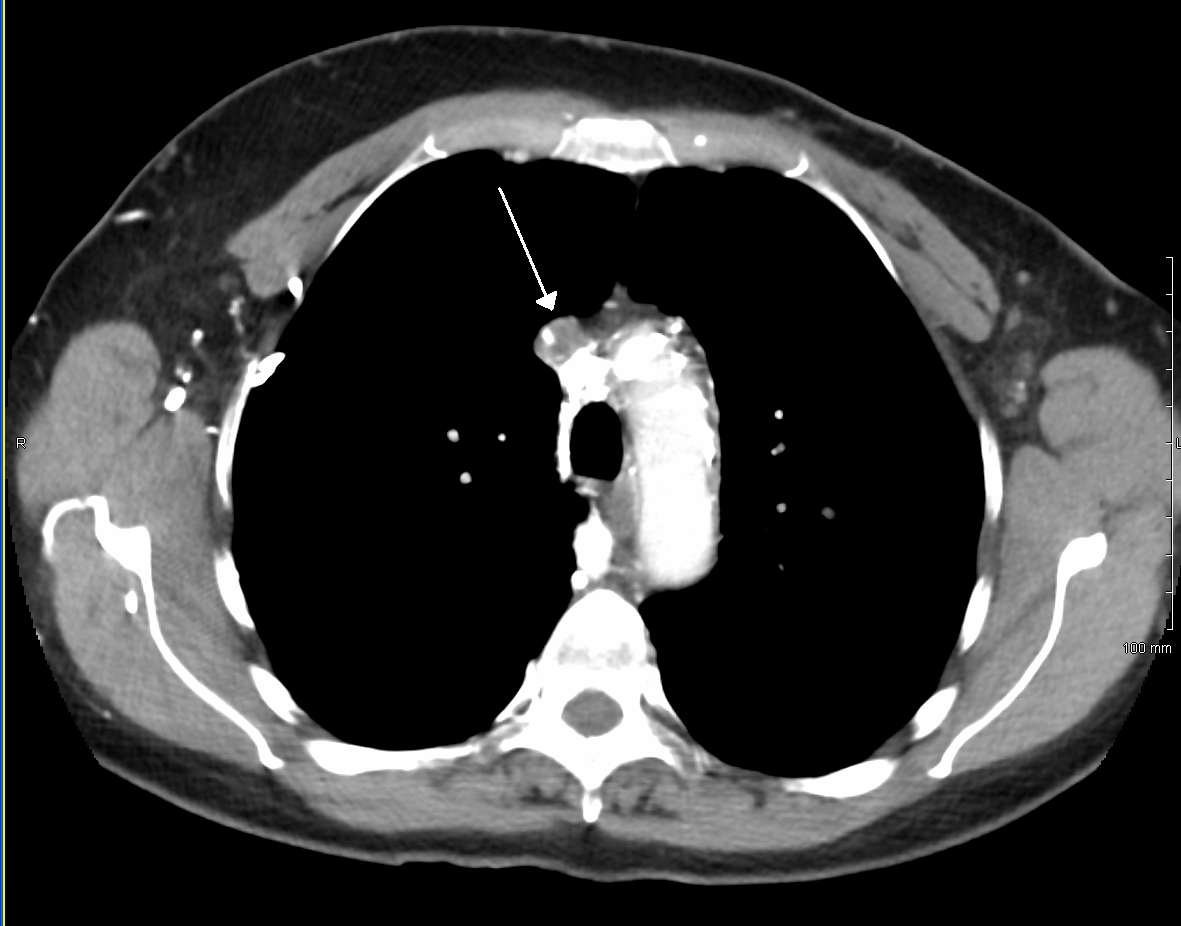
A Pancoast tumor is a tumor of the apex of the lung. It is a type of lung cancer defined primarily by its location situated at the top end of either the right or left lung. It typically spreads to nearby tissues such as the ribs and vertebrae. Most Pancoast tumors are non-small-cell lung cancers. The growing tumor can cause compression of a brachiocephalic vein, subclavian artery, phrenic nerve, recurrent laryngeal nerve, vagus nerve, or, characteristically, compression of a sympathetic ganglion (the stellate ganglion), resulting in a range of symptoms known as Horner's syndrome. Pancoast tumors are named for Henry Pancoast, an American radiologist, who described them in 1924 and 1932. Signs and symptoms Aside from constitutional symptoms of cancer such as malaise, fever, weight loss and fatigue, Pancoast tumor can include a complete Horner's syndrome in severe cases: miosis (constriction of the pupils), anhidrosis (lack of sweating), ptosis (drooping of the eyelid), and pseud ... [...More Info...] [...Related Items...] OR: [Wikipedia] [Google] [Baidu] |
Lung Cancer
Lung cancer, also known as lung carcinoma (since about 98–99% of all lung cancers are carcinomas), is a malignant lung tumor characterized by uncontrolled cell growth in tissue (biology), tissues of the lung. Lung carcinomas derive from transformed, malignant cells that originate as epithelial cells, or from tissues composed of epithelial cells. Other lung cancers, such as the rare sarcomas of the lung, are generated by the malignant transformation of connective tissues (i.e. nerve, fat, muscle, bone), which arise from mesenchymal cells. Lymphomas and melanomas (from lymphoid and melanocyte cell lineages) can also rarely result in lung cancer. In time, this uncontrolled neoplasm, growth can metastasis, metastasize (spreading beyond the lung) either by direct extension, by entering the lymphatic circulation, or via hematogenous, bloodborne spread – into nearby tissue or other, more distant parts of the body. Most cancers that originate from within the lungs, known as primary ... [...More Info...] [...Related Items...] OR: [Wikipedia] [Google] [Baidu] |
Cancer
Cancer is a group of diseases involving abnormal cell growth with the potential to invade or spread to other parts of the body. These contrast with benign tumors, which do not spread. Possible signs and symptoms include a lump, abnormal bleeding, prolonged cough, unexplained weight loss, and a change in bowel movements. While these symptoms may indicate cancer, they can also have other causes. Over 100 types of cancers affect humans. Tobacco use is the cause of about 22% of cancer deaths. Another 10% are due to obesity, poor diet, lack of physical activity or excessive drinking of alcohol. Other factors include certain infections, exposure to ionizing radiation, and environmental pollutants. In the developing world, 15% of cancers are due to infections such as ''Helicobacter pylori'', hepatitis B, hepatitis C, human papillomavirus infection, Epstein–Barr virus and human immunodeficiency virus (HIV). These factors act, at least partly, by changing the genes of ... [...More Info...] [...Related Items...] OR: [Wikipedia] [Google] [Baidu] |
Superior Thoracic Aperture
The superior thoracic aperture, also known as the thoracic outlet, or thoracic inlet refers to the opening at the top of the thoracic cavity. It is also clinically referred to as the thoracic outlet, in the case of thoracic outlet syndrome. A lower thoracic opening is the ''inferior thoracic aperture''. Structure The superior thoracic aperture is essentially a hole surrounded by a bony ring, through which several vital structures pass. It is bounded by: the first thoracic vertebra (T1) ''posteriorly''; the first pair of ribs ''laterally'', forming lateral C-shaped curves posterior to anterior; and the costal cartilage of the first rib and the superior border of the manubrium ''anteriorly''. Dimensions The adult thoracic outlet is around 6.5 cm antero-posteriorly and 11 cm transversely. Because of the obliquity of the first pair of ribs, the aperture slopes antero-inferiorly. Relations The clavicle articulates with the manubrium to form the anterior border of the thoracic out ... [...More Info...] [...Related Items...] OR: [Wikipedia] [Google] [Baidu] |
Cyanosis
Cyanosis is the change of body tissue color to a bluish-purple hue as a result of having decreased amounts of oxygen bound to the hemoglobin in the red blood cells of the capillary bed. Body tissues that show cyanosis are usually in locations where the skin is thinner, including the mucous membranes, lips, nail beds, and ear lobes. Some medications containing amiodarone or silver, Mongolian spots, large birth marks, and the consumption of food products with blue or purple dyes can also result in the bluish skin tissue discoloration and may be mistaken for cyanosis. Cyanosis is further classified into central cyanosis vs. peripheral cyanosis. Pathophysiology The mechanism behind cyanosis is different depending on whether it is central or peripheral. Central cyanosis Central cyanosis is caused by a decrease in arterial oxygen saturation (SaO2) and begins to show once the concentration of deoxyhemoglobin in the blood reaches a concentration of ≥ 5.0 g/dL (≥ 3.1 mmol/L ... [...More Info...] [...Related Items...] OR: [Wikipedia] [Google] [Baidu] |
Superior Vena Cava
The superior vena cava (SVC) is the superior of the two venae cavae, the great venous trunks that return deoxygenated blood from the systemic circulation to the right atrium of the heart. It is a large-diameter (24 mm) short length vein that receives venous return from the upper half of the body, above the diaphragm. Venous return from the lower half, below the diaphragm, flows through the inferior vena cava. The SVC is located in the anterior right superior mediastinum. It is the typical site of central venous access via a central venous catheter or a peripherally inserted central catheter. Mentions of "the cava" without further specification usually refer to the SVC. Structure The superior vena cava is formed by the left and right brachiocephalic veins, which receive blood from the upper limbs, head and neck, behind the lower border of the first right costal cartilage. It passes vertically downwards behind first intercostal space and receives azygos vein just before it p ... [...More Info...] [...Related Items...] OR: [Wikipedia] [Google] [Baidu] |
Superior Vena Cava Syndrome
Superior vena cava syndrome (SVCS), is a group of symptoms caused by obstruction of the superior vena cava ("SVC"), a short, wide vessel carrying circulating blood into the heart. The majority of cases are caused by malignant tumors within the mediastinum, most commonly lung cancer and non-Hodgkin's lymphoma, directly compressing or invading the SVC wall. Non-malignant causes are increasing in prevalence due to expanding use of intravascular devices (such as permanent central venous catheters and leads for pacemakers and defibrillators), which can result in thrombosis. Other non-malignant causes include benign mediastinal tumors, aortic aneurysm, infections, and fibrosing mediastinitis. Characteristic features are edema (swelling due to excess fluid) of the face and arms and development of swollen collateral veins on the front of the chest wall. Shortness of breath and coughing are quite common symptoms; difficulty swallowing is reported in 11% of cases, headache in 6% and ... [...More Info...] [...Related Items...] OR: [Wikipedia] [Google] [Baidu] |
Hoarse Voice
A hoarse voice, also known as dysphonia or hoarseness, is when the voice involuntarily sounds breathy, raspy, or strained, or is softer in volume or lower in pitch. A hoarse voice, can be associated with a feeling of unease or scratchiness in the throat. Hoarseness is often a symptom of problems in the vocal folds of the larynx. It may be caused by laryngitis, which in turn may be caused by an upper respiratory infection, a cold, or allergies. Cheering at sporting events, speaking loudly in noisy situations, talking for too long without resting one's voice, singing loudly, or speaking with a voice that's too high or too low can also cause temporary hoarseness. A number of other causes for losing one's voice exist, and treatment is generally by resting the voice and treating the underlying cause. If the cause is misuse or overuse of the voice, drinking plenty of water may alleviate the problems. It appears to occur more commonly in females and the elderly. Furthermore, certain occup ... [...More Info...] [...Related Items...] OR: [Wikipedia] [Google] [Baidu] |
Hand
A hand is a prehensile, multi-fingered appendage located at the end of the forearm or forelimb of primates such as humans, chimpanzees, monkeys, and lemurs. A few other vertebrates such as the koala (which has two opposable thumbs on each "hand" and fingerprints extremely similar to human fingerprints) are often described as having "hands" instead of paws on their front limbs. The raccoon is usually described as having "hands" though opposable thumbs are lacking. Some evolutionary anatomists use the term ''hand'' to refer to the appendage of digits on the forelimb more generally—for example, in the context of whether the three digits of the bird hand involved the same homologous loss of two digits as in the dinosaur hand. The human hand usually has five digits: four fingers plus one thumb; these are often referred to collectively as five fingers, however, whereby the thumb is included as one of the fingers. It has 27 bones, not including the sesamoid bone, the number o ... [...More Info...] [...Related Items...] OR: [Wikipedia] [Google] [Baidu] |
Weakness
Weakness is a symptom of a number of different conditions. The causes are many and can be divided into conditions that have true or perceived muscle weakness. True muscle weakness is a primary symptom of a variety of skeletal muscle diseases, including muscular dystrophy and inflammatory myopathy. It occurs in neuromuscular junction disorders, such as myasthenia gravis. Pathophysiology Muscle cells work by detecting a flow of electrical impulses from the brain, which signals them to contract through the release of calcium by the sarcoplasmic reticulum. Fatigue (reduced ability to generate force) may occur due to the nerve, or within the muscle cells themselves. New research from scientists at Columbia University suggests that muscle fatigue is caused by calcium leaking out of the muscle cell. This makes less calcium available for the muscle cell. In addition, the Columbia researchers propose that an enzyme activated by this released calcium eats away at muscle fibers. Substr ... [...More Info...] [...Related Items...] OR: [Wikipedia] [Google] [Baidu] |
Pain
Pain is a distressing feeling often caused by intense or damaging stimuli. The International Association for the Study of Pain defines pain as "an unpleasant sensory and emotional experience associated with, or resembling that associated with, actual or potential tissue damage." In medical diagnosis, pain is regarded as a symptom of an underlying condition. Pain motivates the individual to withdraw from damaging situations, to protect a damaged body part while it heals, and to avoid similar experiences in the future. Most pain resolves once the noxious stimulus is removed and the body has healed, but it may persist despite removal of the stimulus and apparent healing of the body. Sometimes pain arises in the absence of any detectable stimulus, damage or disease. Pain is the most common reason for physician consultation in most developed countries. It is a major symptom in many medical conditions, and can interfere with a person's quality of life and general functioning. Simple ... [...More Info...] [...Related Items...] OR: [Wikipedia] [Google] [Baidu] |
Brachial Plexus
The brachial plexus is a network () of nerves formed by the anterior rami of the lower four cervical nerves and first thoracic nerve ( C5, C6, C7, C8, and T1). This plexus extends from the spinal cord, through the cervicoaxillary canal in the neck, over the first rib, and into the armpit, it supplies afferent and efferent nerve fibers the to chest, shoulder, arm, forearm, and hand. Structure The brachial plexus is divided into five ''roots'', three ''trunks'', six ''divisions'' (three anterior and three posterior), three ''cords'', and five ''branches''. There are five "terminal" branches and numerous other "pre-terminal" or "collateral" branches, such as the subscapular nerve, the thoracodorsal nerve, and the long thoracic nerve, that leave the plexus at various points along its length. A common structure used to identify part of the brachial plexus in cadaver dissections is the M or W shape made by the musculocutaneous nerve, lateral cord, median nerve, medial cord, and ... [...More Info...] [...Related Items...] OR: [Wikipedia] [Google] [Baidu] |


