|
Palatoglossus Muscle
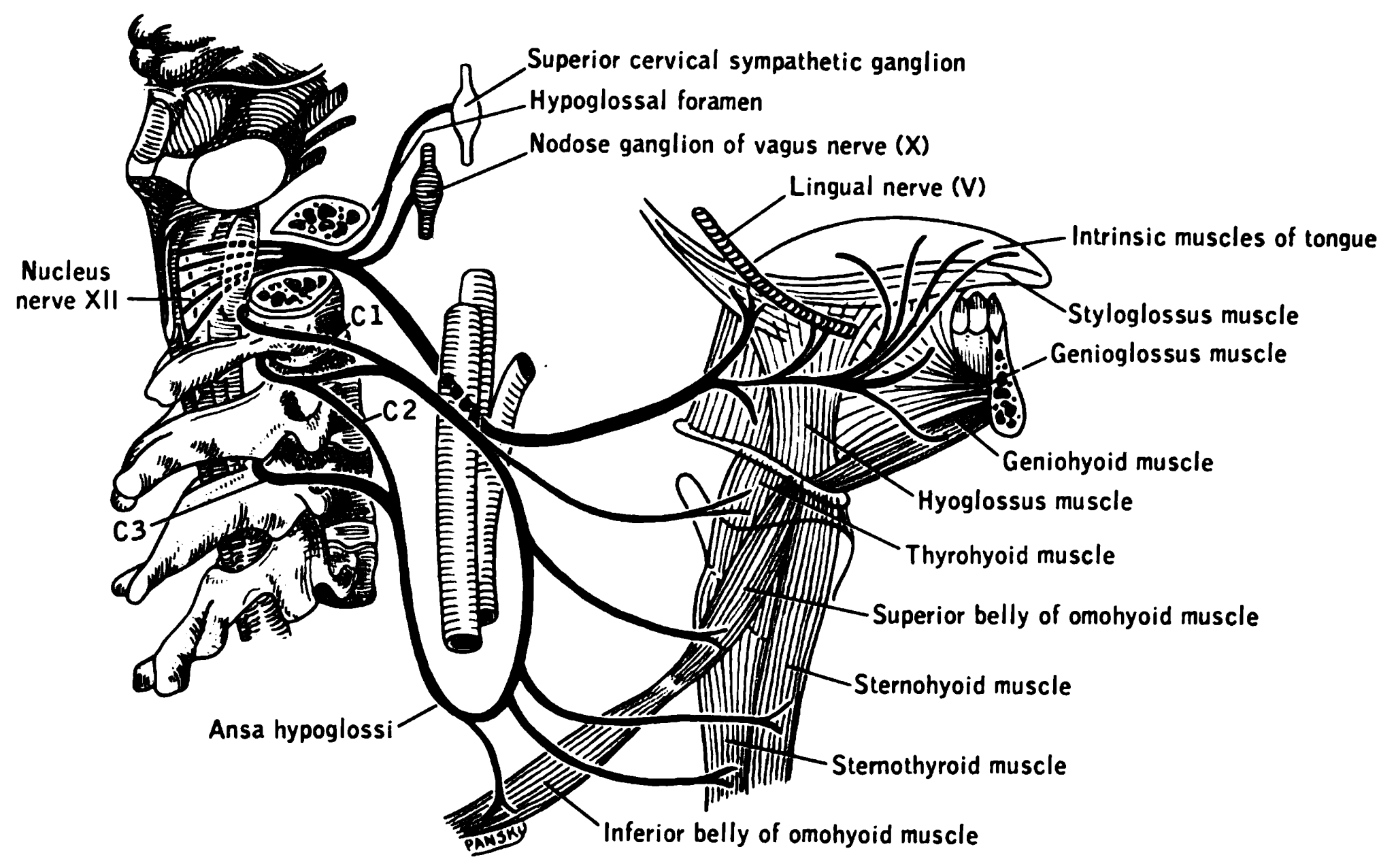
The palatoglossus or palatoglossal muscle is a muscle of the soft palate and extrinsic muscle of the tongue. Its surface is covered by oral mucosa and forms the visible palatoglossal arch. Structure Palatoglossus arises from the palatine aponeurosis of the soft palate, where it is continuous with the muscle of the opposite side, and passing downward, forward, and lateralward in front of the palatine tonsil, is inserted into the side of the tongue, some of its fibers spreading over the dorsum, and others passing deeply into the substance of the organ to intermingle with the transverse muscle of tongue. Innervation Palatoglossus is the only muscle of the tongue that is ''not'' innervated by the hypoglossal nerve (CN XII). It is innervated by the pharyngeal branch of the vagus nerve (CN X). Controversy Some sources state that the palatoglossus is innervated by fibers from the cranial part of the accessory nerve (CN XI) that travel via the pharyngeal plexus. Other sources state th ... [...More Info...] [...Related Items...] OR: [Wikipedia] [Google] [Baidu] |
Glossopalatine Arch
The palatoglossal arch (glossopalatine arch, anterior pillar of fauces) on either side runs downward, Lateral (anatomy), lateral (to the side), and forward to the side of the Posterior tongue, base of the tongue, and is formed by the projection of the glossopalatine muscle with its covering mucous membrane. It is the anterior border of the isthmus of the fauces and marks the border between the mouth and the palatopharyngeal arch. The latter marks the beginning of the pharynx. References External links * * Palate {{anatomy-stub ... [...More Info...] [...Related Items...] OR: [Wikipedia] [Google] [Baidu] |
Palatine Aponeurosis
Attached to the posterior border of the hard palate is a thin, firm, fibrous lamella called the palatine aponeurosis, which supports the muscles and gives strength to the soft palate. It is thicker above and narrows on the way down where it becomes very thin and difficult to define. Laterally, it is continuous with the pharyngeal aponeurosis. It serves as the insertion for the tensor veli palatini and levator veli palatini, and the origin for the musculus uvulae, palatopharyngeus, and palatoglossus. It provides support for the soft palate. See also * Aponeurosis An aponeurosis (; plural: ''aponeuroses'') is a type or a variant of the deep fascia, in the form of a sheet of pearly-white fibrous tissue that attaches sheet-like muscles needing a wide area of attachment. Their primary function is to join musc ... References Human head and neck {{musculoskeletal-stub ... [...More Info...] [...Related Items...] OR: [Wikipedia] [Google] [Baidu] |
Tongue
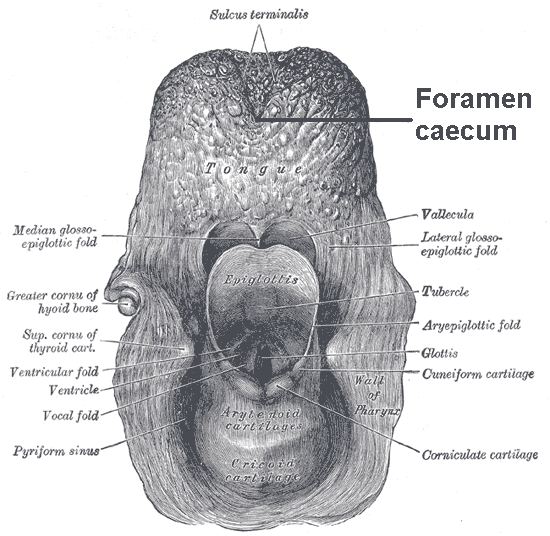
The tongue is a muscular organ (anatomy), organ in the mouth of a typical tetrapod. It manipulates food for mastication and swallowing as part of the digestive system, digestive process, and is the primary organ of taste. The tongue's upper surface (dorsum) is covered by taste buds housed in numerous lingual papillae. It is sensitive and kept moist by saliva and is richly supplied with nerves and blood vessels. The tongue also serves as a natural means of oral hygiene, cleaning the teeth. A major function of the tongue is the enabling of speech in humans and animal communication, vocalization in other animals. The human tongue is divided into two parts, an oral cavity, oral part at the front and a pharynx, pharyngeal part at the back. The left and right sides are also separated along most of its length by a vertical section of connective tissue, fibrous tissue (the lingual septum) that results in a groove, the median sulcus, on the tongue's surface. There are two groups of muscle ... [...More Info...] [...Related Items...] OR: [Wikipedia] [Google] [Baidu] |
Vagus Nerve
The vagus nerve, also known as the tenth cranial nerve, cranial nerve X, or simply CN X, is a cranial nerve that interfaces with the parasympathetic control of the heart, lungs, and digestive tract. It comprises two nerves—the left and right vagus nerves—but they are typically referred to collectively as a single subsystem. The vagus is the longest nerve of the autonomic nervous system in the human body and comprises both sensory and motor fibers. The sensory fibers originate from neurons of the nodose ganglion, whereas the motor fibers come from neurons of the dorsal motor nucleus of the vagus and the nucleus ambiguus. The vagus was also historically called the pneumogastric nerve. Structure Upon leaving the medulla oblongata between the olive and the inferior cerebellar peduncle, the vagus nerve extends through the jugular foramen, then passes into the carotid sheath between the internal carotid artery and the internal jugular vein down to the neck, chest, and abdom ... [...More Info...] [...Related Items...] OR: [Wikipedia] [Google] [Baidu] |
Tongue
The tongue is a muscular organ (anatomy), organ in the mouth of a typical tetrapod. It manipulates food for mastication and swallowing as part of the digestive system, digestive process, and is the primary organ of taste. The tongue's upper surface (dorsum) is covered by taste buds housed in numerous lingual papillae. It is sensitive and kept moist by saliva and is richly supplied with nerves and blood vessels. The tongue also serves as a natural means of oral hygiene, cleaning the teeth. A major function of the tongue is the enabling of speech in humans and animal communication, vocalization in other animals. The human tongue is divided into two parts, an oral cavity, oral part at the front and a pharynx, pharyngeal part at the back. The left and right sides are also separated along most of its length by a vertical section of connective tissue, fibrous tissue (the lingual septum) that results in a groove, the median sulcus, on the tongue's surface. There are two groups of muscle ... [...More Info...] [...Related Items...] OR: [Wikipedia] [Google] [Baidu] |
Palatoglossal Arch
The palatoglossal arch (glossopalatine arch, anterior pillar of fauces) on either side runs downward, lateral (to the side), and forward to the side of the base of the tongue, and is formed by the projection of the glossopalatine muscle with its covering mucous membrane. It is the anterior border of the isthmus of the fauces and marks the border between the mouth and the palatopharyngeal arch. The latter marks the beginning of the pharynx The pharynx (plural: pharynges) is the part of the throat behind the mouth and nasal cavity, and above the oesophagus and trachea (the tubes going down to the stomach and the lungs). It is found in vertebrates and invertebrates, though its struc .... References External links * * Palate {{anatomy-stub ... [...More Info...] [...Related Items...] OR: [Wikipedia] [Google] [Baidu] |
Palatine Aponeurosis
Attached to the posterior border of the hard palate is a thin, firm, fibrous lamella called the palatine aponeurosis, which supports the muscles and gives strength to the soft palate. It is thicker above and narrows on the way down where it becomes very thin and difficult to define. Laterally, it is continuous with the pharyngeal aponeurosis. It serves as the insertion for the tensor veli palatini and levator veli palatini, and the origin for the musculus uvulae, palatopharyngeus, and palatoglossus. It provides support for the soft palate. See also * Aponeurosis An aponeurosis (; plural: ''aponeuroses'') is a type or a variant of the deep fascia, in the form of a sheet of pearly-white fibrous tissue that attaches sheet-like muscles needing a wide area of attachment. Their primary function is to join musc ... References Human head and neck {{musculoskeletal-stub ... [...More Info...] [...Related Items...] OR: [Wikipedia] [Google] [Baidu] |
Soft Palate
The soft palate (also known as the velum, palatal velum, or muscular palate) is, in mammals, the soft tissue constituting the back of the roof of the mouth. The soft palate is part of the palate of the mouth; the other part is the hard palate. The soft palate is distinguished from the hard palate at the front of the mouth in that it does not contain bone. Structure Muscles The five muscles of the soft palate play important roles in swallowing and breathing. The muscles are: # Tensor veli palatini, which is involved in swallowing # Palatoglossus, involved in swallowing # Palatopharyngeus, involved in breathing # Levator veli palatini, involved in swallowing # Musculus uvulae, which moves the uvula These muscles are innervated by the pharyngeal plexus via the vagus nerve, with the exception of the tensor veli palatini. The tensor veli palatini is innervated by the mandibular division of the trigeminal nerve (V3). Function The soft palate is moveable, consisting of muscle f ... [...More Info...] [...Related Items...] OR: [Wikipedia] [Google] [Baidu] |
Transverse Muscle Of Tongue
The transverse muscle of tongue (transversus linguae) is an intrinsic muscle of the tongue. It consists of fibers which arise from the median fibrous septum. It passes laterally to insert into the submucous fibrous tissue at the sides of the tongue. It is supplied by the hypoglossal nerve (CN XII). It moves the tongue. Structure The transverse muscle of the tongue is an intrinsic muscle of the tongue. It consists of fibers which arise from the median fibrous septum. It passes laterally to insert into the submucous fibrous tissue at the sides of the tongue. Nerve supply The transverse lingual muscle is supplied by the hypoglossal nerve (CN XII). Function The transverse muscle of the tongue muscle moves the tongue The tongue is a muscular organ (anatomy), organ in the mouth of a typical tetrapod. It manipulates food for mastication and swallowing as part of the digestive system, digestive process, and is the primary organ of taste. The tongue's upper surfa .... It narr ... [...More Info...] [...Related Items...] OR: [Wikipedia] [Google] [Baidu] |
Hypoglossal Nerve
The hypoglossal nerve, also known as the twelfth cranial nerve, cranial nerve XII, or simply CN XII, is a cranial nerve that innervates all the extrinsic and intrinsic muscles of the tongue except for the palatoglossus, which is innervated by the vagus nerve. CN XII is a nerve with a solely motor function. The nerve arises from the hypoglossal nucleus in the medulla as a number of small rootlets, passes through the hypoglossal canal and down through the neck, and eventually passes up again over the tongue muscles it supplies into the tongue. The nerve is involved in controlling tongue movements required for speech and swallowing, including sticking out the tongue and moving it from side to side. Damage to the nerve or the neural pathways which control it can affect the ability of the tongue to move and its appearance, with the most common sources of damage being injury from trauma or surgery, and motor neuron disease. The first recorded description of the nerve is by Herophil ... [...More Info...] [...Related Items...] OR: [Wikipedia] [Google] [Baidu] |
Accessory Nerve
The accessory nerve, also known as the eleventh cranial nerve, cranial nerve XI, or simply CN XI, is a cranial nerve that supplies the sternocleidomastoid and trapezius muscles. It is classified as the eleventh of twelve pairs of cranial nerves because part of it was formerly believed to originate in the brain. The sternocleidomastoid muscle tilts and rotates the head, whereas the trapezius muscle, connecting to the scapula, acts to shrug the shoulder. Traditional descriptions of the accessory nerve divide it into a spinal part and a cranial part. The cranial component rapidly joins the vagus nerve, and there is ongoing debate about whether the cranial part should be considered part of the accessory nerve proper. Consequently, the term "accessory nerve" usually refers only to nerve supplying the sternocleidomastoid and trapezius muscles, also called the spinal accessory nerve. Strength testing of these muscles can be measured during a neurological examination to assess funct ... [...More Info...] [...Related Items...] OR: [Wikipedia] [Google] [Baidu] |
Pharyngeal Plexus Of Vagus Nerve
The pharyngeal plexus is a network of nerve fibers innervating most of the palate and pharynx. (The larynx, which is innervated by the superior and recurrent laryngeal nerves from vagus nerve (CN X), is not included.) It is located on the surface of the middle pharyngeal constrictor muscle. Sources Although the ''Terminologia Anatomica'' name of the plexus has "vagus nerve" in the title, other nerves make contributions to the plexus. It has the following sources: * CN IX – pharyngeal branches of glossopharyngeal nerve – sensory * CN X – pharyngeal branch of vagus nerve – motor * superior cervical ganglion sympathetic fibers – vasomotor Because the cranial part of accessory nerve (CN XI) leaves the jugular foramen as a part of the CN X, it is sometimes considered part of the plexus as well. Innervation Sensory The pharyngeal plexus provides sensory innervation of the oropharynx and laryngopharynx from CN IX and CN X. (The nasopharynx above the pharyngotympanic tube ... [...More Info...] [...Related Items...] OR: [Wikipedia] [Google] [Baidu] |


