|
Paget–Schroetter Disease
Paget–Schroetter disease (also known as venous thoracic outlet syndrome) is a form of upper extremity deep vein thrombosis (DVT), a medical condition in which blood clots form in the deep veins of the arms. These DVTs typically occur in the axillary and/or subclavian veins. Signs and symptoms The condition is relatively rare. It usually presents in young and otherwise healthy patients, and also occurs more often in males than females. The syndrome also became known as "effort-induced thrombosis" in the 1960s, as it has been reported to occur after vigorous activity, though it can also occur due to anatomic abnormality such as clavicle impingement or spontaneously. It may develop as a sequela of thoracic outlet syndrome. It is differentiated from secondary causes of upper extremity thrombosis caused by intravascular catheters. Paget–Schroetter syndrome was described once for a viola player who suddenly increased practice time 10-fold, creating enough repetitive pressure again ... [...More Info...] [...Related Items...] OR: [Wikipedia] [Google] [Baidu] |
Deep Vein Thrombosis
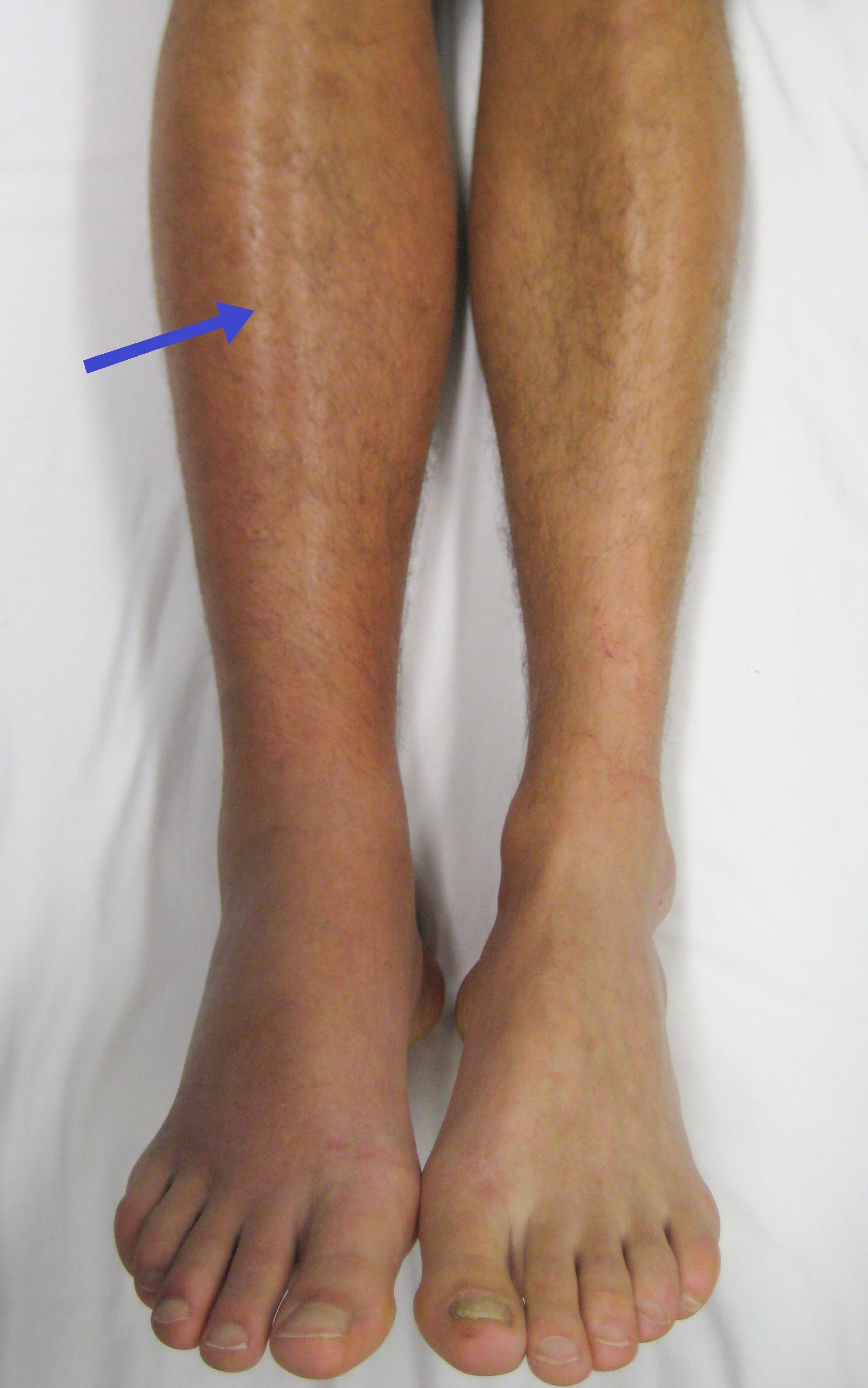
Deep vein thrombosis (DVT) is a type of venous thrombosis involving the formation of a blood clot in a deep vein, most commonly in the legs or pelvis. A minority of DVTs occur in the arms. Symptoms can include pain, swelling, redness, and enlarged veins in the affected area, but some DVTs have no symptoms. The most common life-threatening concern with DVT is the potential for a clot to embolize (detach from the veins), travel as an embolus through the right side of the heart, and become lodged in a pulmonary artery that supplies blood to the lungs. This is called a pulmonary embolism (PE). DVT and PE comprise the cardiovascular disease of venous thromboembolism (VTE). About two-thirds of VTE manifests as DVT only, with one-third manifesting as PE with or without DVT. The most frequent long-term DVT complication is post-thrombotic syndrome, which can cause pain, swelling, a sensation of heaviness, itching, and in severe cases, ulcers. Recurrent VTE occurs in about 30% of those i ... [...More Info...] [...Related Items...] OR: [Wikipedia] [Google] [Baidu] |
Blood Clot
A thrombus (plural thrombi), colloquially called a blood clot, is the final product of the blood coagulation step in hemostasis. There are two components to a thrombus: aggregated platelets and red blood cells that form a plug, and a mesh of cross-linked fibrin protein. The substance making up a thrombus is sometimes called cruor. A thrombus is a healthy response to injury intended to stop and prevent further bleeding, but can be harmful in thrombosis, when a clot obstructs blood flow through healthy blood vessels in the circulatory system. In the microcirculation consisting of the very small and smallest blood vessels the capillaries, tiny thrombi known as microclots can obstruct the flow of blood in the capillaries. This can cause a number of problems particularly affecting the alveoli in the lungs of the respiratory system resulting from reduced oxygen supply. Microclots have been found to be a characteristic feature in severe cases of COVID-19, and in long COVID. Mural thr ... [...More Info...] [...Related Items...] OR: [Wikipedia] [Google] [Baidu] |
Vein
Veins are blood vessels in humans and most other animals that carry blood towards the heart. Most veins carry deoxygenated blood from the tissues back to the heart; exceptions are the pulmonary and umbilical veins, both of which carry oxygenated blood to the heart. In contrast to veins, arteries carry blood away from the heart. Veins are less muscular than arteries and are often closer to the skin. There are valves (called ''pocket valves'') in most veins to prevent backflow. Structure Veins are present throughout the body as tubes that carry blood back to the heart. Veins are classified in a number of ways, including superficial vs. deep, pulmonary vs. systemic, and large vs. small. * Superficial veins are those closer to the surface of the body, and have no corresponding arteries. *Deep veins are deeper in the body and have corresponding arteries. *Perforator veins drain from the superficial to the deep veins. These are usually referred to in the lower limbs and feet. *Communic ... [...More Info...] [...Related Items...] OR: [Wikipedia] [Google] [Baidu] |
Axillary Vein
In human anatomy, the axillary vein is a large blood vessel that conveys blood from the lateral aspect of the thorax, axilla (armpit) and upper limb toward the heart. There is one axillary vein on each side of the body. Structure Its origin is at the lower margin of the teres major muscle and a continuation of the brachial vein. This large vein is formed by the brachial vein and the basilic vein. At its terminal part, it is also joined by the cephalic vein. Other tributaries include the subscapular vein, circumflex humeral vein, lateral thoracic vein and thoraco-acromial vein. It terminates at the lateral margin of the first rib, at which it becomes the subclavian vein. It is accompanied along its course by a similarly named artery, the axillary artery In human anatomy, the axillary artery is a large blood vessel that conveys oxygenated blood to the lateral aspect of the thorax, the axilla (armpit) and the upper limb. Its origin is at the lateral margin of the first ... [...More Info...] [...Related Items...] OR: [Wikipedia] [Google] [Baidu] |
Subclavian Vein
The subclavian vein is a paired large vein, one on either side of the body, that is responsible for draining blood from the upper extremities, allowing this blood to return to the heart. The left subclavian vein plays a key role in the absorption of lipids, by allowing products that have been carried by lymph in the thoracic duct to enter the bloodstream. The diameter of the subclavian veins is approximately 1–2 cm, depending on the individual. Structure Each subclavian vein is a continuation of the axillary vein and runs from the outer border of the first rib to the medial border of anterior scalene muscle. From here it joins with the internal jugular vein to form the brachiocephalic vein (also known as "innominate vein"). The angle of union is termed the venous angle. The subclavian vein follows the subclavian artery and is separated from the subclavian artery by the insertion of anterior scalene. Thus, the subclavian vein lies anterior to the anterior scalene while the su ... [...More Info...] [...Related Items...] OR: [Wikipedia] [Google] [Baidu] |
Central Venous Catheter
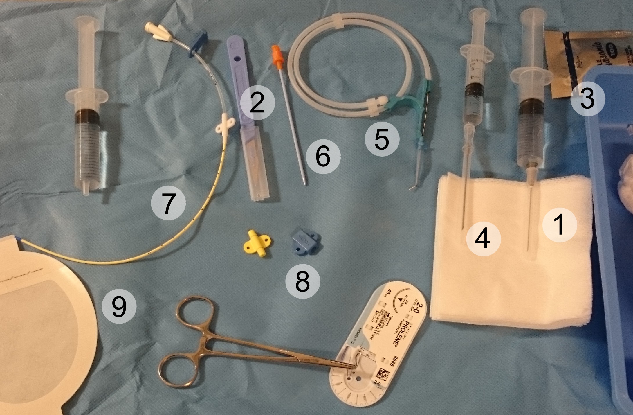
A central venous catheter (CVC), also known as a central line(c-line), central venous line, or central venous access catheter, is a catheter placed into a large vein. It is a form of venous access. Placement of larger catheters in more centrally located veins is often needed in critically ill patients, or in those requiring prolonged intravenous therapies, for more reliable vascular access. These catheters are commonly placed in veins in the neck (internal jugular vein), chest (subclavian vein or axillary vein), groin (femoral vein), or through veins in the arms (also known as a Peripherally inserted central catheter, PICC line, or peripherally inserted central catheters). Central lines are used to administer medication or fluids that are unable to be taken by mouth or would harm a smaller Peripheral vascular system, peripheral vein, obtain blood tests (specifically the "central venous oxygen saturation"), administer fluid or blood products for large volume resuscitation, and m ... [...More Info...] [...Related Items...] OR: [Wikipedia] [Google] [Baidu] |
Viola
The viola ( , also , ) is a string instrument that is bow (music), bowed, plucked, or played with varying techniques. Slightly larger than a violin, it has a lower and deeper sound. Since the 18th century, it has been the middle or alto voice of the violin family, between the violin (which is tuned a perfect fifth above) and the cello (which is tuned an octave below). The strings from low to high are typically tuned to scientific pitch notation, C3, G3, D4, and A4. In the past, the viola varied in size and style, as did its names. The word viola originates from the Italian language. The Italians often used the term viola da braccio meaning literally: 'of the arm'. "Brazzo" was another Italian word for the viola, which the Germans adopted as ''Bratsche''. The French had their own names: ''cinquiesme'' was a small viola, ''haute contre'' was a large viola, and ''taile'' was a tenor. Today, the French use the term ''alto'', a reference to its range. The viola was popular in the heyd ... [...More Info...] [...Related Items...] OR: [Wikipedia] [Google] [Baidu] |
Pulmonary Embolism
Pulmonary embolism (PE) is a blockage of an pulmonary artery, artery in the lungs by a substance that has moved from elsewhere in the body through the bloodstream (embolism). Symptoms of a PE may include dyspnea, shortness of breath, chest pain particularly upon breathing in, and coughing up blood. Symptoms of a deep vein thrombosis, blood clot in the leg may also be present, such as a erythema, red, warm, swollen, and painful leg. Signs of a PE include low blood oxygen saturation, oxygen levels, tachypnea, rapid breathing, tachycardia, rapid heart rate, and sometimes a mild fever. Severe cases can lead to Syncope (medicine), passing out, shock (circulatory), abnormally low blood pressure, obstructive shock, and cardiac arrest, sudden death. PE usually results from a blood clot in the leg that travels to the lung. The risk of blood clots is increased by advanced age, cancer, prolonged bed rest and immobilization, smoking, stroke, long-haul travel over 4 hours, certain genetics, g ... [...More Info...] [...Related Items...] OR: [Wikipedia] [Google] [Baidu] |
Anticoagulation
Anticoagulants, commonly known as blood thinners, are chemical substances that prevent or reduce coagulation of blood, prolonging the clotting time. Some of them occur naturally in blood-eating animals such as leeches and mosquitoes, where they help keep the bite area unclotted long enough for the animal to obtain some blood. As a class of medications, anticoagulants are used in therapy for thrombotic disorders. Oral anticoagulants (OACs) are taken by many people in pill or tablet form, and various intravenous anticoagulant dosage forms are used in hospitals. Some anticoagulants are used in medical equipment, such as sample tubes, blood transfusion bags, heart–lung machines, and dialysis equipment. One of the first anticoagulants, warfarin, was initially approved as a rodenticide. Anticoagulants are closely related to antiplatelet drugs and thrombolytic drugs by manipulating the various pathways of blood coagulation. Specifically, antiplatelet drugs inhibit platelet aggre ... [...More Info...] [...Related Items...] OR: [Wikipedia] [Google] [Baidu] |
Alteplase
Alteplase (t-PA), a biosynthetic form of human tissue-type plasminogen activator (t-PA), is a thrombolytic medication, used to treat acute ischemic stroke, acute ST elevation myocardial infarction, ST-elevation myocardial infarction (a type of heart attack), pulmonary embolism associated with low blood pressure, and blocked central venous catheter. It is given by injection into a vein or artery. Alteplase is the same as the normal Tissue plasminogen activator, human plasminogen activator produced in Endothelium, vascular endothelial cells and is synthesized via recombinant DNA technology in Chinese hamster ovary cells (CHO). Alteplase causes the breakdown of a clot by inducing fibrinolysis. Medical uses Alteplase is mainly used to treat acute ischemic stroke, acute myocardial infarction, acute massive pulmonary embolism, and blocked catheters. Similar to other Thrombolysis, thrombolytic drugs, alteplase is used to Thrombolysis, dissolve clots to restore tissue perfusion, but ... [...More Info...] [...Related Items...] OR: [Wikipedia] [Google] [Baidu] |
James Paget
Sir James Paget, 1st Baronet FRS HFRSE (11 January 1814 – 30 December 1899) (, rhymes with "gadget") was an English surgeon and pathologist who is best remembered for naming Paget's disease and who is considered, together with Rudolf Virchow, as one of the founders of scientific medical pathology. His famous works included ''Lectures on Tumours'' (1851) and ''Lectures on Surgical Pathology'' (1853). There are several medical conditions which were described by, and later named after, Paget: * Paget's disease of bone * Paget's disease of the nipple (a form of intraductal breast cancer spreading into the skin around the nipple) ** Extramammary Paget's disease refers to a group of similar, more rare skin lesions discovered by Radcliffe Crocker in 1889 which affect the male and female genitalia. * Paget–Schroetter disease * Paget's abscess, an abscess that recurs at the site of a former abscess which had resolved. Life Paget was born in Great Yarmouth, England, on 11 January ... [...More Info...] [...Related Items...] OR: [Wikipedia] [Google] [Baidu] |


