|
P Wave (electrocardiography)
The P wave on the ECG represents atrial depolarization, which results in atrial contraction, or atrial systole. Physiology The P wave is a summation wave generated by the depolarization front as it transits the atria. Normally the right atrium depolarizes slightly earlier than left atrium since the depolarization wave originates in the sinoatrial node, in the high right atrium and then travels to and through the left atrium. The depolarization front is carried through the atria along semi-specialized conduction pathways including Bachmann's bundle resulting in uniform shaped waves. Depolarization originating elsewhere in the atria (atrial ectopics) result in P waves with a different morphology from normal. Pathology Peaked P waves (> 0.25 mV) suggest right atrial enlargement, cor pulmonale, (''P pulmonale'' rhythm), but have a low predictive value (~20%). A P wave with increased amplitude can indicate hypokalemia. It can also indicate right atrial enlargement. A P wave ... [...More Info...] [...Related Items...] OR: [Wikipedia] [Google] [Baidu] |
Normal P Wave (ECG)
Normal(s) or The Normal(s) may refer to: Film and television * ''Normal'' (2003 film), starring Jessica Lange and Tom Wilkinson * ''Normal'' (2007 film), starring Carrie-Anne Moss, Kevin Zegers, Callum Keith Rennie, and Andrew Airlie * ''Normal'' (2009 film), an adaptation of Anthony Neilson's 1991 play ''Normal: The Düsseldorf Ripper'' * ''Normal!'', a 2011 Algerian film * ''The Normals'' (film), a 2012 American comedy film * "Normal" (''New Girl''), an episode of the TV series Mathematics * Normal (geometry), an object such as a line or vector that is perpendicular to a given object * Normal basis (of a Galois extension), used heavily in cryptography * Normal bundle * Normal cone, of a subscheme in algebraic geometry * Normal coordinates, in differential geometry, local coordinates obtained from the exponential map (Riemannian geometry) * Normal distribution, the Gaussian continuous probability distribution * Normal equations, describing the solution of the linear least sq ... [...More Info...] [...Related Items...] OR: [Wikipedia] [Google] [Baidu] |
Chronic Obstructive Pulmonary Disease
Chronic obstructive pulmonary disease (COPD) is a type of progressive lung disease characterized by long-term respiratory symptoms and airflow limitation. The main symptoms include shortness of breath and a cough, which may or may not produce mucus. COPD progressively worsens, with everyday activities such as walking or dressing becoming difficult. While COPD is incurable, it is preventable and treatable. The two most common conditions of COPD are emphysema and chronic bronchitis and they have been the two classic COPD phenotypes. Emphysema is defined as enlarged airspaces ( alveoli) whose walls have broken down resulting in permanent damage to the lung tissue. Chronic bronchitis is defined as a productive cough that is present for at least three months each year for two years. Both of these conditions can exist without airflow limitation when they are not classed as COPD. Emphysema is just one of the structural abnormalities that can limit airflow and can exist without ai ... [...More Info...] [...Related Items...] OR: [Wikipedia] [Google] [Baidu] |
T Wave
In electrocardiography, the T wave represents the repolarization of the ventricles. The interval from the beginning of the QRS complex to the apex of the T wave is referred to as the ''absolute refractory period''. The last half of the T wave is referred to as the ''relative refractory period'' or ''vulnerable period''. The T wave contains more information than the QT interval. The T wave can be described by its symmetry, skewness, slope of ascending and descending limbs, amplitude and subintervals like the Tpeak–Tend interval. In most leads, the T wave is positive. This is due to the repolarization of the membrane. During ventricle contraction (QRS complex), the heart depolarizes. Repolarization of the ventricle happens in the opposite direction of depolarization and is negative current, signifying the relaxation of the cardiac muscle of the ventricles. But this negative flow causes a positive T wave; although the cell becomes more negatively charged, the net effect is in t ... [...More Info...] [...Related Items...] OR: [Wikipedia] [Google] [Baidu] |
ST Segment
In electrocardiography, the ST segment connects the QRS complex and the T wave and has a duration of 0.005 to 0.150 sec (5 to 150 ms). It starts at the J point (junction between the QRS complex and ST segment) and ends at the beginning of the T wave. However, since it is usually difficult to determine exactly where the ST segment ends and the T wave begins, the relationship between the ST segment and T wave should be examined together. The typical ST segment duration is usually around 0.08 sec (80 ms). It should be essentially level with the PR and TP segments. The ST segment represents the isoelectric period when the ventricles are in between depolarization and repolarization. Interpretation * The normal ST segment has a slight upward concavity. * Flat, downsloping, or depressed ST segments may indicate coronary ischemia. * ST elevation may indicate transmural myocardial infarction. An elevation of >1mm and longer than 80 milliseconds following the J-point. This measure has a ... [...More Info...] [...Related Items...] OR: [Wikipedia] [Google] [Baidu] |
QT Interval
The QT interval is a measurement made on an electrocardiogram used to assess some of the electrical properties of the heart. It is calculated as the time from the start of the Q wave to the end of the T wave, and approximates to the time taken from when the cardiac ventricles start to contract to when they finish relaxing. An abnormally long or abnormally short QT interval is associated with an increased risk of developing abnormal heart rhythms and sudden cardiac death. Abnormalities in the QT interval can be caused by genetic conditions such as long QT syndrome, by certain medications such as sotalol or pitolisant, by disturbances in the concentrations of certain salts within the blood such as hypokalaemia, or by hormonal imbalances such as hypothyroidism. Measurement The QT interval is most commonly measured in lead II for evaluation of serial ECGs, with leads I and V5 being comparable alternatives to lead II. Leads III, aVL and V1 are generally avoided for measuremen ... [...More Info...] [...Related Items...] OR: [Wikipedia] [Google] [Baidu] |
QRS Complex
The QRS complex is the combination of three of the graphical deflections seen on a typical electrocardiogram (ECG or EKG). It is usually the central and most visually obvious part of the tracing. It corresponds to the depolarization of the right and left ventricles of the heart and contraction of the large ventricular muscles. In adults, the QRS complex normally lasts ; in children it may be shorter. The Q, R, and S waves occur in rapid succession, do not all appear in all leads, and reflect a single event and thus are usually considered together. A Q wave is any downward deflection immediately following the P wave. An R wave follows as an upward deflection, and the S wave is any downward deflection after the R wave. The T wave follows the S wave, and in some cases, an additional U wave follows the T wave. To measure the QRS interval start at the end of the PR interval (or beginning of the Q wave) to the end of the S wave. Normally this interval is 0.08 to 0.10 seconds. When ... [...More Info...] [...Related Items...] OR: [Wikipedia] [Google] [Baidu] |
PR Interval
In electrocardiography, the PR interval is the period, measured in milliseconds, that extends from the beginning of the P wave (the onset of atrial depolarization) until the beginning of the QRS complex (the onset of ventricular depolarization); it is normally between 120 and 200 ms in duration. The PR interval is sometimes termed the PQ interval. Interpretation Variations in the PQ interval can be associated with certain medical conditions: * Duration ** A long PR interval (of over 200 ms) indicates a slowing of conduction between the atria and ventricles, usually due to slow conduction through the atrioventricular node (AV node). This is known as first degree heart block. Prolongation can be associated with fibrosis of the AV node, high vagal tone, medications that slow the AV node such as beta-blockers, hypokalemia, acute rheumatic fever, or carditis associated with Lyme disease. ** A short PR interval (of less than 120ms) may be associated with a Pre-excitation syndromes ... [...More Info...] [...Related Items...] OR: [Wikipedia] [Google] [Baidu] |
Lewis Lead
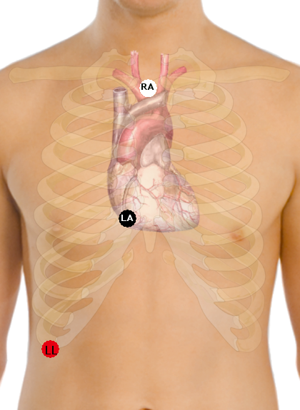
A Lewis Lead (also called the S5 lead) is a modified ECG lead used to detect atrial flutter waves when atrial flutter is suspected clinically, based on signs and symptoms, but is not definitely demonstrated on the standard 12 lead ECG. In order to create the Lewis Lead, the right arm electrode is moved to the manubrium adjacent to the sternum. Then the left arm electrode is moved to the right, fifth intercostal space adjacent to the sternum. The left leg electrode is placed on the right lower costal margin. The Lewis Lead is then read as Lead I on the ECG and, since in most patients it will be roughly perpendicular to the wave of ventricular depolarization In biology, depolarization or hypopolarization is a change within a cell, during which the cell undergoes a shift in electric charge distribution, resulting in less negative charge inside the cell compared to the outside. Depolarization is ess ..., atrial flutter waves may be more apparent. References * Goldman's ''Principl ... [...More Info...] [...Related Items...] OR: [Wikipedia] [Google] [Baidu] |
Ventricular Escape Beat
In cardiology, a ventricular escape beat is a self-generated electrical discharge initiated by, and causing contraction of the ventricles of the heart; normally the heart rhythm is begun in the atria of the heart and is subsequently transmitted to the ventricles. The ventricular escape beat follows a long pause in ventricular rhythm and acts to prevent cardiac arrest. It indicates a failure of the electrical conduction system of the heart to stimulate the ventricles (which would lead to the absence of heartbeats, unless ventricular escape beats occur). Causes Ventricular escape beats occur when the rate of electrical discharge reaching the ventricles (normally initiated by the heart's sinoatrial node (SA node), transmitted to the atrioventricular node (AV node), and then further transmitted to the ventricles) falls below the base rate determined by the rate of Phase 4 spontaneous depolarisation of ventricular pacemaker cells. An escape beat usually occurs 2–3 seconds after an ... [...More Info...] [...Related Items...] OR: [Wikipedia] [Google] [Baidu] |
Sinoatrial Arrest
Sinoatrial arrest is a medical condition wherein the sinoatrial node of the heart transiently ceases to generate the electrical impulses that normally stimulate the myocardial tissues to contract and thus the heart to beat. It is defined as lasting from 2.0 seconds to several minutes. Since the heart contains multiple pacemakers, this interruption of the cardiac cycle generally lasts only a few seconds before another part of the heart, such as the atrio-ventricular junction or the ventricles, begins pacing and restores the heart action. This condition can be detected on an electrocardiogram (ECG) as a brief period of irregular length with no electrical activity before either the sinoatrial node resumes normal pacing, or another pacemaker begins pacing. If a pacemaker other than the sinoatrial node is pacing the heart, this condition is known as an escape rhythm. If no other pacemaker begins pacing during an episode of sinus arrest it becomes a cardiac arrest. This condition i ... [...More Info...] [...Related Items...] OR: [Wikipedia] [Google] [Baidu] |
Atrial Flutter
Atrial flutter (AFL) is a common abnormal heart rhythm that starts in the atrial chambers of the heart. When it first occurs, it is usually associated with a fast heart rate and is classified as a type of supraventricular tachycardia. Atrial flutter is characterized by a sudden-onset (usually) regular abnormal heart rhythm on an electrocardiogram (ECG) in which the heart rate is fast. Symptoms may include a feeling of the heart beating too fast, too hard, or skipping beats, chest discomfort, difficulty breathing, a feeling as if one's stomach has dropped, a feeling of being light-headed, or loss of consciousness. Although this abnormal heart rhythm typically occurs in individuals with cardiovascular disease (e.g. high blood pressure, coronary artery disease, and cardiomyopathy) and diabetes mellitus, it may occur spontaneously in people with otherwise normal hearts. It is typically not a stable rhythm, and often degenerates into atrial fibrillation (AF). But rarely does it ... [...More Info...] [...Related Items...] OR: [Wikipedia] [Google] [Baidu] |


