|
Psoas Major
The psoas major ( or ; from grc, ψόᾱ, psóā, muscles of the loins) is a long fusiform muscle located in the lateral lumbar region between the vertebral column and the brim of the lesser pelvis. It joins the iliacus muscle to form the iliopsoas. In animals, this muscle is equivalent to the tenderloin. Structure The psoas major is divided into a superficial and a deep part. The deep part originates from the transverse processes of lumbar vertebrae L1–L5. The superficial part originates from the lateral surfaces of the last thoracic vertebra, lumbar vertebrae L1–L4, and the neighboring intervertebral discs. The lumbar plexus lies between the two layers. Together, the iliacus muscle and the psoas major form the iliopsoas, which is surrounded by the iliac fascia. The iliopsoas runs across the iliopubic eminence through the muscular lacuna to its insertion on the lesser trochanter of the femur. The iliopectineal bursa separates the tendon of the iliopsoas muscle from th ... [...More Info...] [...Related Items...] OR: [Wikipedia] [Google] [Baidu] |
Transverse Processes
The spinal column, a defining synapomorphy shared by nearly all vertebrates,Hagfish are believed to have secondarily lost their spinal column is a moderately flexible series of vertebrae (singular vertebra), each constituting a characteristic irregular bone whose complex structure is composed primarily of bone, and secondarily of hyaline cartilage. They show variation in the proportion contributed by these two tissue types; such variations correlate on one hand with the cerebral/caudal rank (i.e., location within the vertebral column, backbone), and on the other with phylogenetic differences among the vertebrate taxon, taxa. The basic configuration of a vertebra varies, but the bone is its ''body'', with the central part of the body constituting the ''centrum''. The upper (closer to) and lower (further from), respectively, the cranium and its central nervous system surfaces of the vertebra body support attachment to the intervertebral discs. The posterior part of a vertebra fo ... [...More Info...] [...Related Items...] OR: [Wikipedia] [Google] [Baidu] |
Intervertebral Disc
An intervertebral disc (or intervertebral fibrocartilage) lies between adjacent vertebrae in the vertebral column. Each disc forms a fibrocartilaginous joint (a symphysis), to allow slight movement of the vertebrae, to act as a ligament to hold the vertebrae together, and to function as a shock absorber for the spine. Structure Intervertebral discs consist of an outer fibrous ring, the anulus fibrosus disci intervertebralis, which surrounds an inner gel-like center, the nucleus pulposus. The ''anulus fibrosus'' consists of several layers (laminae) of fibrocartilage made up of both type I and type II collagen. Type I is concentrated toward the edge of the ring, where it provides greater strength. The stiff laminae can withstand compressive forces. The fibrous intervertebral disc contains the ''nucleus pulposus'' and this helps to distribute pressure evenly across the disc. This prevents the development of stress concentrations which could cause damage to the underlying vertebrae ... [...More Info...] [...Related Items...] OR: [Wikipedia] [Google] [Baidu] |
List Of Flexors Of The Human Body
A flexor is a muscle that flexes a joint. In anatomy, flexion (from the Latin verb ''flectere'', to bend) is a joint movement that decreases the angle between the bones that converge at the joint. For example, one’s elbow joint flexes when one brings their hand closer to the shoulder. Flexion is typically instigated by muscle contraction of a flexor. Flexors Upper limb *of the humerus bone (the bone in the upper arm) at the shoulder **Pectoralis major ** Anterior deltoid **Coracobrachialis **Biceps brachii * of the forearm at the elbow **Brachialis **Brachioradialis **Biceps brachii *of carpus (the carpal bones) at the wrist **flexor carpi radialis **flexor carpi ulnaris **palmaris longus *of the hand **flexor pollicis longus muscle **flexor pollicis brevis muscle **flexor digitorum profundus muscle **flexor digitorum superficialis muscle Lower limb Hip The hip flexors are (in descending order of importance to the action of flexing the hip joint):Platzer (2004), p 246 *Col ... [...More Info...] [...Related Items...] OR: [Wikipedia] [Google] [Baidu] |
Supine Position
The supine position ( or ) means lying horizontally with the face and torso facing up, as opposed to the prone position, which is face down. When used in surgical procedures, it grants access to the peritoneal, thoracic and pericardial regions; as well as the head, neck and extremities. Using anatomical terms of location, the dorsal side is down, and the ventral side is up, when supine. Semi-supine In scientific literature "semi-supine" commonly refers to positions where the upper body is tilted (at 45° or variations) and not completely horizontal. Relation to sudden infant death syndrome The decline in death due to sudden infant death syndrome (SIDS) is said to be attributable to having babies sleep in the supine position. The realization that infants sleeping face down, or in a prone position, had an increased mortality rate re-emerged into medical awareness at the end of the 1980s when two researchers, Susan Beal in Australia and Gus De Jonge in the Netherlands, indep ... [...More Info...] [...Related Items...] OR: [Wikipedia] [Google] [Baidu] |
Flexion
Motion, the process of movement, is described using specific anatomical terms. Motion includes movement of organs, joints, limbs, and specific sections of the body. The terminology used describes this motion according to its direction relative to the anatomical position of the body parts involved. Anatomists and others use a unified set of terms to describe most of the movements, although other, more specialized terms are necessary for describing unique movements such as those of the hands, feet, and eyes. In general, motion is classified according to the anatomical plane it occurs in. ''Flexion'' and ''extension'' are examples of ''angular'' motions, in which two axes of a joint are brought closer together or moved further apart. ''Rotational'' motion may occur at other joints, for example the shoulder, and are described as ''internal'' or ''external''. Other terms, such as ''elevation'' and ''depression'', describe movement above or below the horizontal plane. Many anatomica ... [...More Info...] [...Related Items...] OR: [Wikipedia] [Google] [Baidu] |
Myocyte
A muscle cell is also known as a myocyte when referring to either a cardiac muscle cell (cardiomyocyte), or a smooth muscle cell as these are both small cells. A skeletal muscle cell is long and threadlike with many nuclei and is called a muscle fiber. Muscle cells (including myocytes and muscle fibers) develop from embryonic precursor cells called myoblasts. Myoblasts fuse to form multinucleated skeletal muscle cells known as syncytia in a process known as myogenesis. Skeletal muscle cells and cardiac muscle cells both contain myofibrils and sarcomeres and form a striated muscle tissue. Cardiac muscle cells form the cardiac muscle in the walls of the heart chambers, and have a single central nucleus. Cardiac muscle cells are joined to neighboring cells by intercalated discs, and when joined in a visible unit they are described as a ''cardiac muscle fiber''. Smooth muscle cells control involuntary movements such as the peristalsis contractions in the esophagus and stomach. Sm ... [...More Info...] [...Related Items...] OR: [Wikipedia] [Google] [Baidu] |
Fast-twitching
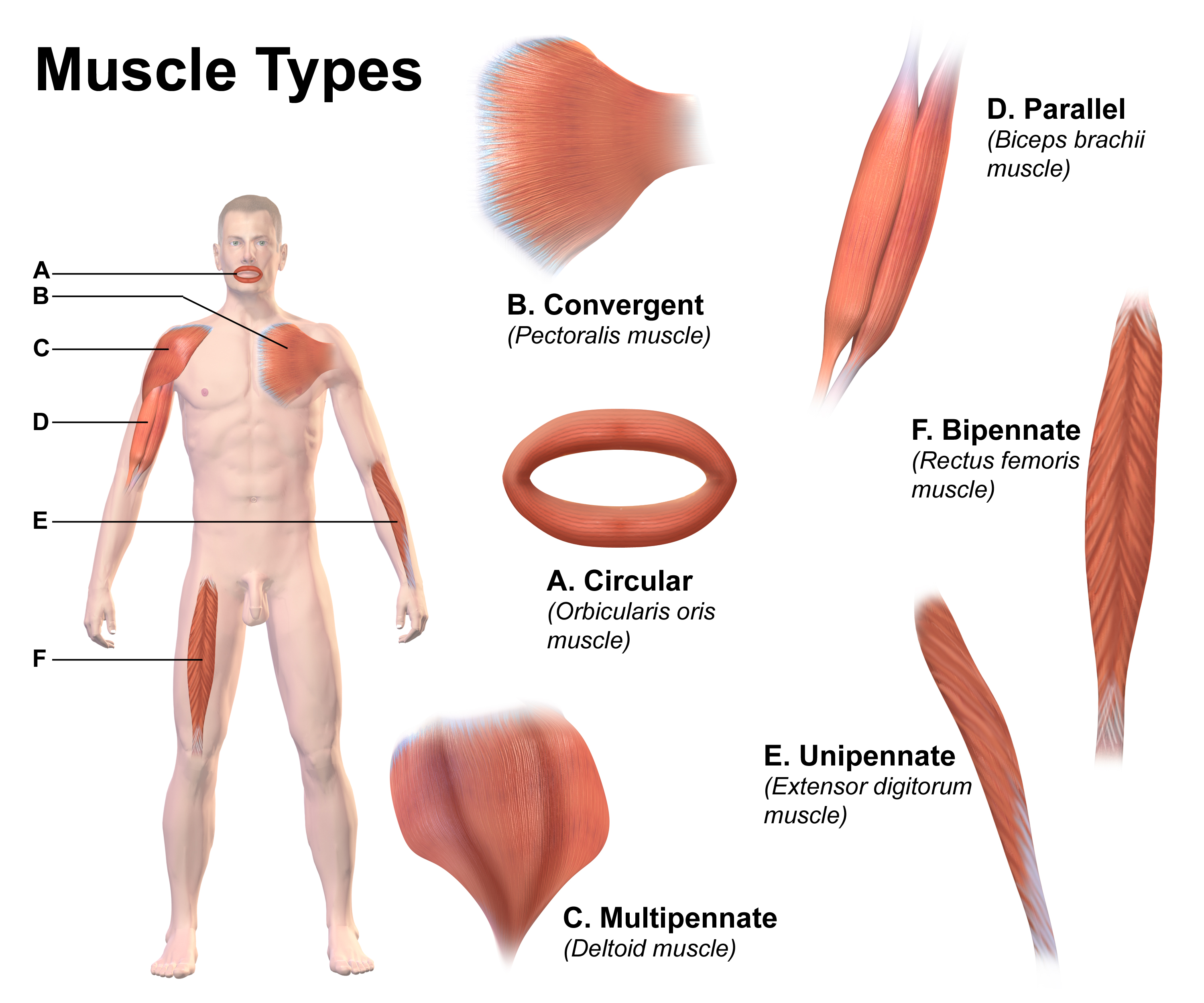
Skeletal muscles (commonly referred to as muscles) are organs of the vertebrate muscular system and typically are attached by tendons to bones of a skeleton. The muscle cells of skeletal muscles are much longer than in the other types of muscle tissue, and are often known as muscle fibers. The muscle tissue of a skeletal muscle is striated – having a striped appearance due to the arrangement of the sarcomeres. Skeletal muscles are voluntary muscles under the control of the somatic nervous system. The other types of muscle are cardiac muscle which is also striated and smooth muscle which is non-striated; both of these types of muscle tissue are classified as involuntary, or, under the control of the autonomic nervous system. A skeletal muscle contains multiple fascicles – bundles of muscle fibers. Each individual fiber, and each muscle is surrounded by a type of connective tissue layer of fascia. Muscle fibers are formed from the fusion of developmental myoblasts in a pro ... [...More Info...] [...Related Items...] OR: [Wikipedia] [Google] [Baidu] |
Journal Of Anatomy
The ''Journal of Anatomy'' is a monthly peer-reviewed scientific journal published by Wiley on behalf of the Anatomical Society. It covers all aspects of anatomy and morphology. The journal was first published in 1867 and was originally known as the ''Journal of Anatomy and Physiology'', obtaining its current title in October 1916. The editors-in-chief are Phil Cox (Hull York Medical School), Thomas H. Gillingwater (University of Edinburgh), Stefan Milz (Ludwig-Maximilians University), and Neil Vargesson (University of Aberdeen). According to the ''Journal Citation Reports'', the journal has a 2021 impact factor of 2.921. In conjunction with their centennial in 2009, the international Special Libraries Association included the ''Journal of Anatomy'' as one of the 100 most influential journals in biology and medicine over the past 100 years. History The journal was established in 1867 as the ''Journal of Anatomy and Physiology''. The journal was conceived at the 1866 meeting of the ... [...More Info...] [...Related Items...] OR: [Wikipedia] [Google] [Baidu] |
Psoas Minor
The psoas minor muscle ( or ; from grc, ψόᾱ, psóā, muscles of the loins) is a long, slender skeletal muscle. When present, it is located anterior to the psoas major muscle.Tank (2005), p 93Gray (2008), p 1372 Structure The psoas minor muscle originates from the vertical fascicles inserted on the last thoracic and first lumbar vertebrae. From there, it passes down onto the medial border of the psoas major, and is inserted to the innominate line and the iliopectineal eminence. Additionally, it attaches to and stretches the deep surface of the iliac fascia and occasionally its lowermost fibers reach the inguinal ligament.Bendavid (2001), p 58 It is posteriolateral to the iliopsoas muscle. Variations occur, however, and the insertion on the iliopubic eminence sometimes radiates into the iliopectineal arch.Platzer (2004), p 234 The psoas minor muscle receives oxygenated blood from the four lumbar arteries (inferior to the subcostal artery) and the lumbar branch of the iliolum ... [...More Info...] [...Related Items...] OR: [Wikipedia] [Google] [Baidu] |
Capsule Of Hip Joint
The articular capsule (capsular ligament) is strong and dense. Anterosuperiorly, it is attached to the margin of the acetabulum 5 to 6 mm. beyond the labrum behind; but in front, it is attached to the outer margin of the labrum, and, opposite to the notch where the margin of the cavity is deficient, it is connected to the transverse ligament, and by a few fibers to the edge of the obturator foramen. It surrounds the neck of the femur, and is attached, in front, to the intertrochanteric line; above, to the base of the neck; behind, to the neck, about 1.25 cm. above the intertrochanteric crest; below, to the lower part of the neck, close to the lesser trochanter. From its femoral attachment some of the fibers are reflected upward along the neck as longitudinal bands, termed ''retinacula''. The capsule is much thicker at the upper and forepart of the joint, where the most resistance is required; behind and below, it is thin and loose. It consists of two sets of fibers, ... [...More Info...] [...Related Items...] OR: [Wikipedia] [Google] [Baidu] |
Iliopectineal Bursa
The iliopectineal bursa or the iliopsoas bursa is a large synovial bursa that separates the external surface of the hip joint capsule from the normally just the tendon of the iliopsoas muscle. The most proximal of part the iliopectineal bursa lies on the iliopubic eminence of the superior pubic ramus. The iliopectineal bursa passes across the front of the capsule of the hip joint and extends distally downwards almost as far as to the lesser trochanter. The iliopectineal bursa frequently communicates by a circular aperture with the cavity of the hip joint. In 13% of all cases the iliopectineal bursa is partly separated by a septum into two cavities. Here the tendon of the psoas major muscle passes over the medial chamber and the tendon of the iliacus muscle runs over the lateral chamber. Inflammation of the iliopectineal bursa is called iliopectineal bursitis or iliopsoas bursitis Iliopsoas bursitis is inflammation of a bursa (synovial sac) lying between iliopsoas muscle and h ... [...More Info...] [...Related Items...] OR: [Wikipedia] [Google] [Baidu] |
Muscular Lacuna
The muscular lacuna (Latin: ''lacuna musculorum'') is the lateral compartment of the thigh inferior to the inguinal ligament, for the passage of the iliopsoas muscle, the femoral nerve and the lateral cutaneous nerve of the thigh; it is separated by the iliopectineal arch from the vascular lacuna The vascular lacuna (Latin: ''lacuna vasorum'') is the compartment beneath the inguinal ligament which allows for passage of the femoral vessels, lymph vessels and lymph nodes. Its boundaries are the iliopectineal arch, the inguinal ligament, .... Muscular system {{musculoskeletal-stub ... [...More Info...] [...Related Items...] OR: [Wikipedia] [Google] [Baidu] |


