|
Proliferative Index
Proliferation, as one of the Hallmarks of cancer, hallmarks and most fundamental biological processes in tumors, is associated with tumor progression, response to therapy, and cancer patient survival. Consequently, the evaluation of a tumor proliferative index (or growth fraction) has clinical significance in characterizing many solid tumors and hematologic malignancies. This has led investigators to develop different technologies to evaluate the proliferation index in tumor samples. The most commonly used methods in evaluating a proliferative index include mitotic indexing, thymidine-labeling index, bromodeoxyuridine assay, the determination of fraction of cells in various phases of cell cycle, and the Immunohistochemistry, immunohistochemical evaluation of cell cycle-associated proteins. Mitotic index (also called mitotic count) Mitotic indexing is the oldest method of assessing proliferation and is determined by counting the number of mitotic figures (cells undergoing mitosis) ... [...More Info...] [...Related Items...] OR: [Wikipedia] [Google] [Baidu] |
Hallmarks Of Cancer
The hallmarks of cancer were originally six biological capabilities acquired during the multistep development of human tumors and have since been increased to eight capabilities and two enabling capabilities. The idea was coined by Douglas Hanahan and Robert Weinberg (biologist), Robert Weinberg in their paper "The Hallmarks of Cancer" published January 2000 in ''Cell (journal), Cell''. These hallmarks constitute an organizing principle for rationalizing the complexities of neoplastic disease. They include sustaining proliferative signaling, evading growth suppressors, resisting cell death, enabling replicative immortality, inducing angiogenesis, and activating invasion and metastasis. Underlying these hallmarks are genome instability, which generates the genetic diversity that expedites their acquisition, and inflammation, which fosters multiple hallmark functions. In addition to cancer cells, tumors exhibit another dimension of complexity: they incorporate a community of recrui ... [...More Info...] [...Related Items...] OR: [Wikipedia] [Google] [Baidu] |
Mitotic Index
Mitotic index is defined as the ratio between the number of a population's cells undergoing mitosis to its total number of cells. Purpose The mitotic index is a measure of cellular proliferation. It is defined as the percentage of cells undergoing mitosis in a given population of cells. Mitosis is the division of somatic cells into two daughter cells. Durations of the cell cycle and mitosis vary in different cell types. An elevated mitotic index indicates more cells are dividing. In cancer cells, the mitotic index may be elevated compared to normal growth of tissues or cellular repair of the site of an injury. The mitotic index is therefore an important prognostic factor predicting both overall survival and response to chemotherapy in most types of cancer. It may lose much of its predictive value for elderly populations. For example, a low mitotic index loses any prognostic value for women over 70 years old with breast cancer. Calculation The mitotic index is the number of c ... [...More Info...] [...Related Items...] OR: [Wikipedia] [Google] [Baidu] |
Thymidine
Thymidine (symbol dT or dThd), also known as deoxythymidine, deoxyribosylthymine, or thymine deoxyriboside, is a pyrimidine deoxynucleoside. Deoxythymidine is the DNA nucleoside T, which pairs with deoxyadenosine (A) in double-stranded DNA. In cell biology it is used to synchronize the cells in G1/early S phase. The prefix deoxy- is often left out since there are no precursors of thymine nucleotides involved in RNA synthesis. Before the boom in thymidine use caused by the need for thymidine in the production of the antiretroviral drug azidothymidine (AZT), much of the world's thymidine production came from herring sperm. Thymidine occurs almost exclusively in DNA but it also occurs in the T-loop of tRNA. Structure and properties In its composition, deoxythymidine is a nucleoside composed of deoxyribose (a pentose sugar) joined to the pyrimidine base thymine. Deoxythymidine can be phosphorylated with one, two or three phosphoric acid groups, creating dTMP (deoxythymidin ... [...More Info...] [...Related Items...] OR: [Wikipedia] [Google] [Baidu] |
Bromodeoxyuridine
Bromodeoxyuridine (5-bromo-2'-deoxyuridine, BrdU, BUdR, BrdUrd, broxuridine) is a synthetic nucleoside analogue with a chemical structure similar to thymidine. BrdU is commonly used to study cell proliferation in living tissues and has been studied as a radiosensitizer and diagnostic tool in people with cancer. During S phase of the cell cycle (when DNA replication occurs), BrdU can be incorporated in place of thymidine in newly synthesized DNA molecules of dividing cells. Cells that have recently performed DNA replication or DNA repair can be detected with antibodies specific for BrdU using techniques such as immunohistochemistry or immunofluorescence. BrdU-labelled cells in humans can be detected up to two years after BrdU infusion. Because BrdU can replace thymidine during DNA replication, it can cause mutations, and its use is therefore potentially a health hazard. However, because it is neither radioactive nor myelotoxic at labeling concentrations, it is widely preferred ... [...More Info...] [...Related Items...] OR: [Wikipedia] [Google] [Baidu] |
Immunohistochemistry
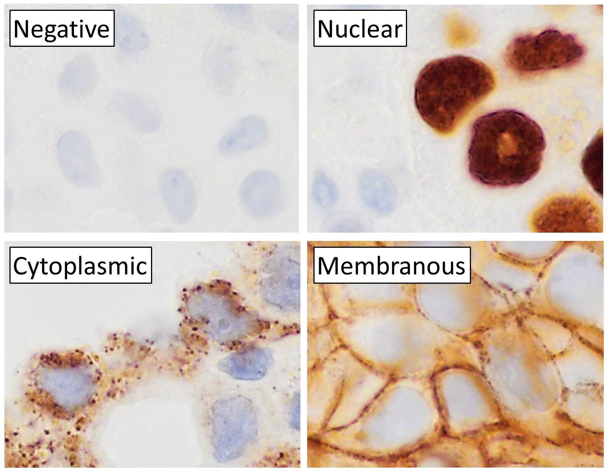
Immunohistochemistry (IHC) is the most common application of immunostaining. It involves the process of selectively identifying antigens (proteins) in cells of a tissue section by exploiting the principle of antibodies binding specifically to antigens in biological tissues. IHC takes its name from the roots "immuno", in reference to antibodies used in the procedure, and "histo", meaning tissue (compare to immunocytochemistry). Albert Coons conceptualized and first implemented the procedure in 1941. Visualising an antibody-antigen interaction can be accomplished in a number of ways, mainly either of the following: * ''Chromogenic immunohistochemistry'' (CIH), wherein an antibody is conjugated to an enzyme, such as peroxidase (the combination being termed immunoperoxidase), that can catalyse a colour-producing reaction. * ''Immunofluorescence'', where the antibody is tagged to a fluorophore, such as fluorescein or rhodamine. Immunohistochemical staining is widely used in the ... [...More Info...] [...Related Items...] OR: [Wikipedia] [Google] [Baidu] |
Mitoses In Neuroendocrine Tumor
In cell biology, mitosis () is a part of the cell cycle in which replicated chromosomes are separated into two new nuclei. Cell division by mitosis gives rise to genetically identical cells in which the total number of chromosomes is maintained. Therefore, mitosis is also known as equational division. In general, mitosis is preceded by S phase of interphase (during which DNA replication occurs) and is often followed by telophase and cytokinesis; which divides the cytoplasm, organelles and cell membrane of one cell into two new cells containing roughly equal shares of these cellular components. The different stages of mitosis altogether define the mitotic (M) phase of an animal cell cycle—the division of the mother cell into two daughter cells genetically identical to each other. The process of mitosis is divided into stages corresponding to the completion of one set of activities and the start of the next. These stages are preprophase (specific to plant cells), prophase, ... [...More Info...] [...Related Items...] OR: [Wikipedia] [Google] [Baidu] |
H&E Stain
Hematoxylin and eosin stain ( or haematoxylin and eosin stain or hematoxylin-eosin stain; often abbreviated as H&E stain or HE stain) is one of the principal tissue stains used in histology. It is the most widely used stain in medical diagnosis and is often the gold standard. For example, when a pathologist looks at a biopsy of a suspected cancer, the histological section is likely to be stained with H&E. H&E is the combination of two histological stains: hematoxylin and eosin. The hematoxylin stains cell nuclei a purplish blue, and eosin stains the extracellular matrix and cytoplasm pink, with other structures taking on different shades, hues, and combinations of these colors. Hence a pathologist can easily differentiate between the nuclear and cytoplasmic parts of a cell, and additionally, the overall patterns of coloration from the stain show the general layout and distribution of cells and provides a general overview of a tissue sample's structure. Thus, pattern recognit ... [...More Info...] [...Related Items...] OR: [Wikipedia] [Google] [Baidu] |
Autoradiograph
An autoradiograph is an image on an X-ray film or nuclear emulsion produced by the pattern of decay emissions (e.g., beta particles or gamma rays) from a distribution of a radioactive substance. Alternatively, the autoradiograph is also available as a digital image (digital autoradiography), due to the recent development of scintillation gas detectors or rare earth phosphorimaging systems. The film or emulsion is apposed to the labeled tissue section to obtain the autoradiograph (also called an autoradiogram). The ''auto-'' prefix indicates that the radioactive substance is within the sample, as distinguished from the case of historadiography or microradiography, in which the sample is marked using an external source. Some autoradiographs can be examined microscopically for localization of silver grains (such as on the interiors or exteriors of cells or organelles) in which the process is termed micro-autoradiography. For example, micro-autoradiography was used to examine whethe ... [...More Info...] [...Related Items...] OR: [Wikipedia] [Google] [Baidu] |
Radiolabeled
A radioactive tracer, radiotracer, or radioactive label is a chemical compound in which one or more atoms have been replaced by a radionuclide so by virtue of its radioactive decay it can be used to explore the mechanism of chemical reactions by tracing the path that the radioisotope follows from reactants to products. Radiolabeling or radiotracing is thus the radioactive form of isotopic labeling. In biological contexts, use of radioisotope tracers are sometimes called radioisotope feeding experiments. Radioisotopes of hydrogen, carbon, phosphorus, sulfur, and iodine have been used extensively to trace the path of biochemical reactions. A radioactive tracer can also be used to track the distribution of a substance within a natural system such as a cell or tissue, or as a flow tracer to track fluid flow. Radioactive tracers are also used to determine the location of fractures created by hydraulic fracturing in natural gas production.Reis, John C. (1976). ''Environmental Control i ... [...More Info...] [...Related Items...] OR: [Wikipedia] [Google] [Baidu] |
Histogram
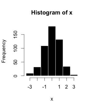
A histogram is an approximate representation of the frequency distribution, distribution of numerical data. The term was first introduced by Karl Pearson. To construct a histogram, the first step is to "Data binning, bin" (or "Data binning, bucket") the range of values—that is, divide the entire range of values into a series of intervals—and then count how many values fall into each interval. The bins are usually specified as consecutive, non-overlapping interval (mathematics), intervals of a variable. The bins (intervals) must be adjacent and are often (but not required to be) of equal size. If the bins are of equal size, a bar is drawn over the bin with height proportional to the Frequency (statistics), frequency—the number of cases in each bin. A histogram may also be normalization (statistics), normalized to display "relative" frequencies showing the proportion of cases that fall into each of several Categorization, categories, with the sum of the heights equaling 1. ... [...More Info...] [...Related Items...] OR: [Wikipedia] [Google] [Baidu] |
Ki-67 (protein)
Antigen KI-67, also known as Ki-67, Ki-67 or MKI67 (Marker Of Proliferation Ki-67), is a protein that in humans is encoded by the ''MKI67'' gene (antigen identified by monoclonal antibody Ki-67). Function Antigen KI-67 is a nuclear protein that is associated with cellular proliferation. Altering Ki-67 expression levels did not significantly affect cell proliferation in vivo. Ki-67 mutant mice developed normally and cells lacking Ki-67 proliferated efficiently. Furthermore, it is associated with ribosomal RNA transcription. Inactivation of antigen KI-67 leads to inhibition of ribosomal RNA synthesis. Use as a marker of proliferating cells The Ki-67 protein (also known as MKI67) is a cellular marker for proliferation, and can be used in immunohistochemistry. It is strictly associated with cell proliferation. During interphase, the Ki-67 antigen can be exclusively detected within the cell nucleus, whereas in mitosis most of the protein is relocated to the surface of the chromo ... [...More Info...] [...Related Items...] OR: [Wikipedia] [Google] [Baidu] |
Proliferating Cell Nuclear Antigen
Proliferating cell nuclear antigen (PCNA) is a DNA clamp that acts as a processivity factor for DNA polymerase δ in eukaryotic cells and is essential for replication. PCNA is a homotrimer and achieves its processivity by encircling the DNA, where it acts as a scaffold to recruit proteins involved in DNA replication, DNA repair, chromatin remodeling and epigenetics. Many proteins interact with PCNA via the two known PCNA-interacting motifs PCNA-interacting peptide (PIP) box and AlkB homologue 2 PCNA interacting motif (APIM). Proteins binding to PCNA via the PIP-box are mainly involved in DNA replication whereas proteins binding to PCNA via APIM are mainly important in the context of genotoxic stress. Function The protein encoded by this gene is found in the nucleus and is a cofactor of DNA polymerase delta. The encoded protein acts as a homotrimer and helps increase the processivity of leading strand synthesis during DNA replication. In response to DNA damage, this protein i ... [...More Info...] [...Related Items...] OR: [Wikipedia] [Google] [Baidu] |


