|
Primary Familial Brain Calcification
Primary Indiana familial brain calcification Initial Posting: April 18, 2004; Last Update: August 24, 2017. (PFBC), also known as familial idiopathic basal ganglia calcification (FIBGC) and Fahr's disease, is a rare, genetically dominant, inherited neurological disorder characterized by abnormal deposits of calcium in areas of the brain that control movement. Through the use of CT scans, calcifications are seen primarily in the basal ganglia and in other areas such as the cerebral cortex. Signs and symptoms Symptoms of this disease include deterioration of motor functions and speech, seizures, and other involuntary movement. Other symptoms are headaches, dementia, and vision impairment. Characteristics of Parkinson's Disease are also similar to PFBC. The disease usually manifests itself in the third to fifth decade of life but may appear in childhood or later in life.Sobrido MJ, Hopfer S, Geschwind DH (2007)Familial idiopathic basal ganglia calcification" In: Pagon RA, Bird TD ... [...More Info...] [...Related Items...] OR: [Wikipedia] [Google] [Baidu] |
CT Scan
A computed tomography scan (CT scan; formerly called computed axial tomography scan or CAT scan) is a medical imaging technique used to obtain detailed internal images of the body. The personnel that perform CT scans are called radiographers or radiology technologists. CT scanners use a rotating X-ray tube and a row of detectors placed in a gantry (medical), gantry to measure X-ray Attenuation#Radiography, attenuations by different tissues inside the body. The multiple X-ray measurements taken from different angles are then processed on a computer using tomographic reconstruction algorithms to produce Tomography, tomographic (cross-sectional) images (virtual "slices") of a body. CT scans can be used in patients with metallic implants or pacemakers, for whom magnetic resonance imaging (MRI) is Contraindication, contraindicated. Since its development in the 1970s, CT scanning has proven to be a versatile imaging technique. While CT is most prominently used in medical diagnosis, ... [...More Info...] [...Related Items...] OR: [Wikipedia] [Google] [Baidu] |
Chromosome 22
Chromosome 22 is one of the 23 pairs of chromosomes in human cell (biology), cells. Humans normally have two copies of chromosome 22 in each cell. Chromosome 22 is the second smallest human chromosome, spanning about 49 million DNA base pairs and representing between 1.5 and 2% of the total DNA in cell (biology), cells. In 1999, researchers working on the Human Genome Project announced they had determined the sequence of base pairs that make up this chromosome. Chromosome 22 was the first human chromosome to be fully sequenced. Human chromosomes are numbered by their apparent size in the karyotype, with Chromosome 1 being the largest and Chromosome 22 having originally been identified as the smallest. However, genome sequencing has revealed that Chromosome 21 is actually smaller than Chromosome 22. Genes Number of genes The following are some of the gene count estimates of human chromosome 22. Because researchers use different approaches to genome annotation, their predictions ... [...More Info...] [...Related Items...] OR: [Wikipedia] [Google] [Baidu] |
Dentate Nucleus
The dentate nucleus is a cluster of neurons, or nerve cells, in the central nervous system that has a dentate – tooth-like or serrated – edge. It is located within the deep white matter of each cerebellar hemisphere, and it is the largest single structure linking the cerebellum to the rest of the brain.Sultan, F., Hamodeh, S., & Baizer, J. S. (2010). THE HUMAN DENTATE NUCLEUS: A COMPLEX SHAPE UNTANGLED. rticle Neuroscience, 167(4), 965–968. It is the largest and most lateral, or farthest from the midline, of the four pairs of deep cerebellar nuclei, the others being the globose and emboliform nuclei, which together are referred to as the interposed nucleus, and the fastigial nucleus. The dentate nucleus is responsible for the planning, initiation and control of voluntary movements. The dorsal region of the dentate nucleus contains output channels involved in motor function, which is the movement of skeletal muscle, while the ventral region contains output channels involv ... [...More Info...] [...Related Items...] OR: [Wikipedia] [Google] [Baidu] |
Caudate Nucleus
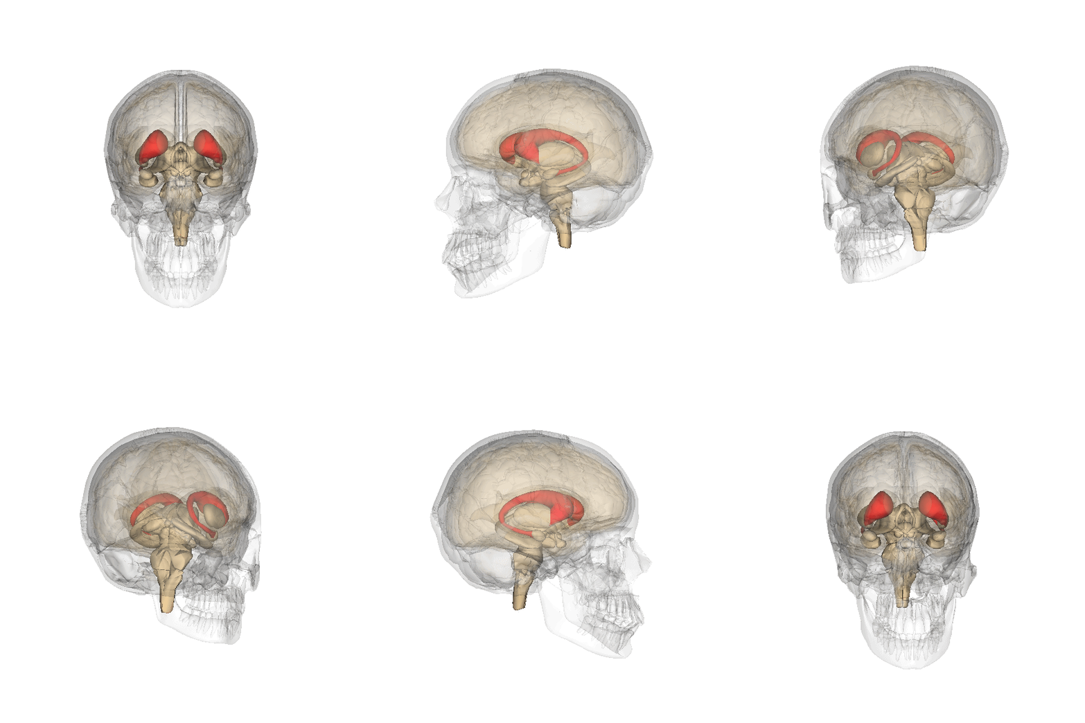
The caudate nucleus is one of the structures that make up the corpus striatum, which is a component of the basal ganglia in the human brain. While the caudate nucleus has long been associated with motor processes due to its role in Parkinson's disease, it plays important roles in various other nonmotor functions as well, including procedural learning, associative learning and inhibitory control of action, among other functions. The caudate is also one of the brain structures which compose the reward system and functions as part of the cortico–basal ganglia– thalamic loop. Structure Together with the putamen, the caudate forms the dorsal striatum, which is considered a single functional structure; anatomically, it is separated by a large white matter tract, the internal capsule, so it is sometimes also referred to as two structures: the medial dorsal striatum (the caudate) and the lateral dorsal striatum (the putamen). In this vein, the two are functionally distinct not a ... [...More Info...] [...Related Items...] OR: [Wikipedia] [Google] [Baidu] |
Globus Pallidus
The globus pallidus (GP), also known as paleostriatum or dorsal pallidum, is a subcortical structure of the brain. It consists of two adjacent segments, one external, known in rodents simply as the globus pallidus, and one internal, known in rodents as the entopeduncular nucleus. It is part of the telencephalon, but retains close functional ties with the subthalamus in the diencephalon – both of which are part of the extrapyramidal motor system. The globus pallidus is a major component of the basal ganglia, with principal inputs from the striatum, and principal direct outputs to the thalamus and the substantia nigra. The latter is made up of similar neuronal elements, has similar afferents from the striatum, similar projections to the thalamus, and has a similar synaptology. Neither receives direct cortical afferents, and both receive substantial additional inputs from the intralaminar thalamus. Globus pallidus is Latin for "pale globe". Structure Pallidal nuclei are ma ... [...More Info...] [...Related Items...] OR: [Wikipedia] [Google] [Baidu] |
Lenticular Nucleus
The lentiform nucleus, or lenticular nucleus, comprises the putamen and the globus pallidus within the basal ganglia. With the caudate nucleus, it forms the dorsal striatum. It is a large, lens-shaped mass of gray matter just lateral to the internal capsule. Structure When divided horizontally, it exhibits, to some extent, the appearance of a biconvex lens, while a coronal section of its central part presents a somewhat triangular outline. It is shorter than the caudate nucleus and does not extend as far forward. Boundaries It is lateral to the caudate nucleus and thalamus, and is seen only in sections of the hemisphere. It is bounded laterally by a lamina of white substance called the external capsule, and lateral to this is a thin layer of gray substance termed the claustrum. Its anterior end is continuous with the lower part of the head of the caudate nucleus and with the anterior perforated substance. Components In a coronal section through the middle of the lentiform nu ... [...More Info...] [...Related Items...] OR: [Wikipedia] [Google] [Baidu] |
OCLN
Occludin is an enzyme ( EC 1.6) that oxidizes NADH. It was first identified in epithelial cells as a 65 kDa integral plasma-membrane protein localized at the tight junctions. Together with Claudins, and zonula occludens-1 (ZO-1), occludin has been considered a staple of tight junctions, and although it was shown to regulate the formation, maintenance, and function of tight junctions, its precise mechanism of action remained elusive and most of its actions were initially attributed to conformational changes following selective phosphorylation, and its redox-sensitive dimerization. However, mounting evidence demonstrated that occludin is not only present in epithelial/endothelial cells, but is also expressed in large quantities in cells that do not have tight junctions but have very active metabolism: pericytes, neurons and astrocytes, oligodendrocytes, dendritic cells, monocytes/macrophages lymphocytes, and myocardium. Recent work, using molecular modeling, supported by biochemical ... [...More Info...] [...Related Items...] OR: [Wikipedia] [Google] [Baidu] |
JAM3
Junctional adhesion molecule C is a protein that in humans is encoded by the ''JAM3'' gene. Gene This gene is located on the long arm of chromosome 11 (11q25) on the Watson strand. It is 83,077 bases in length. The encoded protein is 310 amino acids long with a predicted molecular weight of 35.02 kiloDaltons. Function Tight junctions represent one mode of cell-to-cell adhesion in epithelial or endothelial cell sheets, forming continuous seals around cells and serving as a physical barrier to prevent solutes and water from passing freely through the paracellular space. The protein encoded by this immunoglobulin superfamily gene member is localized in the tight junctions between high endothelial cells. Unlike other proteins in this family, this protein is unable to adhere to leukocyte cell lines and only forms weak homotypic interactions. The encoded protein is a member of the junctional adhesion molecule protein family and acts as a receptor for another member of this family. ... [...More Info...] [...Related Items...] OR: [Wikipedia] [Google] [Baidu] |
JAM2
Junctional adhesion molecule B is a protein that in humans is encoded by the ''JAM2'' gene. JAM2 has also been designated as CD322 (cluster of differentiation 322). Function Tight junctions represent one mode of cell-to-cell adhesion in endothelial cell sheets, forming continuous seals around cells and serving as a physical barrier to prevent solutes and water from passing freely through the paracellular space. The protein encoded by this immunoglobulin superfamily gene member is localized in the tight junctions between high endothelial cells. It acts as an adhesive ligand for interacting with a variety of immune cell types and may play a role in lymphocyte homing to secondary lymphoid organs. It is purported to promote lymphocyte transendothelial migration. It might also be involved with endothelial cell polarity, by associating to cell polarity protein PAR-3, together with JAM3. Interactions JAM2 has been shown to interact with PARD3. It also interacts with the integr ... [...More Info...] [...Related Items...] OR: [Wikipedia] [Google] [Baidu] |
Chromosome 9
Chromosome 9 is one of the 23 pairs of chromosomes in humans. Humans normally have two copies of this chromosome, as they normally do with all chromosomes. Chromosome 9 spans about 138 million base pairs of nucleic acids (the building blocks of DNA) and represents between 4.0 and 4.5% of the total DNA in cells. Genes Number of genes These are some of the gene count estimates of human chromosome 9. Because researchers use different approaches to genome annotation, their predictions of the number of genes on each chromosome varies (for technical details, see gene prediction). Among various projects, the collaborative consensus coding sequence project ( CCDS) takes an extremely conservative strategy. So CCDS's gene number prediction represents a lower bound on the total number of human protein-coding genes. Gene list The following is a partial list of genes on human chromosome 9. For a complete list, see the link in the infobox on the right. Diseases and disorders The follow ... [...More Info...] [...Related Items...] OR: [Wikipedia] [Google] [Baidu] |
Chromosome 1
Chromosome 1 is the designation for the largest human chromosome. Humans have two copies of chromosome 1, as they do with all of the autosomes, which are the non-sex chromosomes. Chromosome 1 spans about 249 million nucleotide base pairs, which are the basic units of information for DNA.http://vega.sanger.ac.uk/Homo_sapiens/mapview?chr=1 Chromosome size and number of genes derived from this database, retrieved 2012-03-11. It represents about 8% of the total DNA in human cells. It was the last completed chromosome, sequenced two decades after the beginning of the Human Genome Project. Genes Number of genes The following are some of the gene count estimates of human chromosome 1. Because researchers use different approaches to genome annotation their predictions of the number of genes on each chromosome varies (for technical details, see gene prediction). Among various projects, the collaborative consensus coding sequence project ( CCDS) takes an extremely conservative strategy. So ... [...More Info...] [...Related Items...] OR: [Wikipedia] [Google] [Baidu] |


