|
Preferential Hyperacuity Perimetry
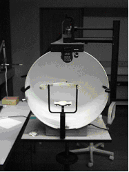
Preferential hyperacuity perimetry (PHP) is a psychophysical test used to identify and quantify visual abnormalities such as metamorphopsia and scotoma. It is a type of perimetry. Background Vision abnormalities such as metamorphopsia (distortions) and scotoma are symptoms of retinal diseases such as macular degeneration. In advanced stages of the disease, photoreceptor cells may be irreversibly damaged. Hence, if not treated, macular degeneration may lead to blindness. Awareness to early changes in vision, especially in high risk patients, leads to early treatment (such as intravitreal injection of anti-VEGF factors, e.g. bevacizumab or ranibizumab) and prevents loss of vision. Because of complex brain mechanisms such as filling-in, patients with small and peripheral defects in their vision are often unaware of such changes until late stages in the disease. Another problem is that minute visual aberrations can be normal and therefore should be distinguished from genuine visual ab ... [...More Info...] [...Related Items...] OR: [Wikipedia] [Google] [Baidu] |
Metamorphopsia
Metamorphopsia (from , ) is a type of distorted vision in which a grid of straight lines appears wavy and parts of the grid may appear blank. People can first notice they suffer with the condition when looking at mini-blinds in their home. For example, straight lines might be wavy or bendy. Things may appear closer or further than they are. Initially characterized in the 1800s, metamorphopsia was described as one of the primary and most notable indications of myopic and senile maculopathies. Metamorphopsia can present itself as unbalanced vision, resulting from small unintentional movements of the eye as it tries to stabilize the field of vision. Metamorphopsia can also lead to the misrepresentation of an object’s size or shape. It is mainly associated with macular degeneration, particularly age-related macular degeneration with choroidal neovascularization. 80%)—deposition of yellowish extracellular material in and between Bruch's membrane and retinal pigment epithelium (“ ... [...More Info...] [...Related Items...] OR: [Wikipedia] [Google] [Baidu] |
Scotoma
A scotoma is an area of partial alteration in the field of vision consisting of a partially diminished or entirely degenerated visual acuity that is surrounded by a field of normal – or relatively well-preserved – vision. Every normal mammalian eye has a scotoma in its field of vision, usually termed its blind spot. This is a location with no photoreceptor cells, where the retinal ganglion cell axons that compose the optic nerve exit the retina. This location is called the optic disc. There is no direct conscious awareness of visual scotomas. They are simply regions of reduced information within the visual field. Rather than recognizing an incomplete image, patients with scotomas report that things "disappear" on them. The presence of the blind spot scotoma can be demonstrated subjectively by covering one eye, carefully holding fixation with the open eye, and placing an object (such as one's thumb) in the lateral and horizontal visual field, about 15 degrees from fix ... [...More Info...] [...Related Items...] OR: [Wikipedia] [Google] [Baidu] |
Perimetry
A visual field test is an eye examination that can detect dysfunction in central and peripheral vision which may be caused by various medical conditions such as glaucoma, stroke, pituitary disease, brain tumours or other neurological deficits. Visual field testing can be performed clinically by keeping the subject's gaze fixed while presenting objects at various places within their visual field. Simple manual equipment can be used such as in the tangent screen test or the Amsler grid. When dedicated machinery is used it is called a perimeter. The exam may be performed by a technician in one of several ways. The test may be performed by a technician directly, with the assistance of a machine, or completely by an automated machine. Machine-based tests aid diagnostics by allowing a detailed printout of the patient's visual field. Other names for this test may include perimetry, Tangent screen exam, Automated perimetry exam, Goldmann visual field exam, or brand names such as Hen ... [...More Info...] [...Related Items...] OR: [Wikipedia] [Google] [Baidu] |
Macular Degeneration
Macular degeneration, also known as age-related macular degeneration (AMD or ARMD), is a medical condition which may result in blurred or no vision in the center of the visual field. Early on there are often no symptoms. Over time, however, some people experience a gradual worsening of vision that may affect one or both eyes. While it does not result in complete blindness, loss of central vision can make it hard to recognize faces, drive, read, or perform other activities of daily life. Visual hallucinations may also occur. Macular degeneration typically occurs in older people. Genetic factors and smoking also play a role. It is due to damage to the macula of the retina. Diagnosis is by a complete eye exam. The severity is divided into early, intermediate, and late types. The late type is additionally divided into "dry" and "wet" forms with the dry form making up 90% of cases. The difference between the two forms is the change of macula. Those with dry form AMD have drusen, ce ... [...More Info...] [...Related Items...] OR: [Wikipedia] [Google] [Baidu] |
Photoreceptor Cell
A photoreceptor cell is a specialized type of neuroepithelial cell found in the retina that is capable of visual phototransduction. The great biological importance of photoreceptors is that they convert light (visible electromagnetic radiation) into signals that can stimulate biological processes. To be more specific, photoreceptor proteins in the cell absorb photons, triggering a change in the cell's membrane potential. There are currently three known types of photoreceptor cells in mammalian eyes: rods, cones, and intrinsically photosensitive retinal ganglion cells. The two classic photoreceptor cells are rods and cones, each contributing information used by the visual system to form an image of the environment, sight. Rods primarily mediate scotopic vision (dim conditions) whereas cones primarily mediate to photopic vision (bright conditions), but the processes in each that supports phototransduction is similar. A third class of mammalian photoreceptor cell was discovered ... [...More Info...] [...Related Items...] OR: [Wikipedia] [Google] [Baidu] |
Bevacizumab
Bevacizumab, sold under the brand name Avastin among others, is a medication used to treat a number of types of cancers and a specific eye disease. For cancer, it is given by slow injection into a vein (intravenous) and used for colon cancer, lung cancer, glioblastoma, and renal-cell carcinoma. In many of these diseases it is used as a first-line therapy. For age-related macular degeneration it is given by injection into the eye (intravitreal). Common side effects when used for cancer include nose bleeds, headache, high blood pressure, and rash. Other severe side effects include gastrointestinal perforation, bleeding, allergic reactions, blood clots, and an increased risk of infection. When used for eye disease side effects can include vision loss and retinal detachment. Bevacizumab is a monoclonal antibody that functions as an angiogenesis inhibitor. It works by slowing the growth of new blood vessels by inhibiting vascular endothelial growth factor A (VEGF-A), in other word ... [...More Info...] [...Related Items...] OR: [Wikipedia] [Google] [Baidu] |
Ranibizumab
Ranibizumab, sold under the brand name Lucentis among others, is a monoclonal antibody fragment ( Fab) created from the same parent mouse antibody as bevacizumab. It is an anti-angiogenic that is approved to treat the "wet" type of age-related macular degeneration (AMD, also ARMD), diabetic retinopathy, and macular edema due to branch retinal vein occlusion or central retinal vein occlusion. Ranibizumab was developed by Genentech and marketed by them in the United States, and elsewhere by Novartis, under the brand name Lucentis. Ranibizumab (Lucentis) was approved for medical use in the United States in June 2006. Ranibizumab (Susvimo) was approved for medical use in the United States in October 2021. Medical uses In the United States, ranibizumab is indicated for the treatment of neovascular (wet) age-related macular degeneration, macular edema following retinal vein occlusion, diabetic macular edema, diabetic retinopathy, and myopic choroidal neovascularization. In the Europ ... [...More Info...] [...Related Items...] OR: [Wikipedia] [Google] [Baidu] |
Filling-in
In vision, filling-in phenomena are those responsible for the completion of missing information across the physiological blind spot, and across natural and artificial scotomata. There is also evidence for similar mechanisms of completion in normal visual analysis. Classical demonstrations of perceptual filling-in involve filling in at the blind spot in monocular vision, and images stabilized on the retina either by means of special lenses, or under certain conditions of steady fixation. For example, naturally in monocular vision at the physiological blind spot, the percept is not a hole in the visual field, but the content is “filled-in” based on information from the surrounding visual field. When a textured stimulus is presented centered on but extending beyond the region of the blind spot, a continuous texture is perceived. This partially inferred percept is paradoxically considered more reliable than a percept based on external input. (Ehinger ''et al.'' 2017). A second ty ... [...More Info...] [...Related Items...] OR: [Wikipedia] [Google] [Baidu] |
Vernier Acuity
Vernier acuity (from the term "vernier scale", named after astronomer Pierre Vernier) is a type of visual acuity – more precisely of hyperacuity – that measures the ability to discern a disalignment among two line segments or gratings. A subject's vernier (IPA: ) acuity is the smallest visible offset between the stimuli that can be detected. Because the disalignments are often much smaller than the diameter and spacing of retinal receptors, vernier acuity requires neural processing and "pooling" to detect it. Because vernier acuity exceeds acuity by far, the phenomenon has been termed hyperacuity. Vernier acuity develops rapidly during infancy and continues to slowly develop throughout childhood. At approximately three to twelve months old, it surpasses grating acuity in foveal vision in humans. However, vernier acuity decreases more quickly than grating acuity in peripheral vision. Vernier acuity was first explained by Ewald Hering in 1899, based on earlier data by Alfred V ... [...More Info...] [...Related Items...] OR: [Wikipedia] [Google] [Baidu] |
Visual Acuity
Visual acuity (VA) commonly refers to the clarity of vision, but technically rates an examinee's ability to recognize small details with precision. Visual acuity is dependent on optical and neural factors, i.e. (1) the sharpness of the retinal image within the eye, (2) the health and functioning of the retina, and (3) the sensitivity of the interpretative faculty of the brain. The most commonly referred visual acuity is the far acuity (e.g. 6/6 or 20/20 acuity), which describes the examinee's ability to recognize small details at a far distance, and is relevant to people with myopia; however, for people with hyperopia, the near acuity is used instead to describe the examinee's ability to recognize small details at a near distance. A common cause of low visual acuity is refractive error (ametropia), errors in how the light is refracted in the eyeball, and errors in how the retinal image is interpreted by the brain. The latter is the primary cause for low vision in people with a ... [...More Info...] [...Related Items...] OR: [Wikipedia] [Google] [Baidu] |
Macula
The macula (/ˈmakjʊlə/) or macula lutea is an oval-shaped pigmented area in the center of the retina of the human eye and in other animals. The macula in humans has a diameter of around and is subdivided into the umbo, foveola, foveal avascular zone, fovea, parafovea, and perifovea areas. The anatomical macula at a size of is much larger than the clinical macula which, at a size of , corresponds to the anatomical fovea. The macula is responsible for the central, high-resolution, color vision that is possible in good light; and this kind of vision is impaired if the macula is damaged, for example in macular degeneration. The clinical macula is seen when viewed from the pupil, as in ophthalmoscopy or retinal photography. The term macula lutea comes from Latin ''macula'', "spot", and ''lutea'', "yellow". Structure The macula is an oval-shaped pigmented area in the center of the retina of the human eye and other animal eyes. Its center is shifted slightly away from the ... [...More Info...] [...Related Items...] OR: [Wikipedia] [Google] [Baidu] |
Footnotes
A note is a string of text placed at the bottom of a page in a book or document or at the end of a chapter, volume, or the whole text. The note can provide an author's comments on the main text or citations of a reference work in support of the text. Footnotes are notes at the foot of the page while endnotes are collected under a separate heading at the end of a chapter, volume, or entire work. Unlike footnotes, endnotes have the advantage of not affecting the layout of the main text, but may cause inconvenience to readers who have to move back and forth between the main text and the endnotes. In some editions of the Bible, notes are placed in a narrow column in the middle of each page between two columns of biblical text. Numbering and symbols In English, a footnote or endnote is normally flagged by a superscripted number immediately following that portion of the text the note references, each such footnote being numbered sequentially. Occasionally, a number between brack ... [...More Info...] [...Related Items...] OR: [Wikipedia] [Google] [Baidu] |
.png)


