|
Pneumatosis
Pneumatosis is the abnormal presence of air or other gas within tissues. In the lungs, emphysema involves enlargement of the distal airspaces,page 64 in: and is a major feature of (COPD). Other pneumatoses in the lungs are focal (localized) blebs and bullae, pulmonary cysts and cavities. |
Pneumatosis Intestinalis
Pneumatosis intestinalis (also called intestinal pneumatosis, pneumatosis cystoides intestinalis, pneumatosis coli, or intramural bowel gas) is pneumatosis of an intestine, that is, gas cysts in the bowel wall. As a radiological sign it is highly suggestive for necrotizing enterocolitis. This is in contrast to gas in the intestinal lumen (which is relieved by flatulence). In newborns, pneumatosis intestinalis is considered diagnostic for necrotizing enterocolitis, and the gas is produced by bacteria in the bowel wall. The pathogenesis of pneumatosis intestinalis is poorly understood and is likely multifactorial. PI itself is not a disease, but rather a clinical sign. In some cases, PI is an incidental finding, whereas in others, it portends a life-threatening intra-abdominal condition. __TOC__ Additional images File:Pneumatosis intestinalis CT LF Darmischaemie cor.jpg, Pneumatosis intestinalis at computed tomography in intestinal ischemia. Lung window for better representation of ... [...More Info...] [...Related Items...] OR: [Wikipedia] [Google] [Baidu] |
Pneumatosis Cystoides Intestinalis - Low Mag
Pneumatosis is the abnormal presence of air or other gas within tissues. In the lungs, emphysema involves enlargement of the distal airspaces,page 64 in: and is a major feature of (COPD). Other pneumatoses in the lungs are focal (localized) blebs and bullae, pulmonary cysts and cavities. |
Emphysema
Emphysema, or pulmonary emphysema, is a lower respiratory tract disease, characterised by air-filled spaces ( pneumatoses) in the lungs, that can vary in size and may be very large. The spaces are caused by the breakdown of the walls of the alveoli and they replace the spongy lung parenchyma. This reduces the total alveolar surface available for gas exchange leading to a reduction in oxygen supply for the blood. Emphysema usually affects the middle aged or older population because it takes time to develop with the effects of tobacco smoking, and other risk factors. Alpha-1 antitrypsin deficiency is a genetic risk factor that may lead to the condition presenting earlier. When associated with significant airflow limitation, emphysema is a major subtype of chronic obstructive pulmonary disease ( COPD), a progressive lung disease characterized by long-term breathing problems and poor airflow. Without COPD, the finding of emphysema on a CT lung scan still confers a higher m ... [...More Info...] [...Related Items...] OR: [Wikipedia] [Google] [Baidu] |
Pneumothorax
A pneumothorax is an abnormal collection of air in the pleural space between the lung and the chest wall. Symptoms typically include sudden onset of sharp, one-sided chest pain and shortness of breath. In a minority of cases, a one-way valve is formed by an area of damaged tissue, and the amount of air in the space between chest wall and lungs increases; this is called a tension pneumothorax. This can cause a steadily worsening oxygen shortage and low blood pressure. This leads to a type of shock called obstructive shock, which can be fatal unless reversed. Very rarely, both lungs may be affected by a pneumothorax. It is often called a "collapsed lung", although that term may also refer to atelectasis. A primary spontaneous pneumothorax is one that occurs without an apparent cause and in the absence of significant lung disease. A secondary spontaneous pneumothorax occurs in the presence of existing lung disease. Smoking increases the risk of primary spontaneous pneumot ... [...More Info...] [...Related Items...] OR: [Wikipedia] [Google] [Baidu] |
Peptic Ulcer
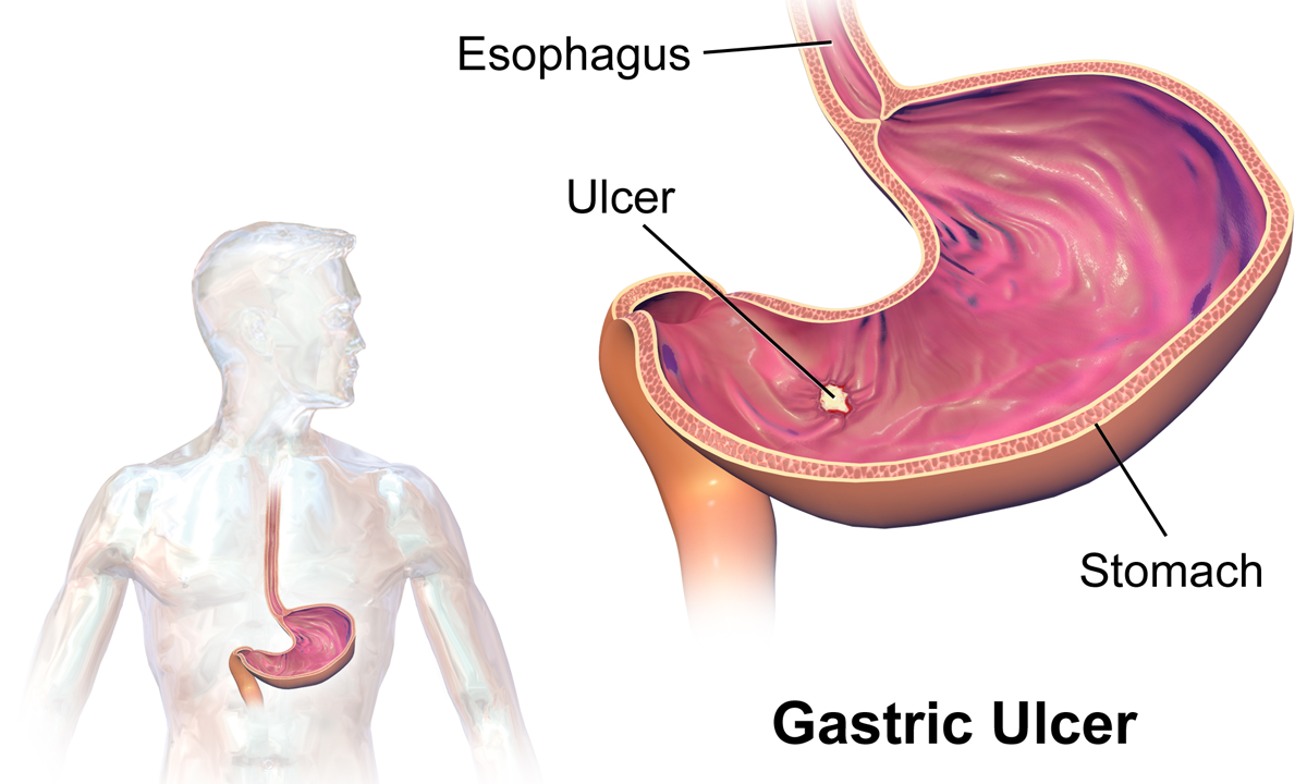
Peptic ulcer disease (PUD) is a break in the inner lining of the stomach, the first part of the small intestine, or sometimes the lower esophagus. An ulcer in the stomach is called a gastric ulcer, while one in the first part of the intestines is a duodenal ulcer. The most common symptoms of a duodenal ulcer are waking at night with upper abdominal pain and upper abdominal pain that improves with eating. With a gastric ulcer, the pain may worsen with eating. The pain is often described as a burning or dull ache. Other symptoms include belching, vomiting, weight loss, or poor appetite. About a third of older people have no symptoms. Complications may include bleeding, perforation, and blockage of the stomach. Bleeding occurs in as many as 15% of cases. Common causes include the bacteria ''Helicobacter pylori'' and non-steroidal anti-inflammatory drugs (NSAIDs). Other, less common causes include tobacco smoking, stress as a result of other serious health conditions, Behçet ... [...More Info...] [...Related Items...] OR: [Wikipedia] [Google] [Baidu] |
Joint Cracking
Joint cracking is the manipulation of joints to produce a sound and related "popping" sensation. It is sometimes performed by physical therapists, chiropractors, osteopaths, and masseurs in Turkish baths pursuing a variety of outcomes. The cracking of joints, especially knuckles, was long believed to lead to arthritis and other joint problems. However, this is not supported by medical research. The cracking mechanism and the resulting sound is caused by dissolved gas (nitrogen gas) cavitation bubbles suddenly collapsing inside the joints. This happens when the joint cavity is stretched beyond its normal size. The pressure inside the joint cavity drops and the dissolved gas suddenly comes out of solution and takes gaseous form which makes a distinct popping noise. To be able to crack the same knuckle again requires waiting about 15 minutes before the bubbles dissolve back into the synovial fluid and will be able to form again. It is possible for voluntary joint cracking by an ... [...More Info...] [...Related Items...] OR: [Wikipedia] [Google] [Baidu] |
Sternoclavicular Joint
The sternoclavicular joint or sternoclavicular articulation is a synovial saddle joint between the manubrium of the sternum, and the clavicle, as well as the first rib. The joint possesses a joint capsule, and an articular disk, and is reinforced by multiple ligaments. Structure The joint is structurally classed as a synovial plane joint and functionally classed as a diarthrosis and multiaxial joint. It is composed of two portions separated by an articular disc of fibrocartilage. The joint is formed by the sternal end of the clavicle, the clavicular notch (the superior and lateral part of the sternum), and (the superior surface of) the cartilage of the first rib (visible from the outside as the suprasternal notch). The articular surface of the clavicle is larger than that of the sternum, and is invested with a layer of cartilage, which is considerably thicker than that of the sternum. The joint receives arterial supply via branches of the internal thoracic artery, an ... [...More Info...] [...Related Items...] OR: [Wikipedia] [Google] [Baidu] |
Spondylosis
Spondylosis is the degeneration of the vertebral column from any cause. In the more narrow sense it refers to spinal osteoarthritis, the age-related wear and tear of the spinal column, which is the most common cause of spondylosis. The degenerative process in osteoarthritis chiefly affects the vertebral bodies, the neural foramina and the facet joints ( facet syndrome). If severe, it may cause pressure on the spinal cord or nerve roots with subsequent sensory or motor disturbances, such as pain, paresthesia, imbalance, and muscle weakness in the limbs. When the space between two adjacent vertebrae narrows, compression of a nerve root emerging from the spinal cord may result in radiculopathy (sensory and motor disturbances, such as severe pain in the neck, shoulder, arm, back, or leg, accompanied by muscle weakness). Less commonly, direct pressure on the spinal cord (typically in the cervical spine) may result in myelopathy, characterized by global weakness, gait dysfunction, l ... [...More Info...] [...Related Items...] OR: [Wikipedia] [Google] [Baidu] |
Osteoarthritis
Osteoarthritis (OA) is a type of degenerative joint disease that results from breakdown of joint cartilage and underlying bone which affects 1 in 7 adults in the United States. It is believed to be the fourth leading cause of disability in the world. The most common symptoms are joint pain and stiffness. Usually the symptoms progress slowly over years. Initially they may occur only after exercise but can become constant over time. Other symptoms may include joint swelling, decreased range of motion, and, when the back is affected, weakness or numbness of the arms and legs. The most commonly involved joints are the two near the ends of the fingers and the joint at the base of the thumbs; the knee and hip joints; and the joints of the neck and lower back. Joints on one side of the body are often more affected than those on the other. The symptoms can interfere with work and normal daily activities. Unlike some other types of arthritis, only the joints, not internal organs, are ... [...More Info...] [...Related Items...] OR: [Wikipedia] [Google] [Baidu] |
Radiodensity
Radiodensity (or radiopacity) is opacity to the radio wave and X-ray portion of the electromagnetic spectrum: that is, the relative inability of those kinds of electromagnetic radiation to pass through a particular material. Radiolucency or hypodensity indicates greater passage (greater transradiancy) to X-ray photonsNovelline, Robert. ''Squire's Fundamentals of Radiology''. Harvard University Press. 5th edition. 1997. . and is the analogue of transparency and translucency with visible light. Materials that inhibit the passage of electromagnetic radiation are called radiodense or radiopaque, while those that allow radiation to pass more freely are referred to as radiolucent. Radiopaque volumes of material have white appearance on radiographs, compared with the relatively darker appearance of radiolucent volumes. For example, on typical radiographs, bones look white or light gray (radiopaque), whereas muscle and skin look black or dark gray, being mostly invisible (radiolucent). Th ... [...More Info...] [...Related Items...] OR: [Wikipedia] [Google] [Baidu] |
Radiography
Radiography is an imaging technique using X-rays, gamma rays, or similar ionizing radiation and non-ionizing radiation to view the internal form of an object. Applications of radiography include medical radiography ("diagnostic" and "therapeutic") and industrial radiography. Similar techniques are used in airport security (where "body scanners" generally use backscatter X-ray). To create an image in conventional radiography, a beam of X-rays is produced by an X-ray generator and is projected toward the object. A certain amount of the X-rays or other radiation is absorbed by the object, dependent on the object's density and structural composition. The X-rays that pass through the object are captured behind the object by a detector (either photographic film or a digital detector). The generation of flat two dimensional images by this technique is called projectional radiography. In computed tomography (CT scanning) an X-ray source and its associated detectors rotate around ... [...More Info...] [...Related Items...] OR: [Wikipedia] [Google] [Baidu] |
Joint
A joint or articulation (or articular surface) is the connection made between bones, ossicles, or other hard structures in the body which link an animal's skeletal system into a functional whole.Saladin, Ken. Anatomy & Physiology. 7th ed. McGraw-Hill Connect. Webp.274/ref> They are constructed to allow for different degrees and types of movement. Some joints, such as the knee, elbow, and shoulder, are self-lubricating, almost frictionless, and are able to withstand compression and maintain heavy loads while still executing smooth and precise movements. Other joints such as sutures between the bones of the skull permit very little movement (only during birth) in order to protect the brain and the sense organs. The connection between a tooth and the jawbone is also called a joint, and is described as a fibrous joint known as a gomphosis. Joints are classified both structurally and functionally. Classification The number of joints depends on if sesamoids are included, age of ... [...More Info...] [...Related Items...] OR: [Wikipedia] [Google] [Baidu] |


