|
Piskacek's Sign
In medicine, Piskaçek's sign is a physical indication of pregnancy. It is defined as asymmetry of the enlarged uterus, palpable during pelvic examination, after the first few weeks of pregnancy. It is attributed to lateral implantation of the embryo, which can enlarge one uterine horn before the other. It has also been described as focal softening of the uterus, contrasted to the firmness of the area where the placenta is implanted. It is named after obstetrician Ludwig Piskaçek, who described it in Vienna in 1899, though it had already been noted by Robert Latou Dickinson of New York in 1892. A similar physical sign had been described by Carl von Fernwald Braun. It comes from an era when laboratory tests for pregnancy had not been developed, but experience gained in pelvic examination during early pregnancy by western gynecologists led them to publish their physical findings, allowing clinical diagnosis of pregnancy. Other such signs of early pregnancy include Goodell, ... [...More Info...] [...Related Items...] OR: [Wikipedia] [Google] [Baidu] |
Pregnancy
Pregnancy is the time during which one or more offspring develops ( gestates) inside a woman's uterus (womb). A multiple pregnancy involves more than one offspring, such as with twins. Pregnancy usually occurs by sexual intercourse, but can also occur through assisted reproductive technology procedures. A pregnancy may end in a live birth, a miscarriage, an induced abortion, or a stillbirth. Childbirth typically occurs around 40 weeks from the start of the last menstrual period (LMP), a span known as the gestational age. This is just over nine months. Counting by fertilization age, the length is about 38 weeks. Pregnancy is "the presence of an implanted human embryo or fetus in the uterus"; implantation occurs on average 8–9 days after fertilization. An '' embryo'' is the term for the developing offspring during the first seven weeks following implantation (i.e. ten weeks' gestational age), after which the term ''fetus'' is used until birth. Signs an ... [...More Info...] [...Related Items...] OR: [Wikipedia] [Google] [Baidu] |
Uterus
The uterus (from Latin ''uterus'', plural ''uteri'') or womb () is the organ in the reproductive system of most female mammals, including humans that accommodates the embryonic and fetal development of one or more embryos until birth. The uterus is a hormone-responsive sex organ that contains glands in its lining that secrete uterine milk for embryonic nourishment. In the human, the lower end of the uterus, is a narrow part known as the isthmus that connects to the cervix, leading to the vagina. The upper end, the body of the uterus, is connected to the fallopian tubes, at the uterine horns, and the rounded part above the openings to the fallopian tubes is the fundus. The connection of the uterine cavity with a fallopian tube is called the uterotubal junction. The fertilized egg is carried to the uterus along the fallopian tube. It will have divided on its journey to form a blastocyst that will implant itself into the lining of the uterus – the endometrium, where it will ... [...More Info...] [...Related Items...] OR: [Wikipedia] [Google] [Baidu] |
Pelvic Examination
A pelvic examination is the physical examination of the external and internal female pelvic organs. It is frequently used in gynecology for the evaluation of symptoms affecting the female reproductive and urinary tract, such as pain, bleeding, discharge, urinary incontinence, or trauma (e.g. sexual assault). It can also be used to assess a woman's anatomy in preparation for procedures. The exam can be done awake in the clinic and emergency department, or under anesthesia in the operating room. The most commonly performed components of the exam are 1) the external exam, to evaluate the external genitalia 2) the internal exam with palpation (commonly called the bimanual exam) to examine the uterus, ovaries, and fallopian tubes, and 3) the internal exam using the speculum to visualize the vaginal walls and cervix. During the pelvic exam, sample of cells and fluids may be collected to screen for sexually transmitted infections or cancer. Some clinicians perform a pelvic exam as part o ... [...More Info...] [...Related Items...] OR: [Wikipedia] [Google] [Baidu] |
Embryo
An embryo is an initial stage of development of a multicellular organism. In organisms that reproduce sexually, embryonic development is the part of the life cycle that begins just after fertilization of the female egg cell by the male sperm cell. The resulting fusion of these two cells produces a single-celled zygote that undergoes many cell divisions that produce cells known as blastomeres. The blastomeres are arranged as a solid ball that when reaching a certain size, called a morula, takes in fluid to create a cavity called a blastocoel. The structure is then termed a blastula, or a blastocyst in mammals. The mammalian blastocyst hatches before implantating into the endometrial lining of the womb. Once implanted the embryo will continue its development through the next stages of gastrulation, neurulation, and organogenesis. Gastrulation is the formation of the three germ layers that will form all of the different parts of the body. Neurulation forms the nervous ... [...More Info...] [...Related Items...] OR: [Wikipedia] [Google] [Baidu] |
Uterine Horns
The uterine horns (cornua of uterus) are the points in the upper uterus where the fallopian tubes exit to meet the ovaries. They are one of the points of attachment for the round ligament of uterus (the other being the mons pubis). They also provide attachment to the ovarian ligament, which is located below the fallopian tube at the back; while the round ligament of uterus is located below the tube at the front. The uterine horns are far more prominent in other animals (such as cows and cats) than they are in humans. In the cat, implantation of the embryo occurs in one of the two uterine horns, not the body of the uterus itself. Occasionally, if a fallopian tube does not connect, the uterine horn will fill with blood each month, and a minor one-day surgery will be performed to remove it. Often, people who are born with this have trouble getting pregnant as both ovaries are functional and either may ovulate. The spare egg An egg is an organic vessel grown by an animal to ... [...More Info...] [...Related Items...] OR: [Wikipedia] [Google] [Baidu] |
Placenta
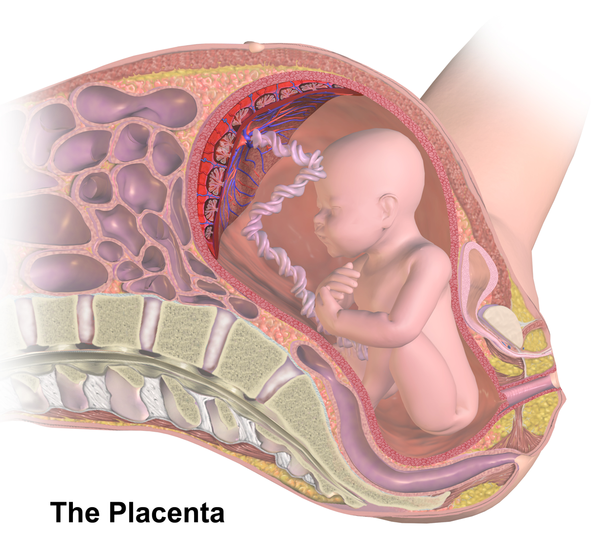
The placenta is a temporary embryonic and later fetal organ that begins developing from the blastocyst shortly after implantation. It plays critical roles in facilitating nutrient, gas and waste exchange between the physically separate maternal and fetal circulations, and is an important endocrine organ, producing hormones that regulate both maternal and fetal physiology during pregnancy. The placenta connects to the fetus via the umbilical cord, and on the opposite aspect to the maternal uterus in a species-dependent manner. In humans, a thin layer of maternal decidual (endometrial) tissue comes away with the placenta when it is expelled from the uterus following birth (sometimes incorrectly referred to as the 'maternal part' of the placenta). Placentas are a defining characteristic of placental mammals, but are also found in marsupials and some non-mammals with varying levels of development. Mammalian placentas probably first evolved about 150 million to 200 million years ... [...More Info...] [...Related Items...] OR: [Wikipedia] [Google] [Baidu] |
Ludwig Piskaçek
Ludwig Piskaçek (16 November 1854, Karcag, Hungary – 19 September 1932, Vienna) was an Austrian obstetrician remembered for describing Piskaçek's sign. He trained in Vienna, gaining his doctorate in 1882. He became apprentice in surgery at the Albert Clinic until 1884, then assistant at the second obstetrical clinic under Josef Späth (1823–1896) and his successor August Breisky Professor August Breisky (25 March 1832, Klattau (Klatovy), Bohemia, Austrian Empire – 25 May 1889) was an Austrian gynecologist and obstetrician. He studied medicine in Prague, obtaining his M.D. degree in 1855. At Prague, he served for ... (1832–1889) until 1888. He became professor of obstetrics in Linz in 1890, and in Vienna in 1901. He was the author of a textbook on midwifery that was published over several editions, ''Lehrbuch für Schülerinnen des Hebammenkurses und Nachschlagebuch für Hebammen''. [...More Info...] [...Related Items...] OR: [Wikipedia] [Google] [Baidu] |
Robert Latou Dickinson
Robert Latou Dickinson (1861–1950) was an American obstetrician and gynecologist, surgeon, maternal health educator, artist, sculptor and medical illustrator, and research scientist. Early life Robert Latou Dickinson was born on February 21, 1861, in Jersey City, New Jersey. He was the son of Horace and Jeannette Latou Dickinson. He became a noted obstetrician, gynecologist, surgeon, research scientist, author, and public health educator. He was an unusually prolific artist, carver and sculptor, who used his skills to illuminate his professional work and delight friends and family. He sketched all his life, including delightful if irreverent sketches in the edges of his school books. According to James Reed, as a boy of ten, Rob Dickinson was trying to beach a boat that he and his father had built. An eddy drove the metal prow into Dickinson's abdomen, gashing it deeply. Holding the two sides of the wound together and some internal organs inside, Dickinson dragged himself ... [...More Info...] [...Related Items...] OR: [Wikipedia] [Google] [Baidu] |
Von Braun-Fernwald's Sign
Von Braun-Fernwald's sign is a clinical sign in which there is an irregular softening and enlargement of the uterine fundus during early pregnancy Pregnancy is the time during which one or more offspring develops ( gestates) inside a woman's uterus (womb). A multiple pregnancy involves more than one offspring, such as with twins. Pregnancy usually occurs by sexual intercourse, but ca .... It occurs at 5–8 weeks gestation.Maternal-Neonatal Nursing Made Incredibly Easy! Page 160. Lippincott Williams & Wilkins, 2007. Google books/ref> The sign is named after Karl von Braun-Fernwald. See also * Piskacek's sign References Medical signs Obstetrics Midwifery {{human-repro-stub ... [...More Info...] [...Related Items...] OR: [Wikipedia] [Google] [Baidu] |
Carl Braun (obstetrician)
Carl Braun (22 March 1822 – 28 March 1891), sometimes Carl Rudolf Braun alternative spelling: Karl Braun, or Karl von Braun-Fernwald, name after knighthood Carl Ritter von Fernwald Braun was an Austrian obstetrician. He was born 22 March 1822 in Zistersdorf, Austria, son of the medical doctor Carl August Braun. Career Carl Braun studied in Vienna from 1841 and, in 1847, took the position of ''Sekundararzt'' (assistant doctor) in the Vienna General Hospital. In 1849 he succeeded Ignaz Semmelweis as assistant to professor Johann Klein at the hospital's first maternity clinic, a position he held until 1853. In 1853, after Braun became a Privatdozent, he was appointed ordinary professor of obstetrics in Trient and vice-director of the Tiroler Landes-Gebär- und Findelanstalt. In November 1856 he was called to Vienna to succeed Johann Klein as professor of obstetrics. On Braun's recommendation, the hospital's first gynaecology clinic was created in 1858, under his direction ... [...More Info...] [...Related Items...] OR: [Wikipedia] [Google] [Baidu] |
Goodell's Sign
In medicine, Goodell's sign is an indication of pregnancy. It is a significant softening of the vaginal portion of the cervix from increased vascularization. This vascularization is a result of hypertrophy and engorgement of the vessels below the growing uterus. This sign occurs at approximately six weeks' gestation. The sign is named after William Goodell (gynecologist) (1829-1874). See also * Chadwick's sign * Hegar's sign * Ladin's sign Ladin's sign is a clinical sign of pregnancy in which there is softening in the midline of the uterus anteriorly at the junction of the uterus and cervix. It occurs and is detectable with a manual examination at about 6 weeks' gestation. Ladin's s ... References {{DEFAULTSORT:Goodell's Sign Medical signs Obstetrics Midwifery ... [...More Info...] [...Related Items...] OR: [Wikipedia] [Google] [Baidu] |
Hegar's Sign
Hegar's sign is a non-sensitive indication of pregnancy in women—its absence does not exclude pregnancy. It pertains to the features of the cervix and the uterine isthmus. It is demonstrated as a softening in the consistency of the uterus, and the uterus and cervix seem to be two separate regions. The sign is usually present from 4–6 weeks until the 12th week of pregnancy. Hegar's sign is more difficult to recognize in multiparous women. Interpretation: On bimanual examination (two fingers in the anterior fornix and two fingers below the uterus per abdomen), the abdominal and vaginal fingers seem to oppose below the body of uterus (examination must be gentle to avoid abortion). This sign was repeatedly demonstrated and described by Ernst Ludwig Alfred Hegar, a German gynecologist, in 1895. Hegar credited Reinl, one of his assistants, who originally described this sign in 1884. See also * Chadwick's sign * Goodell's sign * Ladin's sign Ladin's sign is a clinical sign of pregn ... [...More Info...] [...Related Items...] OR: [Wikipedia] [Google] [Baidu] |


_obstetrician.jpg)