|
Pirogov Triangle
The Pirogov triangle (also Piragoff's triangle) is an area in the human neck formed by the intermediate tendon of the digastric muscle, the posterior border of the mylohyoid muscle, and the hypoglossal nerve. The triangle was named after Russian surgeon and scientist Nikolay Pirogov who performed a first description of that anatomic area of the neck. The lingual artery can be found in the Pirogov triangle underneath the fibers of the hyoglossus The hyoglossus, thin and quadrilateral, arises from the side of the body and from the whole length of the greater cornu of the hyoid bone, and passes almost vertically upward to enter the side of the tongue, between the styloglossus and the inf ... muscle. References Human head and neck {{musculoskeletal-stub ... [...More Info...] [...Related Items...] OR: [Wikipedia] [Google] [Baidu] |
Digastric Muscle
The digastric muscle (also digastricus) (named ''digastric'' as it has two 'bellies') is a small muscle located under the jaw. The term "digastric muscle" refers to this specific muscle. However, other muscles that have two separate muscle bellies include the suspensory muscle of duodenum, omohyoid, occipitofrontalis. It lies below the body of the mandible, and extends, in a curved form, from the mastoid notch to the mandibular symphysis. It belongs to the suprahyoid muscles group. A broad aponeurotic layer is given off from the tendon of the digastric muscle on either side, to be attached to the body and greater cornu of the hyoid bone; this is termed the suprahyoid aponeurosis. Structure The digastricus (digastric muscle) consists of two muscular bellies united by an intermediate rounded tendon. The two bellies of the digastric muscle have different embryological origins, and are supplied by different cranial nerves. Each person has a right and left digastric muscle. ... [...More Info...] [...Related Items...] OR: [Wikipedia] [Google] [Baidu] |
Mylohyoid Muscle
The mylohyoid muscle or diaphragma oris is a paired muscle of the neck. It runs from the mandible to the hyoid bone, forming the floor of the oral cavity of the mouth. It is named after its two attachments near the molar teeth. It forms the floor of the submental triangle. It elevates the hyoid bone and the tongue, important during swallowing and speaking. Structure The mylohyoid muscle is flat and triangular, and is situated immediately superior to the anterior belly of the digastric muscle. It is a pharyngeal muscle (derived from the first pharyngeal arch) and classified as one of the suprahyoid muscles. Together, the paired mylohyoid muscles form a muscular floor for the oral cavity of the mouth. The two mylohyoid muscles arise from the mandible at the mylohyoid line, which extends from the mandibular symphysis in front to the last molar tooth behind. The posterior fibers pass inferomedially and insert at anterior surface of the hyoid bone. The medial fibres of the two ... [...More Info...] [...Related Items...] OR: [Wikipedia] [Google] [Baidu] |
Hypoglossal Nerve
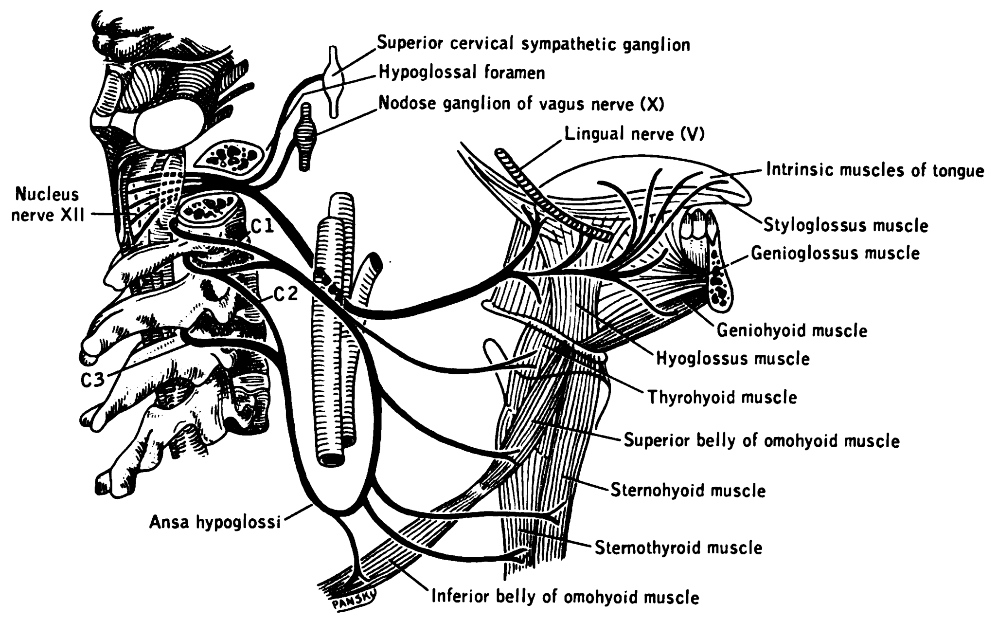
The hypoglossal nerve, also known as the twelfth cranial nerve, cranial nerve XII, or simply CN XII, is a cranial nerve that innervates all the extrinsic and intrinsic muscles of the tongue except for the palatoglossus, which is innervated by the vagus nerve. CN XII is a nerve with a solely motor function. The nerve arises from the hypoglossal nucleus in the medulla as a number of small rootlets, passes through the hypoglossal canal and down through the neck, and eventually passes up again over the tongue muscles it supplies into the tongue. The nerve is involved in controlling tongue movements required for speech and swallowing, including sticking out the tongue and moving it from side to side. Damage to the nerve or the neural pathways which control it can affect the ability of the tongue to move and its appearance, with the most common sources of damage being injury from trauma or surgery, and motor neuron disease. The first recorded description of the nerve is by Herophil ... [...More Info...] [...Related Items...] OR: [Wikipedia] [Google] [Baidu] |
Nikolay Ivanovich Pirogov
Nikolay Ivanovich Pirogov (Russian: Никола́й Ива́нович Пирого́в; — ) was a Russian scientist, medical doctor, pedagogue, public figure, and corresponding member of the Russian Academy of Sciences (1847), one of the most widely recognized Russian physicians. Considered to be the founder of field surgery, he was the first surgeon to use anaesthesia in a field operation (1847) and one of the first surgeons in Europe to use ether as an anaesthetic. He is credited with invention of various kinds of surgical operations and developing his own technique of using plaster casts to treat fractured bones. Biography Childhood and training Nikolay Pirogov was born in Moscow, the 13th of 14 children of Ivan Ivanovich Pirogov (born around 1772), a major in the commissary service and a treasurer at the Moscow Food Depot whose own father came from peasants and served as a soldier in Peter the Great's army before retiring and opening a brewery in Moscow; Pirogov's mot ... [...More Info...] [...Related Items...] OR: [Wikipedia] [Google] [Baidu] |
Lingual Artery
The lingual artery arises from the external carotid artery between the superior thyroid artery and facial artery. It can be located easily in the tongue. Structure The lingual artery first branches off from the external carotid artery. It runs obliquely upward and medially to the greater horns of the hyoid bone. It then curves downward and forward, forming a loop which is crossed by the hypoglossal nerve. It then passes beneath the digastric muscle and stylohyoid muscle running horizontally forward, beneath the hyoglossus. This takes it through the sublingual space. Finally, ascending almost perpendicularly to the tongue, it turns forward on its lower surface as far as the tip of the tongue, now called the deep lingual artery (profunda linguae). Branches The lingual artery gives 4 main branches: the deep lingual artery, the sublingual artery, the suprahyoid branch, and the dorsal lingual branch. Deep lingual artery The deep lingual artery (or ranine artery) is the terminal p ... [...More Info...] [...Related Items...] OR: [Wikipedia] [Google] [Baidu] |
Hyoglossus
The hyoglossus, thin and quadrilateral, arises from the side of the body and from the whole length of the greater cornu of the hyoid bone, and passes almost vertically upward to enter the side of the tongue, between the styloglossus and the inferior longitudinal muscle of the tongue. It forms a part of the floor of submandibular triangle. Structure The fibers arising from the body of the hyoid bone overlap those from the greater cornu. Structures that are medial/deep to the hyoglossus are the glossopharyngeal nerve (cranial nerve 9), the stylohyoid ligament and the lingual artery and lingual vein. The lingual vein passes medial to the hyoglossus, and the lingual artery passes deep to the hyoglossus. Laterally, in between the hyoglossus muscle and the mylohyoid muscle lay several important structures (from upper to lower): sublingual gland, submandibular duct, lingual nerve, vena comitans of hypoglossal nerve, and the hypoglossal nerve. Note, posteriorly, the lingual nerve is ... [...More Info...] [...Related Items...] OR: [Wikipedia] [Google] [Baidu] |


