|
Phototherapeutic Keratectomy
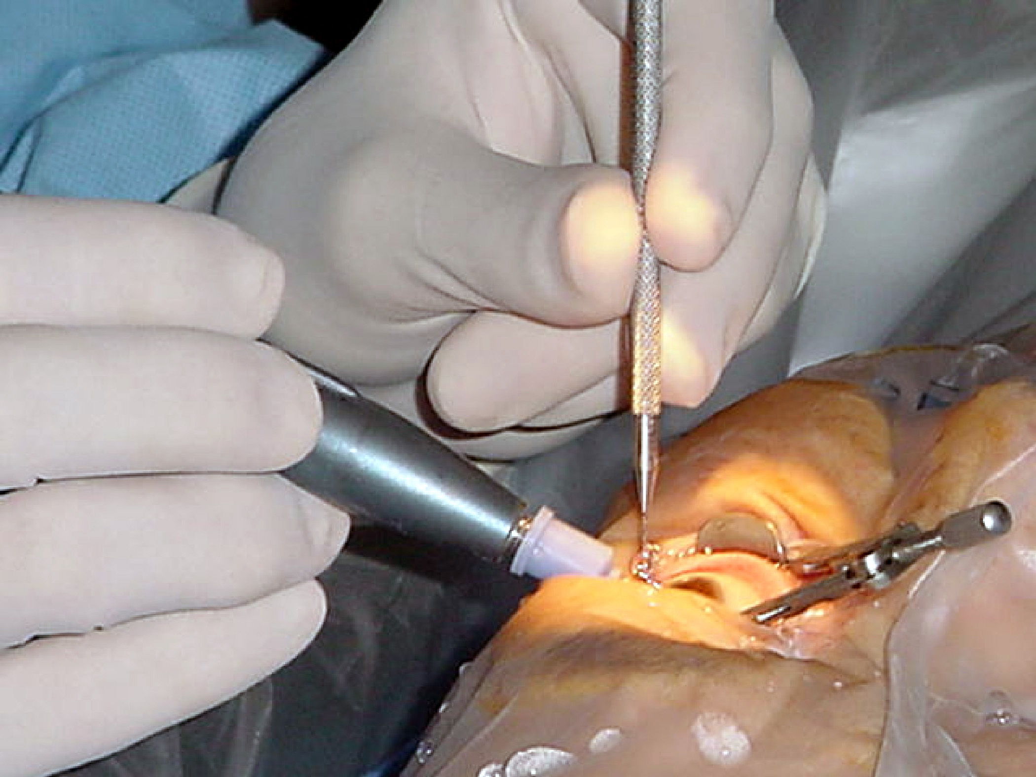
Phototherapeutic keratectomy (PTK) is a type of eye surgery that uses a laser to treat various ocular disorders by removing tissue from the cornea. PTK allows the removal of superficial corneal opacities and surface irregularities. It is similar to photorefractive keratectomy Photorefractive keratectomy (PRK) and laser-assisted sub-epithelial keratectomy (or laser epithelial keratomileusis) (LASEK) are laser eye surgery procedures intended to correct a person's vision, reducing dependency on glasses or contact lenses. ..., which is used for the treatment of refractive conditions. The common indications for PTK are corneal dystrophies, scars, opacities, and bullous keratopathy. TK in the developing worldttp://www.ophthalmologyweb.com/JournalUpdates.aspx?spid=23&jid=15009 References External links "Facts About The Cornea and Corneal Disease" - National Eye Institute {{eye-stub Eye surgery ... [...More Info...] [...Related Items...] OR: [Wikipedia] [Google] [Baidu] |
Corneal Opacity
The human cornea is a transparent membrane which allows light to pass through it. The word corneal opacification literally means loss of normal transparency of cornea. The term corneal opacity is used particularly for the loss of transparency of cornea due to scarring. Transparency of the cornea is dependent on the uniform diameter and the regular spacing and arrangement of the collagen fibrils within the stroma. Alterations in the spacing of collagen fibrils in a variety of conditions including corneal edema, scars, and macular corneal dystrophy is clinically manifested as corneal opacity. The term corneal blindness is commonly used to describe blindness due to corneal opacity. Types Depending on the density, corneal opacity is graded as nebular, macular and leucomatous. Nebular corneal opacity Nebular corneal opacity is a faint opacity which results due to superficial scars involving Bowman's layer and superficial stroma. A nebular corneal opacity allows the details of the iri ... [...More Info...] [...Related Items...] OR: [Wikipedia] [Google] [Baidu] |
Eye Surgery
Eye surgery, also known as ophthalmic or ocular surgery, is surgery performed on the eye or its adnexa, by an ophthalmologist or sometimes, an optometrist. Eye surgery is synonymous with ophthalmology. The eye is a very fragile organ, and requires extreme care before, during, and after a surgical procedure to minimize or prevent further damage. An expert eye surgeon is responsible for selecting the appropriate surgical procedure for the patient, and for taking the necessary safety precautions. Mentions of eye surgery can be found in several ancient texts dating back as early as 1800 BC, with cataract treatment starting in the fifth century BC. Today it continues to be a widely practiced type of surgery, with various techniques having been developed for treating eye problems. Preparation and precautions Since the eye is heavily supplied by nerves, anesthesia is essential. Local anesthesia is most commonly used. Topical anesthesia using lidocaine topical gel is often used fo ... [...More Info...] [...Related Items...] OR: [Wikipedia] [Google] [Baidu] |
Laser
A laser is a device that emits light through a process of optical amplification based on the stimulated emission of electromagnetic radiation. The word "laser" is an acronym for "light amplification by stimulated emission of radiation". The first laser was built in 1960 by Theodore H. Maiman at Hughes Research Laboratories, based on theoretical work by Charles Hard Townes and Arthur Leonard Schawlow. A laser differs from other sources of light in that it emits light which is ''coherent''. Spatial coherence allows a laser to be focused to a tight spot, enabling applications such as laser cutting and lithography. Spatial coherence also allows a laser beam to stay narrow over great distances (collimation), enabling applications such as laser pointers and lidar (light detection and ranging). Lasers can also have high temporal coherence, which allows them to emit light with a very narrow spectrum. Alternatively, temporal coherence can be used to produce ultrashort pulses of ligh ... [...More Info...] [...Related Items...] OR: [Wikipedia] [Google] [Baidu] |
Eye Disease
This is a partial list of human eye diseases and disorders. The World Health Organization publishes a classification of known diseases and injuries, the International Statistical Classification of Diseases and Related Health Problems, or ICD-10. This list uses that classification. H00-H06 Disorders of eyelid, lacrimal system and orbit * (H02.1) Ectropion * (H02.2) Lagophthalmos * (H02.3) Blepharochalasis * (H02.4) Ptosis * (H02.5) Stye, an acne type infection of the sebaceous glands on or near the eyelid. * (H02.6) Xanthelasma of eyelid * (H03.0*) Parasitic infestation of eyelid in diseases classified elsewhere ** Dermatitis of eyelid due to Demodex species ( B88.0+ ) ** Parasitic infestation of eyelid in: *** leishmaniasis ( B55.-+ ) *** loiasis ( B74.3+ ) *** onchocerciasis ( B73+ ) *** phthiriasis ( B85.3+ ) * (H03.1*) Involvement of eyelid in other infectious diseases classified elsewhere ** Involvement of eyelid in: *** herpesviral (herpes simplex) infection ( B00.5+ ) * ... [...More Info...] [...Related Items...] OR: [Wikipedia] [Google] [Baidu] |
Tissue (biology)
In biology, tissue is a biological organizational level between cells and a complete organ. A tissue is an ensemble of similar cells and their extracellular matrix from the same origin that together carry out a specific function. Organs are then formed by the functional grouping together of multiple tissues. The English word "tissue" derives from the French word "tissu", the past participle of the verb tisser, "to weave". The study of tissues is known as histology or, in connection with disease, as histopathology. Xavier Bichat is considered as the "Father of Histology". Plant histology is studied in both plant anatomy and physiology. The classical tools for studying tissues are the paraffin block in which tissue is embedded and then sectioned, the histological stain, and the optical microscope. Developments in electron microscopy, immunofluorescence, and the use of frozen tissue-sections have enhanced the detail that can be observed in tissues. With these tools, the c ... [...More Info...] [...Related Items...] OR: [Wikipedia] [Google] [Baidu] |
Cornea
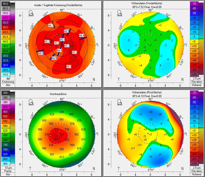
The cornea is the transparent front part of the eye that covers the iris, pupil, and anterior chamber. Along with the anterior chamber and lens, the cornea refracts light, accounting for approximately two-thirds of the eye's total optical power. In humans, the refractive power of the cornea is approximately 43 dioptres. The cornea can be reshaped by surgical procedures such as LASIK. While the cornea contributes most of the eye's focusing power, its focus is fixed. Accommodation (the refocusing of light to better view near objects) is accomplished by changing the geometry of the lens. Medical terms related to the cornea often start with the prefix "'' kerat-''" from the Greek word κέρας, ''horn''. Structure The cornea has unmyelinated nerve endings sensitive to touch, temperature and chemicals; a touch of the cornea causes an involuntary reflex to close the eyelid. Because transparency is of prime importance, the healthy cornea does not have or need blood vessels with ... [...More Info...] [...Related Items...] OR: [Wikipedia] [Google] [Baidu] |
Photorefractive Keratectomy
Photorefractive keratectomy (PRK) and laser-assisted sub-epithelial keratectomy (or laser epithelial keratomileusis) (LASEK) are laser eye surgery procedures intended to correct a person's vision, reducing dependency on glasses or contact lenses. LASEK and PRK permanently change the shape of the anterior central cornea using an excimer laser to ablate (remove by vaporization) a small amount of tissue from the corneal stroma at the front of the eye, just under the corneal epithelium. The outer layer of the cornea is removed prior to the ablation. A computer system tracks the patient's eye position 60 to 4,000 times per second, depending on the specifications of the laser that is used. The computer system redirects laser pulses for precise laser placement. Most modern lasers will automatically center on the patient's visual axis and will pause if the eye moves out of range and then resume ablating at that point after the patient's eye is re-centered. The outer layer of the cornea, ... [...More Info...] [...Related Items...] OR: [Wikipedia] [Google] [Baidu] |
Refractive Error
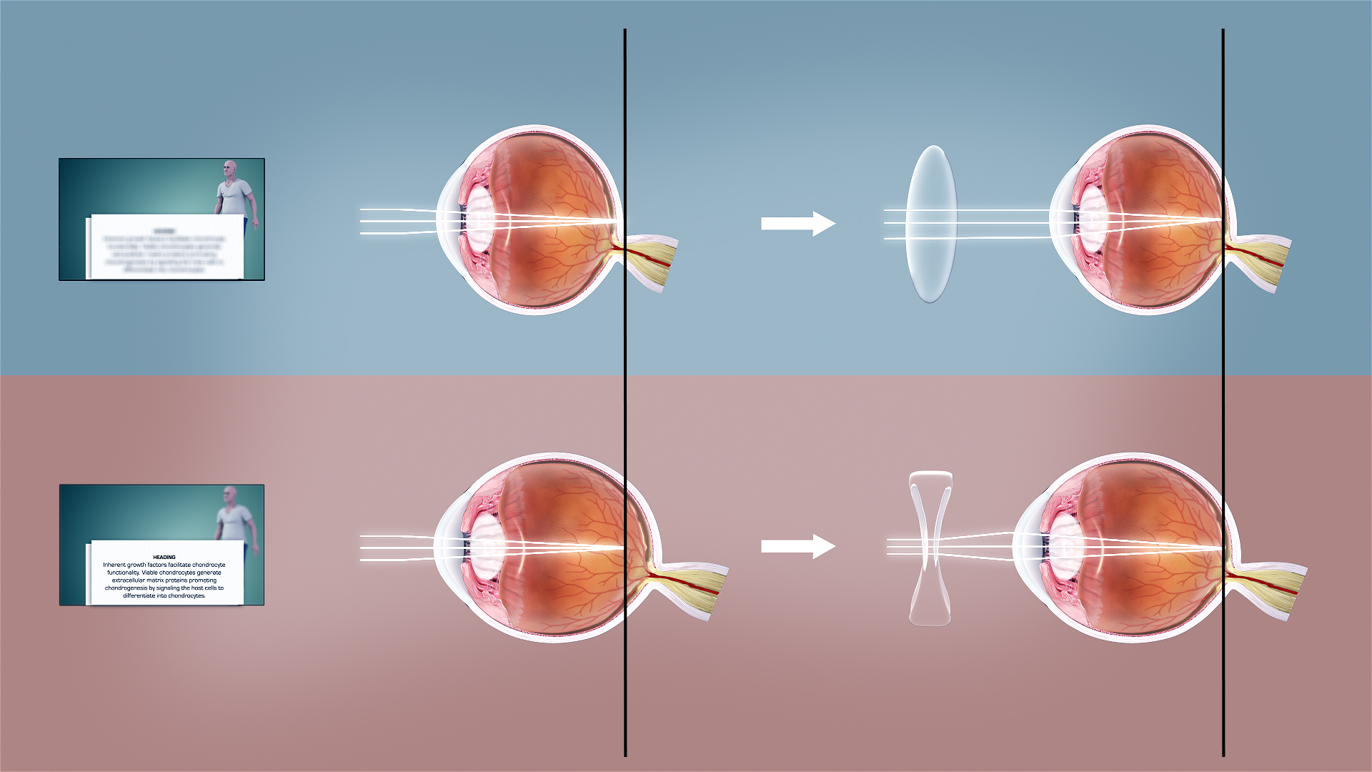
Refractive error, also known as refraction error, is a problem with focus (optics), focusing light accurately on the retina due to the shape of the human eye, eye and or cornea. The most common types of refractive error are myopia, near-sightedness, hyperopia, far-sightedness, astigmatism, and presbyopia. Near-sightedness results in far away objects being blurred vision, blurry, far-sightedness and presbyopia result in close objects being blurry, and astigmatism causes objects to appear stretched out or blurry. Other symptoms may include double vision, headaches, and eye strain. Near-sightedness is due to the length of the eyeball being too long, far-sightedness the eyeball too short, astigmatism the cornea being the wrong shape, and presbyopia aging of the lens of the eye such that it cannot change shape sufficiently. Some refractive errors occur more often among those whose parents are affected. Diagnosis is by eye examination. Refractive errors are corrected with eyeglasses, ... [...More Info...] [...Related Items...] OR: [Wikipedia] [Google] [Baidu] |
Corneal Dystrophy
Corneal dystrophy is a group of rare hereditary disorders characterised by bilateral abnormal deposition of substances in the transparent front part of the eye called the cornea. Signs and symptoms Corneal dystrophy may not significantly affect vision in the early stages. However, it does require proper evaluation and treatment for restoration of optimal vision. Corneal dystrophies usually manifest themselves during the first or second decade but sometimes later. It appears as grayish white lines, circles, or clouding of the cornea. Corneal dystrophy can also have a crystalline appearance. There are over 20 corneal dystrophies that affect all parts of the cornea. These diseases share many traits: * They are usually inherited. * They affect the right and left eyes equally. * They are not caused by outside factors, such as injury or diet. * Most progress gradually. * Most usually begin in one of the five corneal layers and may later spread to nearby layers. * Most do not affect oth ... [...More Info...] [...Related Items...] OR: [Wikipedia] [Google] [Baidu] |
Bullous Keratopathy
Bullous keratopathy, also known as pseudophakic bullous keratopathy (PBK), is a pathological condition in which small vesicles, or '' bullae'', are formed in the cornea due to endothelial dysfunction. In a healthy cornea, endothelial cells keeps the tissue from excess fluid absorption, pumping it back into the aqueous humor. When affected by some reason, such as Fuchs' dystrophy Fuchs dystrophy, also referred to as Fuchs endothelial corneal dystrophy (FECD) and Fuchs endothelial dystrophy (FED), is a slowly progressing corneal dystrophy that usually affects both eyes and is slightly more common in women than in men. Althou ... or a trauma during cataract removal, endothelial cells suffer mortality or damage. The corneal endothelial cells normally do not undergo mitotic cell division, and cell loss results in permanent loss of function. When endothelial cell counts drop too low, the pump starts failing to function and fluid moves anterior into the stroma and epithelium. The excess ... [...More Info...] [...Related Items...] OR: [Wikipedia] [Google] [Baidu] |


