|
Phase Space Measurement With Forward Modeling
Phase space measurement with forward modeling is one approach to address the scattering issue in biomedical imaging. Scattering is one of the biggest problems in biomedical imaging, given that scattered light is eventually defocused, thus resulting in diffused images.Elizabeth M. C. Hillman et al., (2007"Optical brain imaging in vivo: techniques and applications from animal to man"/ref> Instead of removing the scattered light, this approach uses the information of scattered light to reconstruct the original light signals. This approach requires the phase space data of light in imaging system and a forward model to describe scattering events in a turbid medium. Phase space of light can be obtained by using digital micromirror device (DMD)Liu et al., 201"3D imaging in volumetric scattering media using phase-space measurements"/ref> or light field microscopy.Pegard et al., 201"Compressive light-field microscopy for 3D neural activity recording"/ref> Phase space measurement with forward ... [...More Info...] [...Related Items...] OR: [Wikipedia] [Google] [Baidu] |
Scattering
Scattering is a term used in physics to describe a wide range of physical processes where moving particles or radiation of some form, such as light or sound, are forced to deviate from a straight trajectory by localized non-uniformities (including particles and radiation) in the medium through which they pass. In conventional use, this also includes deviation of reflected radiation from the angle predicted by the law of reflection. Reflections of radiation that undergo scattering are often called ''diffuse reflections'' and unscattered reflections are called ''specular'' (mirror-like) reflections. Originally, the term was confined to light scattering (going back at least as far as Isaac Newton in the 17th century). As more "ray"-like phenomena were discovered, the idea of scattering was extended to them, so that William Herschel could refer to the scattering of "heat rays" (not then recognized as electromagnetic in nature) in 1800. John Tyndall, a pioneer in light scattering researc ... [...More Info...] [...Related Items...] OR: [Wikipedia] [Google] [Baidu] |
Digital Micromirror Device
The digital micromirror device, or DMD, is the microoptoelectromechanical system (MOEMS) that is the core of the trademarked DLP projection technology from Texas Instruments (TI). Texas Instrument's DMD was created by solid-state physicist and TI Fellow Emeritus Dr. Larry Hornbeck in 1987. However, the technology goes back to 1973 with Harvey C. Nathanson's (inventor of MEMS c. 1965) use of millions of microscopically small moving mirrors to create a video display of the type now found in digital projectors. History The DMD project began as the deformable mirror device in 1977 using micromechanical analog light modulators. The first analog DMD product was the TI DMD2000 airline ticket printer that used a DMD instead of a laser scanner. Construction and use A DMD chip has on its surface several hundred thousand microscopic mirrors arranged in a rectangular array which correspond to the pixels in the image to be displayed. The mirrors can be individually rotated ±10-12°, to ... [...More Info...] [...Related Items...] OR: [Wikipedia] [Google] [Baidu] |
Light Field Microscopy
Light field microscopy (LFM) is a scanning-free 3-dimensional (3D) microscopic imaging method based on the theory of light field. This technique allows sub-second (~10 Hz) large volumetric imaging ( 0.1 to 1 mmsup>3) with ~1 μm spatial resolution in the condition of weak scattering and semi-transparence, which has never been achieved by other methods. Just as in traditional light field rendering, there are two steps for LFM imaging: light field capture and processing. In most setups, a microlens array is used to capture the light field. As for processing, it can be based on two kinds of representations of light propagation: the ray optics picture and the wave optics picture. The Stanford University Computer Graphics Laboratory published their first prototype LFM in 2006 and has been working on the cutting edge since then. Light field generation A light field is a collection of all the rays flowing through some free space, where each ray can be parameterized with four ... [...More Info...] [...Related Items...] OR: [Wikipedia] [Google] [Baidu] |
Non-negative Least Squares
In mathematical optimization Mathematical optimization (alternatively spelled ''optimisation'') or mathematical programming is the selection of a best element, with regard to some criterion, from some set of available alternatives. It is generally divided into two subfi ..., the problem of non-negative least squares (NNLS) is a type of constrained least squares problem where the coefficients are not allowed to become negative. That is, given a matrix and a (column) vector of response variables , the goal is to find :\operatorname\limits_\mathbf \, \mathbf - \mathbf\, _2^2 subject to . Here means that each component of the vector should be non-negative, and denotes the Euclidean norm. Non-negative least squares problems turn up as subproblems in matrix decomposition, e.g. in algorithms for CP decomposition, PARAFAC and non-negative matrix factorization, non-negative matrix/tensor factorization. The latter can be considered a generalization of NNLS. Another generalizati ... [...More Info...] [...Related Items...] OR: [Wikipedia] [Google] [Baidu] |
Wigner Quasiprobability Distribution
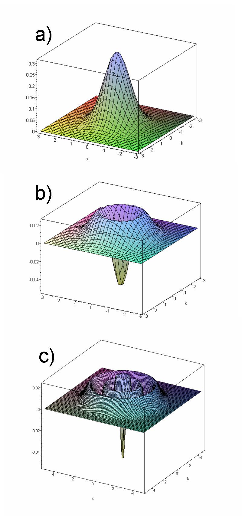
The Wigner quasiprobability distribution (also called the Wigner function or the Wigner–Ville distribution, after Eugene Wigner and Jean-André Ville) is a quasiprobability distribution. It was introduced by Eugene Wigner in 1932 to study quantum corrections to classical statistical mechanics. The goal was to link the wavefunction that appears in Schrödinger's equation to a probability distribution in phase space. It is a generating function for all spatial autocorrelation functions of a given quantum-mechanical wavefunction . Thus, it maps on the quantum density matrix in the map between real phase-space functions and Hermitian operators introduced by Hermann Weyl in 1927, in a context related to representation theory in mathematics (see Weyl quantization). In effect, it is the Wigner–Weyl transform of the density matrix, so the realization of that operator in phase space. It was later rederived by Jean Ville in 1948 as a quadratic (in signal) representation of the lo ... [...More Info...] [...Related Items...] OR: [Wikipedia] [Google] [Baidu] |
Lasso (statistics)
In statistics and machine learning, lasso (least absolute shrinkage and selection operator; also Lasso or LASSO) is a regression analysis method that performs both variable selection and regularization in order to enhance the prediction accuracy and interpretability of the resulting statistical model. It was originally introduced in geophysics, and later by Robert Tibshirani, who coined the term. Lasso was originally formulated for linear regression models. This simple case reveals a substantial amount about the estimator. These include its relationship to ridge regression and best subset selection and the connections between lasso coefficient estimates and so-called soft thresholding. It also reveals that (like standard linear regression) the coefficient estimates do not need to be unique if covariates are collinear. Though originally defined for linear regression, lasso regularization is easily extended to other statistical models including generalized linear models, g ... [...More Info...] [...Related Items...] OR: [Wikipedia] [Google] [Baidu] |
Two-photon Excitation Microscopy
Two-photon excitation microscopy (TPEF or 2PEF) is a fluorescence imaging technique that allows imaging of living tissue up to about one millimeter in thickness, with 0.64 μm lateral and 3.35 μm axial spatial resolution. Unlike traditional fluorescence microscopy, in which the excitation wavelength is shorter than the emission wavelength, two-photon excitation requires simultaneous excitation by two photons with longer wavelength than the emitted light. Two-photon excitation microscopy typically uses near-infrared (NIR) excitation light which can also excite fluorescent dyes. However, for each excitation, two photons of NIR light are absorbed. Using infrared light minimizes scattering in the tissue. Due to the multiphoton absorption, the background signal is strongly suppressed. Both effects lead to an increased penetration depth for this technique. Two-photon excitation can be a superior alternative to confocal microscopy due to its deeper tissue penetration, efficient light d ... [...More Info...] [...Related Items...] OR: [Wikipedia] [Google] [Baidu] |


