|
P-type Calcium Channel
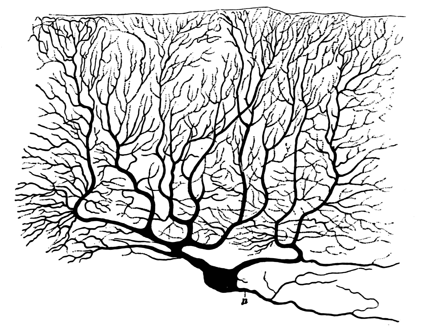
The P-type calcium channel is a type of voltage-dependent calcium channel. Similar to many other high-voltage-gated calcium channels, the α1 subunit determines most of the channel's properties. The 'P' signifies cerebellar Purkinje cells, referring to the channel's initial site of discovery. P-type calcium channels play a similar role to the N-type calcium channel in neurotransmitter release at the presynaptic terminal and in neuronal integration in many neuronal types. History The calcium channel experiments that led to the discovery of P-type calcium channels were initially completed by Rodolfo Llinas, Llinás and Sugimori in 1980. P type calcium channels were named in 1989 because they were discovered within mammalian Purkinje neurons. They were able to use an ''in vitro'' preparation to examine the ionic currents that account for Purkinje cells' electrophysiology, electrophysiological properties. They found that there are calcium dependent action potentials which rise slowly a ... [...More Info...] [...Related Items...] OR: [Wikipedia] [Google] [Baidu] |
Voltage-dependent Calcium Channel
Voltage-gated calcium channels (VGCCs), also known as voltage-dependent calcium channels (VDCCs), are a group of voltage-gated ion channels found in the membrane of excitable cells (''e.g.'', muscle, glial cells, neurons, etc.) with a permeability to the calcium ion Ca2+. These channels are slightly permeable to sodium ions, so they are also called Ca2+-Na+ channels, but their permeability to calcium is about 1000-fold greater than to sodium under normal physiological conditions. At physiologic or resting membrane potential, VGCCs are normally closed. They are activated (''i.e.'': opened) at depolarized membrane potentials and this is the source of the "voltage-gated" epithet. The concentration of calcium (Ca2+ ions) is normally several thousand times higher outside the cell than inside. Activation of particular VGCCs allows a Ca2+ influx into the cell, which, depending on the cell type, results in activation of calcium-sensitive potassium channels, muscular contraction, e ... [...More Info...] [...Related Items...] OR: [Wikipedia] [Google] [Baidu] |
Ribosomal Protein S6
Ribosomal protein S6 (rpS6 or eS6) is a component of the 40S ribosomal subunit and is therefore involved in translation. Mouse model studies have shown that phosphorylation of eS6 is involved in the regulation of cell size, cell proliferation, and glucose homeostasis. Studies show that the p70 ribosomal protein S6 kinases (S6K1 Ribosomal protein S6 kinase beta-1 (S6K1), also known as p70S6 kinase (p70S6K, p70-S6K), is an enzyme (specifically, a protein kinase) that in humans is encoded by the ''RPS6KB1'' gene. It is a serine/threonine kinase that acts downstream of PIP3 ... and S6K2) and p90 ribosomal protein S6 kinases ( RSK) both phosphorylate eS6 and that S6K1 and S6K2 predominate this function. Pathways leading to the induction of human eS6 phosphorylation have been found to enhance IL-8 protein synthesis. This mechanism is dependent on A/U-rich proximal sequences (APS) found in the 3'UTR of IL-8 immediately after the stop codon. References External links * * ... [...More Info...] [...Related Items...] OR: [Wikipedia] [Google] [Baidu] |
Ion Channel
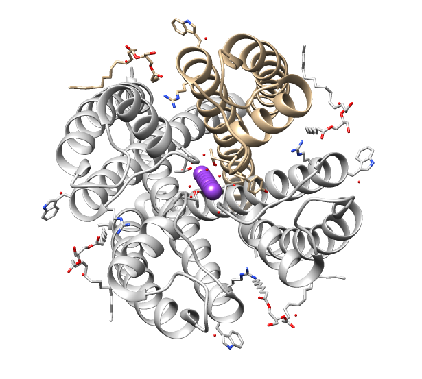
Ion channels are pore-forming membrane proteins that allow ions to pass through the channel pore. Their functions include establishing a resting membrane potential, shaping action potentials and other electrical signals by gating the flow of ions across the cell membrane, controlling the flow of ions across secretory and epithelial cells, and regulating cell volume. Ion channels are present in the membranes of all cells. Ion channels are one of the two classes of ionophoric proteins, the other being ion transporters. The study of ion channels often involves biophysics, electrophysiology, and pharmacology, while using techniques including voltage clamp, patch clamp, immunohistochemistry, X-ray crystallography, fluoroscopy, and RT-PCR. Their classification as molecules is referred to as channelomics. Basic features There are two distinctive features of ion channels that differentiate them from other types of ion transporter proteins: #The rate of ion transport through the ... [...More Info...] [...Related Items...] OR: [Wikipedia] [Google] [Baidu] |
Habenula
In neuroanatomy, habenula (diminutive of Latin ''habena'' meaning rein) originally denoted the stalk of the pineal gland (pineal habenula; pedunculus of pineal body), but gradually came to refer to a neighboring group of nerve cells with which the pineal gland was believed to be associated, the habenular nucleus. The habenular nucleus is a set of well-conserved structures in all vertebrate animals. Currently, this term refers to this separate cell mass in the caudal portion of the dorsal diencephalon, known as the epithalamus, found in all vertebrates on both sides of the third ventricle. It connects the forebrain and midbrain within the epithalamus. It is embedded in the posterior end of the stria medullaris from which it receives most of its afferent fibers. By way of the fasciculus retroflexus (habenulointerpeduncular tract) it projects to the interpeduncular nucleus and other paramedian cell groups of the midbrain tegmentum. Although they were predominantly studied for their ... [...More Info...] [...Related Items...] OR: [Wikipedia] [Google] [Baidu] |
Piriform Cortex
The piriform cortex, or pyriform cortex, is a region in the brain, part of the rhinencephalon situated in the cerebrum. The function of the piriform cortex relates to the sense of smell. Structure The piriform cortex is part of the rhinencephalon situated in the cerebrum. In human anatomy, the piriform cortex has been described as consisting of the cortical amygdala, uncus, and anterior parahippocampal gyrus. More specifically, the human piriform cortex is located between the insula and the temporal lobe, anteriorly and laterally of the amygdala.Howard, J. D., Plailly, J., Grueschow, M., Haynes, J. D., & Gottfried, J. A. (2009). Odor quality coding and categorization in human posterior piriform cortex. Nature neuroscience, 12(7), 932-938. Supplementary material, p.4 Function The function of the piriform cortex relates to olfaction, which is the perception of smell. This has been particularly shown in humans for the posterior piriform cortex. The piriform cortex in rodents and ... [...More Info...] [...Related Items...] OR: [Wikipedia] [Google] [Baidu] |
Entorhinal Cortex
The entorhinal cortex (EC) is an area of the brain's allocortex, located in the medial temporal lobe, whose functions include being a widespread network hub for memory, navigation, and the perception of time.Integrating time from experience in the lateral entorhinal cortex Albert Tsao, Jørgen Sugar, Li Lu, Cheng Wang, James J. Knierim, May-Britt Moser & Edvard I. Moser Naturevolume 561, pages57–62 (2018) The EC is the main interface between the hippocampus and neocortex. The EC-hippocampus system plays an important role in declarative (autobiographical/episodic/semantic) memories and in particular spatial memories including memory formation, memory consolidation, and memory optimization in sleep. The EC is also responsible for the pre-processing (familiarity) of the input signals in the reflex nictitating membrane response of classical trace conditioning; the association of impulses from the eye and the ear occurs in the entorhinal cortex. Structure In rodents, the EC ... [...More Info...] [...Related Items...] OR: [Wikipedia] [Google] [Baidu] |
Brainstem
The brainstem (or brain stem) is the posterior stalk-like part of the brain that connects the cerebrum with the spinal cord. In the human brain the brainstem is composed of the midbrain, the pons, and the medulla oblongata. The midbrain is continuous with the thalamus of the diencephalon through the tentorial notch, and sometimes the diencephalon is included in the brainstem. The brainstem is very small, making up around only 2.6 percent of the brain's total weight. It has the critical roles of regulating cardiac, and respiratory function, helping to control heart rate and breathing rate. It also provides the main motor and sensory nerve supply to the face and neck via the cranial nerves. Ten pairs of cranial nerves come from the brainstem. Other roles include the regulation of the central nervous system and the body's sleep cycle. It is also of prime importance in the conveyance of motor and sensory pathways from the rest of the brain to the body, and from the body back to t ... [...More Info...] [...Related Items...] OR: [Wikipedia] [Google] [Baidu] |
Cerebellum
The cerebellum (Latin for "little brain") is a major feature of the hindbrain of all vertebrates. Although usually smaller than the cerebrum, in some animals such as the mormyrid fishes it may be as large as or even larger. In humans, the cerebellum plays an important role in motor control. It may also be involved in some cognition, cognitive functions such as attention and language as well as emotion, emotional control such as regulating fear and pleasure responses, but its movement-related functions are the most solidly established. The human cerebellum does not initiate movement, but contributes to Motor coordination, coordination, precision, and accurate timing: it receives input from sensory systems of the spinal cord and from other parts of the brain, and integrates these inputs to fine-tune motor activity. Cerebellar damage produces disorders in Fine motor skill, fine movement, Equilibrioception, equilibrium, Human positions, posture, and motor learning in humans. Anatomica ... [...More Info...] [...Related Items...] OR: [Wikipedia] [Google] [Baidu] |
Olfactory Bulb
The olfactory bulb (Latin: ''bulbus olfactorius'') is a grey matter, neural structure of the vertebrate forebrain involved in olfaction, the sense of odor, smell. It sends olfactory information to be further processed in the amygdala, the orbitofrontal cortex (OFC) and the hippocampus where it plays a role in emotion, memory and learning. The bulb is divided into two distinct structures: the main olfactory bulb and the accessory olfactory bulb. The main olfactory bulb connects to the amygdala via the piriform cortex of the primary olfactory cortex and directly projects from the main olfactory bulb to specific amygdala areas. The accessory olfactory bulb resides on the dorsal-posterior region of the main olfactory bulb and forms a parallel pathway. Destruction of the olfactory bulb results in ipsilateral anosmia, while irritative lesions of the uncus can result in olfactory and gustatory hallucinations. Structure In most vertebrates, the olfactory bulb is the most Anatomical term ... [...More Info...] [...Related Items...] OR: [Wikipedia] [Google] [Baidu] |
Periglomerular Cell
Periglomerular cells mediate lateral inhibition in the olfactory system together with granule cells. They have inhibitory synapses on mitral cells and tufted cell Tufted cells are found within the olfactory glomeruli. They receive input from the receptor cells of the olfactory epithelium found in areas of the nose able to sense smell. Both tufted cells and mitral cells are called projection neurons. Projecti ...s. References Olfactory system Neurons {{neuroanatomy-stub ... [...More Info...] [...Related Items...] OR: [Wikipedia] [Google] [Baidu] |
Cell Membrane
The cell membrane (also known as the plasma membrane (PM) or cytoplasmic membrane, and historically referred to as the plasmalemma) is a biological membrane that separates and protects the interior of all cells from the outside environment (the extracellular space). The cell membrane consists of a lipid bilayer, made up of two layers of phospholipids with cholesterols (a lipid component) interspersed between them, maintaining appropriate membrane fluidity at various temperatures. The membrane also contains membrane proteins, including integral proteins that span the membrane and serve as membrane transporters, and peripheral proteins that loosely attach to the outer (peripheral) side of the cell membrane, acting as enzymes to facilitate interaction with the cell's environment. Glycolipids embedded in the outer lipid layer serve a similar purpose. The cell membrane controls the movement of substances in and out of cells and organelles, being selectively permeable to ions a ... [...More Info...] [...Related Items...] OR: [Wikipedia] [Google] [Baidu] |
Smooth Endoplasmic Reticulum
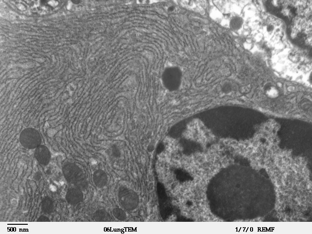
The endoplasmic reticulum (ER) is, in essence, the transportation system of the eukaryotic cell, and has many other important functions such as protein folding. It is a type of organelle made up of two subunits – rough endoplasmic reticulum (RER), and smooth endoplasmic reticulum (SER). The endoplasmic reticulum is found in most eukaryotic cells and forms an interconnected network of flattened, membrane-enclosed sacs known as cisternae (in the RER), and tubular structures in the SER. The membranes of the ER are continuous with the outer nuclear membrane. The endoplasmic reticulum is not found in red blood cells, or spermatozoa. The two types of ER share many of the same proteins and engage in certain common activities such as the synthesis of certain lipids and cholesterol. Different types of cells contain different ratios of the two types of ER depending on the activities of the cell. RER is found mainly toward the nucleus of cell and SER towards the cell membrane or plasma ... [...More Info...] [...Related Items...] OR: [Wikipedia] [Google] [Baidu] |


