|
Osteo-Odonto-Keratoprosthesis
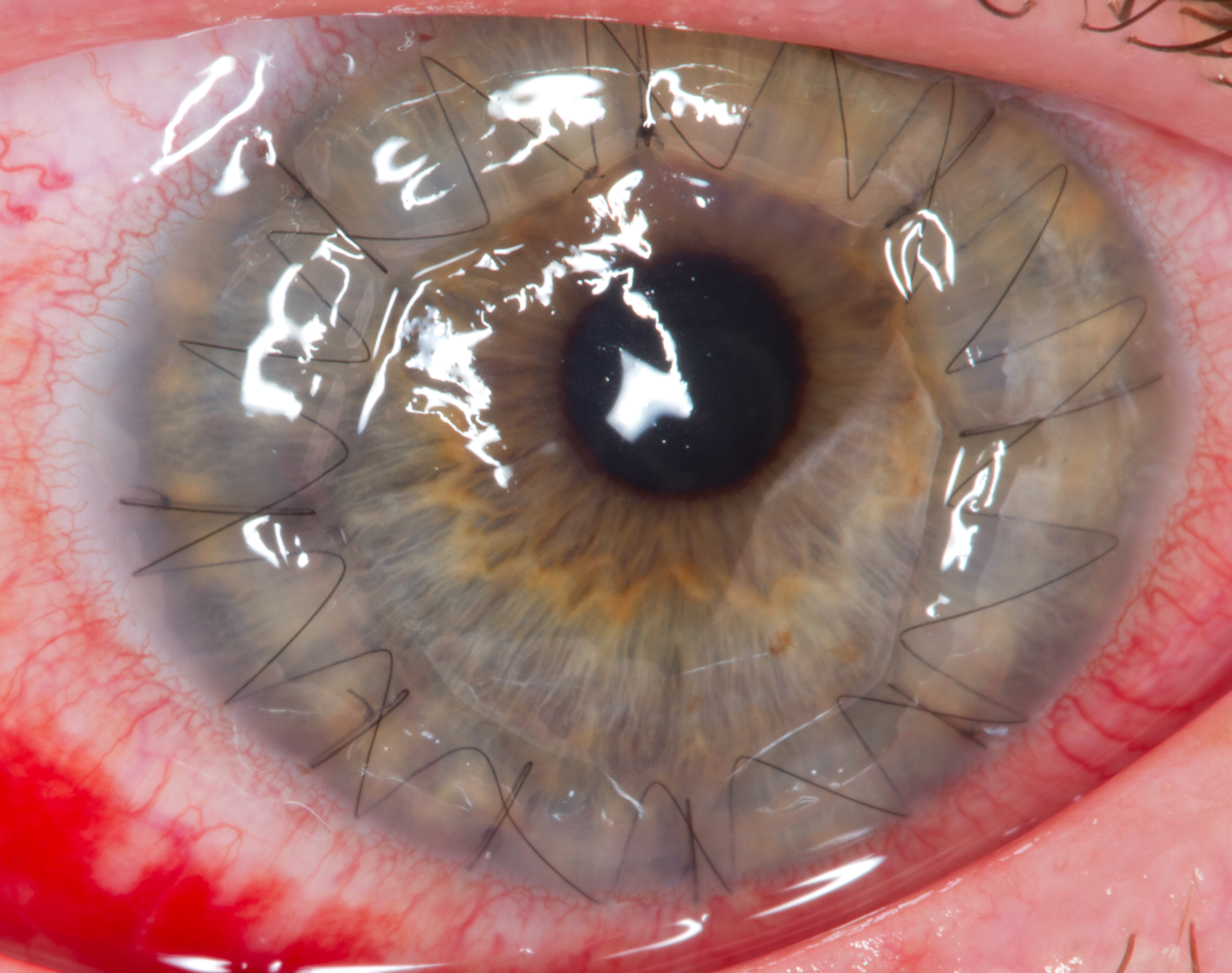
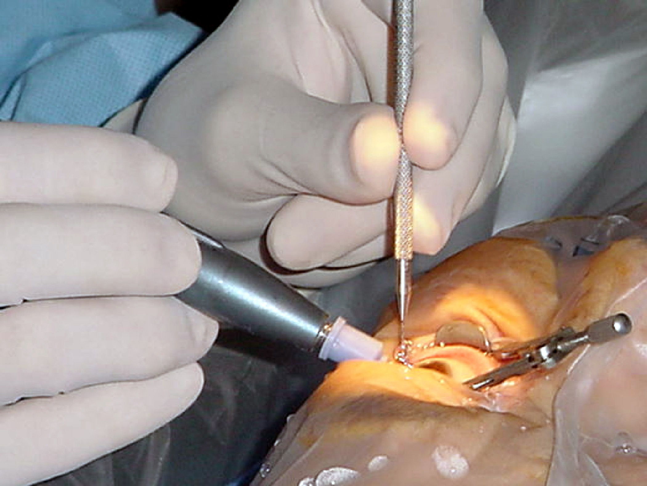
Osteo-odonto-keratoprosthesis (OOKP), also known as "tooth in eye" surgery, is a medical procedure to restore vision in the most severe cases of corneal and ocular surface patients. It includes removal of a tooth from the patient or a donor. After removal, a longitudinal lamina is cut from the tooth and a hole is drilled perpendicular to the lamina. The hole is then fitted with a cylindrical lens. The lamina is grown in the patients' cheek for a period of months and then is implanted upon the eye. The procedure was pioneered by the Italian ophthalmic surgeon Professor Benedetto Strampelli in the early 1960s. Strampelli was a founder-member of the International Intra-Ocular Implant Club (IIIC) in 1966.National Dental Centre - 25 May 2005 TOOTH-IN-EYE (OOKP) SURGERY ... [...More Info...] [...Related Items...] OR: [Wikipedia] [Google] [Baidu] |
Corneal Graft
Corneal transplantation, also known as corneal grafting, is a surgical procedure where a damaged or diseased cornea is replaced by Corneal button, donated corneal tissue (the graft). When the entire cornea is replaced it is known as penetrating keratoplasty and when only part of the cornea is replaced it is known as lamellar keratoplasty. Keratoplasty simply means surgery to the cornea. The graft is taken from a recently deceased individual with no known diseases or other factors that may affect the chance of survival of the donated tissue or the health of the recipient. The cornea is the transparency (optics), transparent front part of the human eye, eye that covers the Iris (anatomy), iris, pupil and anterior chamber. The surgical procedure is performed by Ophthalmology, ophthalmologists, physicians who specialize in eyes, and is often done on an outpatient basis. Donors can be of any age, as is shown in the case of Janis Babson, who donated her eyes after dying at the age of ... [...More Info...] [...Related Items...] OR: [Wikipedia] [Google] [Baidu] |
Lamina (anatomy)
Lamina is a general anatomical term meaning "plate" or "layer". It is used in both gross anatomy and microscopic anatomy to describe structures. Some examples include: * The laminae of the thyroid cartilage: two leaf-like plates of cartilage that make up the walls of the structure. * The vertebral laminae: plates of bone that form the posterior walls of each vertebra, enclosing the spinal cord. * The laminae of the thalamus: the layers of thalamus tissue. * The lamina propria: a connective tissue layer under the epithelium of an organ. * The nuclear lamina: a dense fiber network inside the nucleus of cells. * The lamina affixa: a layer of epithelium growing on the surface of the thalamus The thalamus (from Greek θάλαμος, "chamber") is a large mass of gray matter located in the dorsal part of the diencephalon (a division of the forebrain). Nerve fibers project out of the thalamus to the cerebral cortex in all directions, .... * Lamina cribrosa with two different ... [...More Info...] [...Related Items...] OR: [Wikipedia] [Google] [Baidu] |
Arch
An arch is a vertical curved structure that spans an elevated space and may or may not support the weight above it, or in case of a horizontal arch like an arch dam, the hydrostatic pressure against it. Arches may be synonymous with vaults, but a vault may be distinguished as a continuous arch forming a roof. Arches appeared as early as the 2nd millennium BC in Mesopotamian brick architecture, and their systematic use started with the ancient Romans, who were the first to apply the technique to a wide range of structures. Basic concepts An arch is a pure compression form. It can span a large area by resolving forces into compressive stresses, and thereby eliminating tensile stresses. This is sometimes denominated "arch action". As the forces in the arch are transferred to its base, the arch pushes outward at its base, denominated "thrust". As the rise, i. e. height, of the arch decreases the outward thrust increases. In order to preserve arch action and prevent collapse ... [...More Info...] [...Related Items...] OR: [Wikipedia] [Google] [Baidu] |
Dentistry Procedures
Dentistry, also known as dental medicine and oral medicine, is the branch of medicine focused on the teeth, gums, and mouth. It consists of the study, diagnosis, prevention, management, and treatment of diseases, disorders, and conditions of the mouth, most commonly focused on dentition (the development and arrangement of teeth) as well as the oral mucosa. Dentistry may also encompass other aspects of the craniofacial complex including the temporomandibular joint. The practitioner is called a dentist. The history of dentistry is almost as ancient as the history of humanity and civilization with the earliest evidence dating from 7000 BC to 5500 BC. Dentistry is thought to have been the first specialization in medicine which have gone on to develop its own accredited degree with its own specializations. Dentistry is often also understood to subsume the now largely defunct medical specialty of stomatology (the study of the mouth and its disorders and diseases) for which reason t ... [...More Info...] [...Related Items...] OR: [Wikipedia] [Google] [Baidu] |
Refractive Surgery
Refractive eye surgery is optional eye surgery used to improve the refractive state of the eye and decrease or eliminate dependency on glasses or contact lenses. This can include various methods of surgical remodeling of the cornea (keratomileusis), lens implantation or lens replacement. The most common methods today use excimer lasers to reshape the curvature of the cornea. Refractive eye surgeries are used to treat common vision disorders such as myopia, hyperopia, presbyopia and astigmatism. History The first theoretical work on the potential of refractive surgery was published in 1885 by Hjalmar August Schiøtz, an ophthalmologist from Norway. In 1930, the Japanese ophthalmologist Tsutomu Sato made the first attempts at performing this kind of surgery, hoping to correct the vision of military pilots. His approach was to make radial cuts in the cornea, correcting effects by up to 6 diopters. The procedure unfortunately produced a high rate of corneal degeneration, howeve ... [...More Info...] [...Related Items...] OR: [Wikipedia] [Google] [Baidu] |
Eye Surgery
Eye surgery, also known as ophthalmic or ocular surgery, is surgery performed on the eye or its adnexa, by an ophthalmologist or sometimes, an optometrist. Eye surgery is synonymous with ophthalmology. The eye is a very fragile organ, and requires extreme care before, during, and after a surgical procedure to minimize or prevent further damage. An expert eye surgeon is responsible for selecting the appropriate surgical procedure for the patient, and for taking the necessary safety precautions. Mentions of eye surgery can be found in several ancient texts dating back as early as 1800 BC, with cataract treatment starting in the fifth century BC. Today it continues to be a widely practiced type of surgery, with various techniques having been developed for treating eye problems. Preparation and precautions Since the eye is heavily supplied by nerves, anesthesia is essential. Local anesthesia is most commonly used. Topical anesthesia using lidocaine topical gel is often used fo ... [...More Info...] [...Related Items...] OR: [Wikipedia] [Google] [Baidu] |
Harold Ridley (ophthalmologist)
Sir Nicholas Harold Lloyd Ridley (10 July 1906 – 25 May 2001) was an English ophthalmologist who invented the intraocular lens and pioneered intraocular lens surgery for cataract patients. Early years Nicholas Harold Lloyd Ridley was born in Kibworth Harcourt, Leicestershire, the son of Nicholas Charles Ridley and his wife Margaret, née Parker; he had a younger brother, Olden. Harold had a stammer which he was largely able to manage. As a child he met and sat on the lap of Florence Nightingale, a close friend of his mother. He was educated at Charterhouse School before studying at Pembroke College, Cambridge from 1924 to 1927, and completed his medical training in 1930 at St Thomas' Hospital. Subsequently, he worked as a surgeon at both St Thomas' and Moorfields Eye Hospital in London, specialising in ophthalmology. In 1938 Ridley was appointed full surgeon and consultant at Moorfields Hospital and later appointed consultant surgeon in 1946. Cataract operations and intraocu ... [...More Info...] [...Related Items...] OR: [Wikipedia] [Google] [Baidu] |
Rayner (company)
Rayner designs and manufactures intraocular lenses and proprietary injection devices for use in cataract surgery. With Sir Harold Ridley, they were pioneers in the field from 1949 when Ridley successfully implanted the first intraocular lens (IOL) at St Thomas' Hospital, London. The origin of the company The story of the Rayner Company begins in 1910, when Mr John Baptiste Reiner and Mr Charles Davis Keeler opened their first optician's shop at No 9, Vere Street, London, England. They registered their company as Reiner & Keeler Ltd on 30 October 1910. Before forming the company, J.B. Reiner had completed an apprenticeship in 'the art of an optician and scientific instrument maker' in 1891 and had gone on to work for E.B. Merrowitz Ltd, a branch of a well-known American optical company. In 1915, during the First World War, the company name was changed to Rayner & Keeler Ltd. This was almost certainly a commercial decision of the time as J. B. Reiner retained his name all his lif ... [...More Info...] [...Related Items...] OR: [Wikipedia] [Google] [Baidu] |
Intraocular Lens
Intraocular lens (IOL) is a lens implanted in the eye as part of a treatment for cataracts or myopia. If the natural lens is left in the eye, the IOL is known as phakic, otherwise it is a pseudophakic, or false lens. Such a lens is typically implanted during cataract surgery, after the eye's cloudy natural lens (cataract) has been removed. The pseudophakic IOL provides the same light-focusing function as the natural crystalline lens. The phakic type of IOL is placed over the existing natural lens and is used in refractive surgery to change the eye's optical power as a treatment for myopia (nearsightedness). This is an alternative to LASIK. IOLs usually consist of a small plastic lens with plastic side struts, called haptics, to hold the lens in place in the capsular bag inside the eye. IOLs were conventionally made of an inflexible material (PMMA), although this has largely been superseded by the use of flexible materials, such as silicone. Most IOLs fitted today are fixed mo ... [...More Info...] [...Related Items...] OR: [Wikipedia] [Google] [Baidu] |
Nazareno Strampelli
Nazareno Strampelli (May 29, 1866, in Castelraimondo, Italy – January 23, 1942) was an Italian agronomist and Plant breeding, plant breeder. He was the forerunner of what became known as the Green Revolution of the late 1960s. Strampelli's work allowed Italy to become almost self-sufficient in bread wheat production, relative to Italy's contemporaneous population and consumption patterns, increasing area-based agricultural intensity from an average yield of 1.0 ton, t/hectare, ha at the beginning of the 19th century to about 1.5 t/ha in the 1930s,Gian Tommaso Scarascia MugnozzaThe contribution of Italian wheat geneticists: From Nazareno Strampelli to Francesco D'Amato, 2005. through the Hybrid (biology), hybridization of wheat into high-yield varietals. Early years After graduating in agriculture at the University of Pisa, he taught for a few years at the University of Camerino and in Reggio Calabria. Career In 1900, while in Camerino, he started his research on the Hybrid (biol ... [...More Info...] [...Related Items...] OR: [Wikipedia] [Google] [Baidu] |
Premolar
The premolars, also called premolar teeth, or bicuspids, are transitional teeth located between the canine and molar teeth. In humans, there are two premolars per quadrant in the permanent set of teeth, making eight premolars total in the mouth. They have at least two cusps. Premolars can be considered transitional teeth during chewing, or mastication. They have properties of both the canines, that lie anterior and molars that lie posterior, and so food can be transferred from the canines to the premolars and finally to the molars for grinding, instead of directly from the canines to the molars. Human anatomy The premolars in humans are the maxillary first premolar, maxillary second premolar, mandibular first premolar, and the mandibular second premolar. Premolar teeth by definition are permanent teeth distal to the canines, preceded by deciduous molars. Morphology There is always one large buccal cusp, especially so in the mandibular first premolar. The lower second ... [...More Info...] [...Related Items...] OR: [Wikipedia] [Google] [Baidu] |
Canine Tooth
In mammalian oral anatomy, the canine teeth, also called cuspids, dog teeth, or (in the context of the upper jaw) fangs, eye teeth, vampire teeth, or vampire fangs, are the relatively long, pointed teeth. They can appear more flattened however, causing them to resemble incisors and leading them to be called ''incisiform''. They developed and are used primarily for firmly holding food in order to tear it apart, and occasionally as weapons. They are often the largest teeth in a mammal's mouth. Individuals of most species that develop them normally have four, two in the upper jaw and two in the lower, separated within each jaw by incisors; humans and dogs are examples. In most species, canines are the anterior-most teeth in the maxillary bone. The four canines in humans are the two maxillary canines and the two mandibular canines. Details There are generally four canine teeth: two in the upper (maxillary) and two in the lower (mandibular) arch. A canine is placed laterally to ... [...More Info...] [...Related Items...] OR: [Wikipedia] [Google] [Baidu] |


.jpg)

