|
Orbital Gyri
The inferior or orbital surface of the frontal lobe is concave, and rests on the orbital plate of the frontal bone. It is divided into four orbital gyri by a well-marked H-shaped orbital sulcus. These are named, from their position, the medial, anterior, lateral, and posterior, orbital gyri. The medial orbital gyrus presents a well-marked antero-posterior sulcus, the olfactory sulcus, for the olfactory tract; the portion medial to this is named the straight gyrus, and is continuous with the superior frontal gyrus on the medial surface. Function Bailey and Bremer reported that stimulation to the central end of the vagus nerve The vagus nerve, also known as the tenth cranial nerve, cranial nerve X, or simply CN X, is a cranial nerve that interfaces with the parasympathetic control of the heart, lungs, and digestive tract. It comprises two nerves—the left and right ... caused electrical activity in the inferior orbital surface (http://brain.oxfordjournals.org/cgi/pdf_extract/ ... [...More Info...] [...Related Items...] OR: [Wikipedia] [Google] [Baidu] |
Frontal Lobe
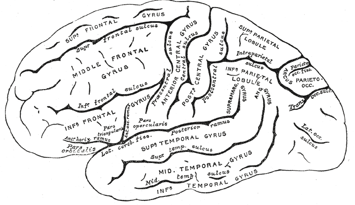
The frontal lobe is the largest of the four major lobes of the brain in mammals, and is located at the front of each cerebral hemisphere (in front of the parietal lobe and the temporal lobe). It is parted from the parietal lobe by a groove between tissues called the central sulcus and from the temporal lobe by a deeper groove called the lateral sulcus (Sylvian fissure). The most anterior rounded part of the frontal lobe (though not well-defined) is known as the frontal pole, one of the three poles of the cerebrum. The frontal lobe is covered by the frontal cortex. The frontal cortex includes the premotor cortex, and the primary motor cortex – parts of the motor cortex. The front part of the frontal cortex is covered by the prefrontal cortex. There are four principal gyri in the frontal lobe. The precentral gyrus is directly anterior to the central sulcus, running parallel to it and contains the primary motor cortex, which controls voluntary movements of specific body parts ... [...More Info...] [...Related Items...] OR: [Wikipedia] [Google] [Baidu] |
Orbital Part Of Frontal Bone
The orbital or horizontal part of the frontal bone (''pars orbitalis'') consists of two thin triangular plates, the orbital plates, which form the vaults of the orbits, and are separated from one another by a median gap, the ethmoidal notch. Surfaces * The inferior surface of each orbital plate is smooth and concave, and presents, laterally, under cover of the zygomatic process, a shallow depression, the lacrimal fossa, for the lacrimal gland; near the nasal part is a depression, the fovea trochlearis, or occasionally a small trochlear spine, for the attachment of the cartilaginous pulley of the obliquus oculi superior. * The superior surface is convex, and marked by depressions for the convolutions of the frontal lobes of the brain, and faint grooves for the meningeal branches of the ethmoidal vessels. ** The ethmoidal notch separates the two orbital plates; it is quadrilateral, and filled, in the articulated skull, by the cribriform plate of the ethmoid. *** The margins of ... [...More Info...] [...Related Items...] OR: [Wikipedia] [Google] [Baidu] |
Orbital Sulcus
The inferior or orbital surface of the frontal lobe is concave, and rests on the orbital plate of the frontal bone The frontal bone is a bone in the human skull. The bone consists of two portions.'' Gray's Anatomy'' (1918) These are the vertically oriented squamous part, and the horizontally oriented orbital part, making up the bony part of the forehead, pa .... It is divided into four orbital gyri by a well-marked H-shaped orbital sulcus Additional Images File:FrontalCaptsBasal.png, Cerebrum. Inferior view. File:Slide2STE.JPG, Cerebrum. Inferior view. Deep dissection. References Sulci (neuroanatomy) Frontal lobe {{neuroanatomy-stub ... [...More Info...] [...Related Items...] OR: [Wikipedia] [Google] [Baidu] |
Olfactory Sulcus
The olfactory tract is a bilateral bundle of afferent nerve fibers from the mitral and tufted cells of the olfactory bulb that connects to several target regions in the brain, including the piriform cortex, amygdala, and entorhinal cortex. It is a narrow white band, triangular on coronal section, the apex being directed upward. Structure The olfactory tract and olfactory bulb lie in the olfactory sulcus a sulcus formed by the medial orbital gyrus on the inferior surface of each frontal lobe. The olfactory tracts lie in the sulci which run closely parallel to the midline. Fibers of the olfactory tract appear to end in the antero-lateral part of the olfactory tubercle, the dorsal and external parts of the anterior olfactory nucleus, the frontal and temporal parts of the prepyriform area, the cortico-medial group of amygdala nuclei and the nucleus of the stria terminalis. The olfactory tract divides posteriorly into a medial and a lateral stria. Caudal to this is the olfacto ... [...More Info...] [...Related Items...] OR: [Wikipedia] [Google] [Baidu] |
Olfactory Tract
The olfactory tract is a bilateral bundle of afferent nerve fibers from the mitral and tufted cells of the olfactory bulb that connects to several target regions in the brain, including the piriform cortex, amygdala, and entorhinal cortex. It is a narrow white band, triangular on coronal section, the apex being directed upward. Structure The olfactory tract and olfactory bulb lie in the olfactory sulcus a sulcus formed by the medial orbital gyrus on the inferior surface of each frontal lobe. The olfactory tracts lie in the sulci which run closely parallel to the midline. Fibers of the olfactory tract appear to end in the antero-lateral part of the olfactory tubercle, the dorsal and external parts of the anterior olfactory nucleus, the frontal and temporal parts of the prepyriform area, the cortico-medial group of amygdala nuclei and the nucleus of the stria terminalis. The olfactory tract divides posteriorly into a medial and a lateral stria. Caudal to this is the olfactor ... [...More Info...] [...Related Items...] OR: [Wikipedia] [Google] [Baidu] |
Straight Gyrus
The portion of the inferior frontal lobe immediately adjacent to the longitudinal fissure (and medial to the medial orbital gyrus and olfactory tract) is named the straight gyrus,(or gyrus rectus) and is continuous with the superior frontal gyrus on the medial surface. A specific function for the straight gyrus has not yet been brought to light; however, in males, greater activation of the straight gyrus within the medial orbitofrontal cortex while observing sexually visual pictures has been strongly linked to HSDD ( hypoactive sexual desire disorder). Additional images File:Straight gyrus animation small2.gif, Animation. Straight gyrus is depicted as red. File:Straight_gyrus_-_inferior_view.png, Basal surface of cerebrum. Straight gyrus is shown in red. File:Gray743 straight gyrus.png, Coronal section of human brain. Straight gyrus depicted as yellow in center bottom. File:Slide2STE.JPG, Cerebrum. Optic and olfactory nerves.Inferior view. Deep dissection. File:Slide2ZEB.JP ... [...More Info...] [...Related Items...] OR: [Wikipedia] [Google] [Baidu] |
Superior Frontal Gyrus
In neuroanatomy, the superior frontal gyrus (SFG, also marginal gyrus) is a gyrus – a ridge on the brain's cerebral cortex – which makes up about one third of the frontal lobe. It is bounded laterally by the superior frontal sulcus. The superior frontal gyrus is one of the frontal gyri. Function Self-awareness In fMRI experiments, Goldberg ''et al.'' have found evidence that the superior frontal gyrus is involved in self-awareness, in coordination with the action of the sensory system. Laughter In 1998, neurosurgeon Itzhak Fried described a 16-year-old female patient (referred to as "patient AK") who laughed when her SFG was stimulated with electric current during treatment for epilepsy. Electrical stimulation was applied to the cortical surface of AK's left frontal lobe while an attempt was made to locate the focus of her epileptic seizures (which were never accompanied by laughter). Fried identified a 2 cm by 2 cm area on the left SFG where stimulation produ ... [...More Info...] [...Related Items...] OR: [Wikipedia] [Google] [Baidu] |
Vagus Nerve
The vagus nerve, also known as the tenth cranial nerve, cranial nerve X, or simply CN X, is a cranial nerve that interfaces with the parasympathetic control of the heart, lungs, and digestive tract. It comprises two nerves—the left and right vagus nerves—but they are typically referred to collectively as a single subsystem. The vagus is the longest nerve of the autonomic nervous system in the human body and comprises both sensory and motor fibers. The sensory fibers originate from neurons of the nodose ganglion, whereas the motor fibers come from neurons of the dorsal motor nucleus of the vagus and the nucleus ambiguus. The vagus was also historically called the pneumogastric nerve. Structure Upon leaving the medulla oblongata between the olive and the inferior cerebellar peduncle, the vagus nerve extends through the jugular foramen, then passes into the carotid sheath between the internal carotid artery and the internal jugular vein down to the neck, chest, and abdom ... [...More Info...] [...Related Items...] OR: [Wikipedia] [Google] [Baidu] |
Cerebrum
The cerebrum, telencephalon or endbrain is the largest part of the brain containing the cerebral cortex (of the two cerebral hemispheres), as well as several subcortical structures, including the hippocampus, basal ganglia, and olfactory bulb. In the human brain, the cerebrum is the uppermost region of the central nervous system. The cerebrum prenatal development, develops prenatally from the forebrain (prosencephalon). In mammals, the Dorsum (biology), dorsal telencephalon, or Pallium (neuroanatomy), pallium, develops into the cerebral cortex, and the ventral telencephalon, or Pallium (neuroanatomy), subpallium, becomes the basal ganglia. The cerebrum is also divided into approximately symmetric Lateralization of brain function, left and right cerebral hemispheres. With the assistance of the cerebellum, the cerebrum controls all voluntary actions in the human body. Structure The cerebrum is the largest part of the brain. Depending upon the position of the animal it lies eithe ... [...More Info...] [...Related Items...] OR: [Wikipedia] [Google] [Baidu] |
Gyri
In neuroanatomy, a gyrus (pl. gyri) is a ridge on the cerebral cortex. It is generally surrounded by one or more sulci (depressions or furrows; sg. ''sulcus''). Gyri and sulci create the folded appearance of the brain in humans and other mammals. Structure The gyri are part of a system of folds and ridges that create a larger surface area for the human brain and other mammalian brains. Because the brain is confined to the skull, brain size is limited. Ridges and depressions create folds allowing a larger cortical surface area, and greater cognitive function, to exist in the confines of a smaller cranium. Development The human brain undergoes gyrification during fetal and neonatal development. In embryonic development, all mammalian brains begin as smooth structures derived from the neural tube. A cerebral cortex without surface convolutions is lissencephalic, meaning 'smooth-brained'. As development continues, gyri and sulci begin to take shape on the fetal brain, with ... [...More Info...] [...Related Items...] OR: [Wikipedia] [Google] [Baidu] |


