|
Optical Projection Tomography
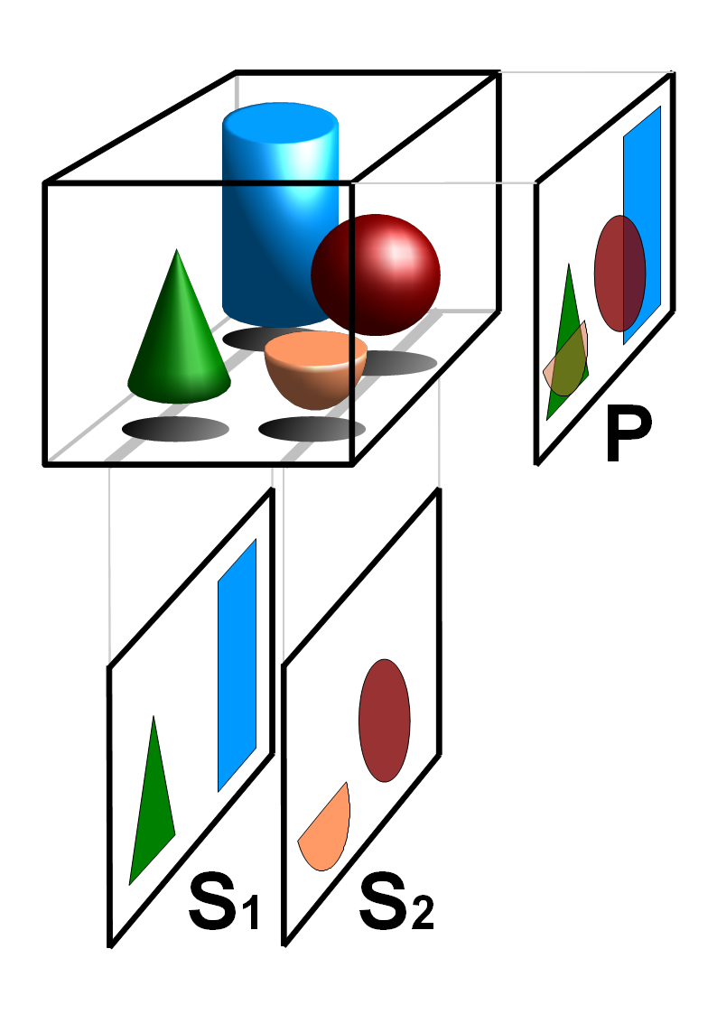
Optical projection tomography is a form of tomography involving optical microscopy. The OPT technique is sometimes referred to as Optical Computed Tomography (optical-CT) and Optical Emission Computed Tomography (optical-ECT) in the literature, to address the fact that the technique bears similarity to X-ray computed tomography (CT) and single-photon emission computed tomography (SPECT). It is in many ways the optical equivalent of X-ray computed tomography or the medical CT scan. OPT differs in the way that it often uses ultraviolet, visible, and near-infrared photons as opposed to X-ray photons. However, essential mathematics and reconstruction algorithms used for CT and OPT are similar; for example, radon transform or iterative reconstruction based on projection data are used in both medical CT scan and OPT for 3D reconstruction. Both medical CT and OPT compute 3D volumes based on transmission of the photon through the material of interest. Given that the tissue is typically opa ... [...More Info...] [...Related Items...] OR: [Wikipedia] [Google] [Baidu] |
Tomography
Tomography is imaging by sections or sectioning that uses any kind of penetrating wave. The method is used in radiology, archaeology, biology, atmospheric science, geophysics, oceanography, plasma physics, materials science, astrophysics, quantum information, and other areas of science. The word ''tomography'' is derived from Ancient Greek τόμος ''tomos'', "slice, section" and γράφω ''graphō'', "to write" or, in this context as well, "to describe." A device used in tomography is called a tomograph, while the image produced is a tomogram. In many cases, the production of these images is based on the mathematical procedure tomographic reconstruction, such as X-ray computed tomography technically being produced from multiple projectional radiographs. Many different reconstruction algorithms exist. Most algorithms fall into one of two categories: filtered back projection (FBP) and iterative reconstruction (IR). These procedures give inexact results: they represent a compr ... [...More Info...] [...Related Items...] OR: [Wikipedia] [Google] [Baidu] |
Optical Microscopy
Optics is the branch of physics that studies the behaviour and properties of light, including its interactions with matter and the construction of instruments that use or detect it. Optics usually describes the behaviour of visible, ultraviolet, and infrared light. Because light is an electromagnetic wave, other forms of electromagnetic radiation such as X-rays, microwaves, and radio waves exhibit similar properties. Most optical phenomena can be accounted for by using the classical electromagnetic description of light. Complete electromagnetic descriptions of light are, however, often difficult to apply in practice. Practical optics is usually done using simplified models. The most common of these, geometric optics, treats light as a collection of rays that travel in straight lines and bend when they pass through or reflect from surfaces. Physical optics is a more comprehensive model of light, which includes wave effects such as diffraction and interference that cannot be ac ... [...More Info...] [...Related Items...] OR: [Wikipedia] [Google] [Baidu] |
X-ray Computed Tomography
An X-ray, or, much less commonly, X-radiation, is a penetrating form of high-energy electromagnetic radiation. Most X-rays have a wavelength ranging from 10 picometers to 10 nanometers, corresponding to frequencies in the range 30 petahertz to 30 exahertz ( to ) and energies in the range 145 eV to 124 keV. X-ray wavelengths are shorter than those of UV rays and typically longer than those of gamma rays. In many languages, X-radiation is referred to as Röntgen radiation, after the German scientist Wilhelm Conrad Röntgen, who discovered it on November 8, 1895. He named it ''X-radiation'' to signify an unknown type of radiation.Novelline, Robert (1997). ''Squire's Fundamentals of Radiology''. Harvard University Press. 5th edition. . Spellings of ''X-ray(s)'' in English include the variants ''x-ray(s)'', ''xray(s)'', and ''X ray(s)''. The most familiar use of X-rays is checking for fractures (broken bones), but X-rays are also used in other ways. Fo ... [...More Info...] [...Related Items...] OR: [Wikipedia] [Google] [Baidu] |
Single-photon Emission Computed Tomography
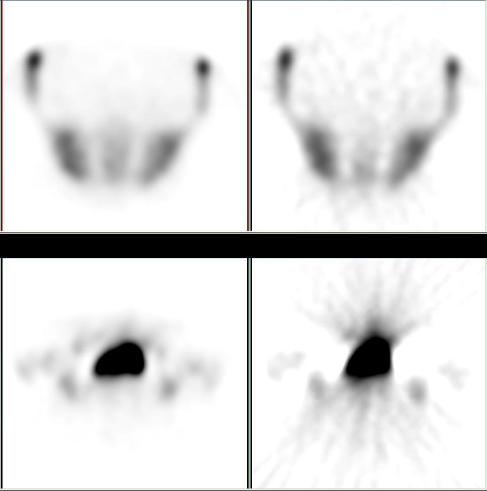
Single-photon emission computed tomography (SPECT, or less commonly, SPET) is a nuclear medicine tomographic imaging technique using gamma rays. It is very similar to conventional nuclear medicine planar imaging using a gamma camera (that is, scintigraphy), but is able to provide true 3D information. This information is typically presented as cross-sectional slices through the patient, but can be freely reformatted or manipulated as required. The technique needs delivery of a gamma-emitting radioisotope (a radionuclide) into the patient, normally through injection into the bloodstream. On occasion, the radioisotope is a simple soluble dissolved ion, such as an isotope of gallium(III). Most of the time, though, a marker radioisotope is attached to a specific ligand to create a radioligand, whose properties bind it to certain types of tissues. This marriage allows the combination of ligand and radiopharmaceutical to be carried and bound to a place of interest in the body, where ... [...More Info...] [...Related Items...] OR: [Wikipedia] [Google] [Baidu] |
Radon Transform
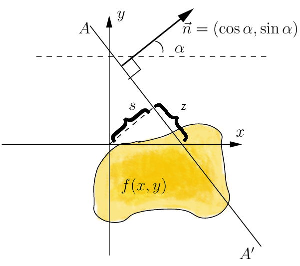
In mathematics, the Radon transform is the integral transform which takes a function ''f'' defined on the plane to a function ''Rf'' defined on the (two-dimensional) space of lines in the plane, whose value at a particular line is equal to the line integral of the function over that line. The transform was introduced in 1917 by Johann Radon, who also provided a formula for the inverse transform. Radon further included formulas for the transform in three dimensions, in which the integral is taken over planes (integrating over lines is known as the X-ray transform). It was later generalized to higher-dimensional Euclidean spaces, and more broadly in the context of integral geometry. The complex analogue of the Radon transform is known as the Penrose transform. The Radon transform is widely applicable to tomography, the creation of an image from the projection data associated with cross-sectional scans of an object. Explanation If a function f represents an unknown density, then t ... [...More Info...] [...Related Items...] OR: [Wikipedia] [Google] [Baidu] |
Iterative Reconstruction
Iterative reconstruction refers to iterative algorithms used to reconstruct 2D and 3D images in certain imaging techniques. For example, in computed tomography an image must be reconstructed from projections of an object. Here, iterative reconstruction techniques are usually a better, but computationally more expensive alternative to the common filtered back projection (FBP) method, which directly calculates the image in a single reconstruction step.Herman, G. T.Fundamentals of computerized tomography: Image reconstruction from projection 2nd edition, Springer, 2009 In recent research works, scientists have shown that extremely fast computations and massive parallelism is possible for iterative reconstruction, which makes iterative reconstruction practical for commercialization. Basic concepts The reconstruction of an image from the acquired data is an inverse problem. Often, it is not possible to exactly solve the inverse problem directly. In this case, a direct algorithm h ... [...More Info...] [...Related Items...] OR: [Wikipedia] [Google] [Baidu] |
Genetically Modified Mouse
A genetically modified mouse or genetically engineered mouse model (GEMM) is a house mouse, mouse (''Mus musculus'') that has had its genome altered through the use of genetic engineering techniques. Genetically modified mice are commonly used for research or as animal models of human diseases, and are also used for research on genes. Together with patient-derived xenografts (PDXs), GEMMs are the most common ''in vivo'' models in cancer research. Both approaches are considered complementary and may be used to recapitulate different aspects of disease. GEMMs are also of great interest for drug development, as they facilitate target validation and the study of response, resistance, toxicity and pharmacodynamics. History In 1974 Beatrice Mintz and Rudolf Jaenisch created the first genetically modified animal by inserting a DNA virus into an early-stage mouse embryo and showing that the inserted genes were present in every cell. However, the mice did not pass the transgene to their offs ... [...More Info...] [...Related Items...] OR: [Wikipedia] [Google] [Baidu] |
Green Fluorescent Protein
The green fluorescent protein (GFP) is a protein that exhibits bright green fluorescence when exposed to light in the blue to ultraviolet range. The label ''GFP'' traditionally refers to the protein first isolated from the jellyfish ''Aequorea victoria'' and is sometimes called ''avGFP''. However, GFPs have been found in other organisms including corals, sea anemones, zoanithids, copepods and lancelets. The GFP from ''A. victoria'' has a major excitation peak at a wavelength of 395 nm and a minor one at 475 nm. Its emission peak is at 509 nm, which is in the lower green portion of the visible spectrum. The fluorescence quantum yield (QY) of GFP is 0.79. The GFP from the sea pansy (''Renilla reniformis'') has a single major excitation peak at 498 nm. GFP makes for an excellent tool in many forms of biology due to its ability to form an internal chromophore without requiring any accessory cofactors, gene products, or enzymes / substrates other than mo ... [...More Info...] [...Related Items...] OR: [Wikipedia] [Google] [Baidu] |
Organ Culture
Organ culture is a development from tissue culture methods of research, the organ culture is able to accurately model functions of an organ in various states and conditions by the use of the actual ''in vitro'' organ itself. Parts of an organ or a whole organ can be cultured in vitro. The main objective is to maintain the architecture of the tissue and direct it towards normal development. In this technique, it is essential that the tissue is never disrupted or damaged. It thus requires careful handling. The media used for a growing organ culture are generally the same as those used for tissue culture. The techniques for organ culture can be classified into (i) those employing a solid medium and (ii) those employing liquid medium. Current progress In April 2006, scientists reported a successful trial of seven bladders grown in-vitro and given to humans. A bladder has been cultured by Anthony Atala of the Wake Forest Institute for Regenerative Medicine in Winston-Salem, North Caro ... [...More Info...] [...Related Items...] OR: [Wikipedia] [Google] [Baidu] |
Diffuse Optical Imaging
Diffuse optical imaging (DOI) is a method of imaging using near-infrared spectroscopy (NIRS) or fluorescence-based methods. When used to create 3D volumetric models of the imaged material DOI is referred to as diffuse optical tomography, whereas 2D imaging methods are classified as diffuse optical imaging. The technique has many applications to neuroscience, sports medicine, wound monitoring, and cancer detection. Typically DOI techniques monitor changes in concentrations of oxygenated and deoxygenated hemoglobin and may additionally measure redox states of cytochromes. The technique may also be referred to as diffuse optical tomography (DOT), near infrared optical tomography (NIROT) or fluorescence diffuse optical tomography (FDOT), depending on the usage. In neuroscience, functional measurements made using NIR wavelengths, DOI techniques may classify as functional near infrared spectroscopy fNIRS. Physical mechanism Biological tissues can be considered strongly diffusive me ... [...More Info...] [...Related Items...] OR: [Wikipedia] [Google] [Baidu] |
Optical Coherence Tomography
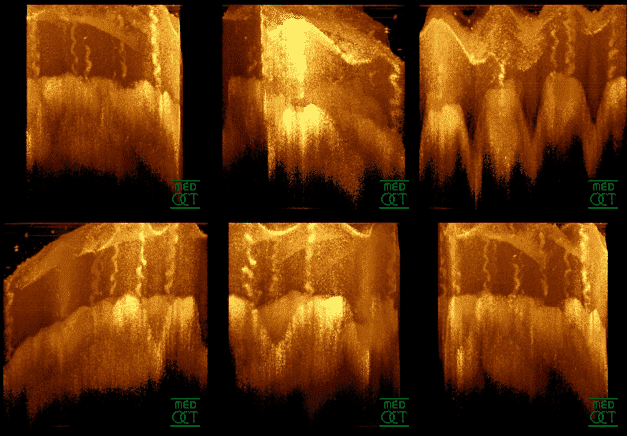
Optical coherence tomography (OCT) is an imaging technique that uses low-coherence light to capture micrometer-resolution, two- and three-dimensional images from within optical scattering media (e.g., biological tissue). It is used for medical imaging and industrial nondestructive testing (NDT). Optical coherence tomography is based on low-coherence interferometry, typically employing near-infrared light. The use of relatively long wavelength light allows it to penetrate into the scattering medium. Confocal microscopy, another optical technique, typically penetrates less deeply into the sample but with higher resolution. Depending on the properties of the light source ( superluminescent diodes, ultrashort pulsed lasers, and supercontinuum lasers have been employed), optical coherence tomography has achieved sub-micrometer resolution (with very wide-spectrum sources emitting over a ~100 nm wavelength range). Optical coherence tomography is one of a class of optical tom ... [...More Info...] [...Related Items...] OR: [Wikipedia] [Google] [Baidu] |


