|
Optic Papillitis
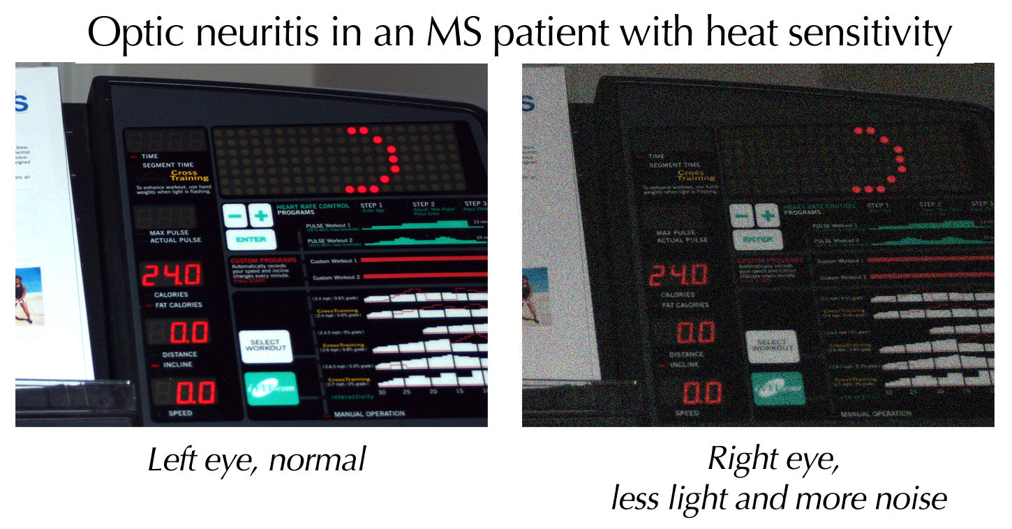
Optic papillitis is a specific type of optic neuritis. Inflammation of the optic nerve head is called "papillitis" or "intraocular optic neuritis"; inflammation of the orbital portion of the nerve is called "retrobulbar optic neuritis" or "orbital optic neuritis". It is often associated with substantial losses in visual fields, pain on moving the globe, and sensitivity to light pressure on the globe. It is often an early sign of multiple sclerosis. Papillitis may have the same appearance as papilledema Papilledema or papilloedema is optic disc swelling that is caused by increased intracranial pressure due to any cause. The swelling is usually bilateral and can occur over a period of hours to weeks. Unilateral presentation is extremely rare. In .... However, papillitis may be unilateral, whereas papilledema is almost always bilateral. Papillitis can be differentiated from papilledema by an afferent pupillary defect (Marcus Gunn pupil), by its greater effect in decreasing visua ... [...More Info...] [...Related Items...] OR: [Wikipedia] [Google] [Baidu] |
Optic Neuritis
Optic neuritis describes any condition that causes inflammation of the optic nerve; it may be associated with demyelinating diseases, or infectious or inflammatory processes. It is also known as optic papillitis (when the head of the optic nerve is involved), neuroretinitis (when there is a combined involvement of the optic disc and surrounding retina in the macular area) and retrobulbar neuritis (when the posterior part of the nerve is involved). Prelaminar optic neuritis describes involvement of the non-myelinated axons in the retina. It is most often associated with multiple sclerosis, and it may lead to complete or partial loss of vision in one or both eyes. Other causes include: # Leber's hereditary optic neuropathy # Parainfectious optic neuritis (associated with viral infections such as measles, mumps, chickenpox, whooping cough and glandular fever) # Infectious optic neuritis (sinus related or associated with cat-scratch fever, tuberculosis, Lyme disease and crypt ... [...More Info...] [...Related Items...] OR: [Wikipedia] [Google] [Baidu] |
Optic Nerve Head
The optic disc or optic nerve head is the point of exit for ganglion cell axons leaving the eye. Because there are no rods or cones overlying the optic disc, it corresponds to a small blind spot in each eye. The ganglion cell axons form the optic nerve after they leave the eye. The optic disc represents the beginning of the optic nerve and is the point where the axons of retinal ganglion cells come together. The optic disc is also the entry point for the major blood vessels that supply the retina. The optic disc in a normal human eye carries 1–1.2 million afferent nerve fibers from the eye towards the brain. Structure The optic disc is placed 3 to 4 mm to the nasal side of the fovea. It is a vertical oval, with average dimensions of 1.76mm horizontally by 1.92mm vertically. There is a central depression, of variable size, called the optic cup. This depression can be a variety of shapes from a shallow indentation to a bean pot—this shape can be significant for dia ... [...More Info...] [...Related Items...] OR: [Wikipedia] [Google] [Baidu] |
Orbit (anatomy)
In anatomy, the orbit is the cavity or socket of the skull in which the eye and its appendages are situated. "Orbit" can refer to the bony socket, or it can also be used to imply the contents. In the adult human, the volume of the orbit is , of which the eye occupies . The orbital contents comprise the eye, the orbital and retrobulbar fascia, extraocular muscles, cranial nerves II, III, IV, V, and VI, blood vessels, fat, the lacrimal gland with its sac and duct, the eyelids, medial and lateral palpebral ligaments, cheek ligaments, the suspensory ligament, septum, ciliary ganglion and short ciliary nerves. Structure The orbits are conical or four-sided pyramidal cavities, which open into the midline of the face and point back into the head. Each consists of a base, an apex and four walls."eye, human."Encyclopædia Britannica from Encyclopædia Britannica 2006 Ultimate Reference Suite DVD 2009 Openings There are two important foramina, or windows, two important fissu ... [...More Info...] [...Related Items...] OR: [Wikipedia] [Google] [Baidu] |
Multiple Sclerosis
Multiple (cerebral) sclerosis (MS), also known as encephalomyelitis disseminata or disseminated sclerosis, is the most common demyelinating disease, in which the insulating covers of nerve cells in the brain and spinal cord are damaged. This damage disrupts the ability of parts of the nervous system to transmit signals, resulting in a range of signs and symptoms, including physical, mental, and sometimes psychiatric problems. Specific symptoms can include double vision, blindness in one eye, muscle weakness, and trouble with sensation or coordination. MS takes several forms, with new symptoms either occurring in isolated attacks (relapsing forms) or building up over time (progressive forms). In the relapsing forms of MS, between attacks, symptoms may disappear completely, although some permanent neurological problems often remain, especially as the disease advances. While the cause is unclear, the underlying mechanism is thought to be either destruction by the immune system ... [...More Info...] [...Related Items...] OR: [Wikipedia] [Google] [Baidu] |
Papilledema
Papilledema or papilloedema is optic disc swelling that is caused by increased intracranial pressure due to any cause. The swelling is usually bilateral and can occur over a period of hours to weeks. Unilateral presentation is extremely rare. In intracranial hypertension, the optic disc swelling most commonly occurs bilaterally. When papilledema is found on fundoscopy, further evaluation is warranted because vision loss can result if the underlying condition is not treated. Further evaluation with a CT or MRI of the brain and/or spine is usually performed. Recent research has shown that point-of-care ultrasound can be used to measure optic nerve sheath diameter for detection of increased intracranial pressure and shows good diagnostic test accuracy compared to CT. Thus, if there is a question of papilledema on fundoscopic examination or if the optic disc cannot be adequately visualized, ultrasound can be used to rapidly assess for increased intracranial pressure and help direct f ... [...More Info...] [...Related Items...] OR: [Wikipedia] [Google] [Baidu] |
Neurological Disorders
A neurological disorder is any disorder of the nervous system. Structural, biochemical or electrical abnormalities in the brain, spinal cord or other nerves can result in a range of symptoms. Examples of symptoms include paralysis, muscle weakness, poor coordination, loss of sensation, seizures, confusion, pain and altered levels of consciousness. There are many recognized neurological disorders, some relatively common, but many rare. They may be assessed by neurological examination, and studied and treated within the specialities of neurology and clinical neuropsychology. Interventions foneurological disordersinclude preventive measures, lifestyle changes, physiotherapy or other therapy, neurorehabilitation, pain management, medication, operations performed by neurosurgeons or a specific diet. The World Health Organization estimated in 2006 that neurological disorders and their sequelae (direct consequences) affect as many as one billion people worldwide, and identified heal ... [...More Info...] [...Related Items...] OR: [Wikipedia] [Google] [Baidu] |


