|
Opponent Color Theory
The opponent process is a color theory that states that the human visual system interprets information about color by processing signals from photoreceptor cells in an antagonistic manner. The opponent-process theory suggests that there are three opponent channels, each comprising an opposing color pair: red versus green, blue versus yellow, and black versus white (luminance). The theory was first proposed in 1892 by the German physiologist Ewald Hering. Color theory Complementary colors When staring at a bright color for awhile (e.g. red), then looking away at a white field, an afterimage is perceived, such that the original color will evoke its complementary color (green, in the case of red input). When complementary colors are combined or mixed, they "cancel each other out" and become neutral (white or gray). That is, complementary colors are never perceived as a mixture; there is no "greenish red" or "yellowish blue", despite Impossible color#Claimed evidence for the ability ... [...More Info...] [...Related Items...] OR: [Wikipedia] [Google] [Baidu] |
Color Theory
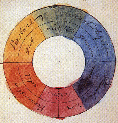
In the visual arts, color theory is the body of practical guidance for color mixing and the visual effects of a specific color combination. Color terminology based on the color wheel and its geometry separates colors into primary color, secondary color, and tertiary color. The understanding of color theory dates to antiquity. Aristotle (d. 322 BCE) and Claudius Ptolemy (d. 168 CE) already discussed which and how colors can be produced by mixing other colors. The influence of light on color was investigated and revealed further by al-Kindi (d. 873) and Ibn al-Haytham (d.1039). Ibn Sina (d. 1037), Nasir al-Din al-Tusi (d. 1274), and Robert Grosseteste (d. 1253) discovered that contrary to the teachings of Aristotle, there are multiple color paths to get from black to white. More modern approaches to color theory principles can be found in the writings of Leone Battista Alberti (c. 1435) and the notebooks of Leonardo da Vinci (c. 1490). A formalization of "color theory" bega ... [...More Info...] [...Related Items...] OR: [Wikipedia] [Google] [Baidu] |
Young–Helmholtz Theory
The Young–Helmholtz theory (based on the work of Thomas Young and Hermann von Helmholtz in the 19th century), also known as the trichromatic theory, is a theory of trichromatic color vision – the manner in which the visual system gives rise to the phenomenological experience of color. In 1802, Young postulated the existence of three types of photoreceptors (now known as cone cells) in the eye, with different but overlapping response to different wavelengths of visible light. Hermann von Helmholtz developed the theory further in 1850: that the three types of cone photoreceptors could be classified as short-preferring ( violet), middle-preferring (green), and long-preferring ( red), according to their response to the wavelengths of light striking the retina. The relative strengths of the signals detected by the three types of cones are interpreted by the brain as a visible color. For instance, yellow light uses different proportions of red and green, but little blue, so any h ... [...More Info...] [...Related Items...] OR: [Wikipedia] [Google] [Baidu] |
Receptive Field
The receptive field, or sensory space, is a delimited medium where some physiological stimuli can evoke a sensory neuronal response in specific organisms. Complexity of the receptive field ranges from the unidimensional chemical structure of odorants to the multidimensional spacetime of human visual field, through the bidimensional skin surface, being a receptive field for touch perception. Receptive fields can positively or negatively alter the membrane potential with or without affecting the rate of action potentials. A sensory space can be dependent of an animal's location. For a particular sound wave traveling in an appropriate transmission medium, by means of sound localization, an auditory space would amount to a reference system that continuously shifts as the animal moves (taking into consideration the space inside the ears as well). Conversely, receptive fields can be largely independent of the animal's location, as in the case of place cells. A sensory space can als ... [...More Info...] [...Related Items...] OR: [Wikipedia] [Google] [Baidu] |
Parvocellular Cell
Parvocellular cells, also called P-cells, are neurons located within the parvocellular layers of the lateral geniculate nucleus (LGN) of the thalamus. "''Parvus''" is Latin for "small", and the name "parvocellular" refers to the small size of the cell compared to the larger magnocellular cells. Phylogenetically, parvocellular neurons are more modern than magnocellular ones. Function The parvocellular neurons of the visual system receive their input from midget cells, a type of retinal ganglion cell, whose axons are exiting the optic tract. These synapses occur in one of the four dorsal parvocellular layers of the lateral geniculate nucleus. The information from each eye is kept separate at this point, and continues to be segregated until processing in the visual cortex. The electrically-encoded visual information leaves the parvocellular cells via relay cells in the optic radiations, traveling to the primary visual cortex layer 4C-β. The parvocellular neurons are sensit ... [...More Info...] [...Related Items...] OR: [Wikipedia] [Google] [Baidu] |
Cell (biology)
The cell is the basic structural and functional unit of life forms. Every cell consists of a cytoplasm enclosed within a membrane, and contains many biomolecules such as proteins, DNA and RNA, as well as many small molecules of nutrients and metabolites.Cell Movements and the Shaping of the Vertebrate Body in Chapter 21 of Molecular Biology of the Cell '' fourth edition, edited by Bruce Alberts (2002) published by Garland Science. The Alberts text discusses how the "cellular building blocks" move to shape developing embryos. It is also common to describe small molecules such as ... [...More Info...] [...Related Items...] OR: [Wikipedia] [Google] [Baidu] |
Magnocellular Cell
Magnocellular cells, also called M-cells, are neurons located within the Adina magnocellular layer of the lateral geniculate nucleus of the thalamus. The cells are part of the visual system. They are termed "magnocellular" since they are characterized by their relatively large size compared to parvocellular cells. Structure The full details of the flow of signaling from the eye to the visual cortex of the brain that result in the experience of vision are incompletely understood. Many aspects are subject to active controversy and the disruption of new evidence. In the visual system, signals mostly travel from the retina to the lateral geniculate nucleus (LGN) and then to the visual cortex. In humans the LGN is normally described as having six distinctive layers. The inner two layers, (1 and 2) are magnocellular cell (M cell) layers, while the outer four layers, (3,4,5 and 6), are parvocellular cell (P cell) layers. An additional set of neurons, known as the koniocellular c ... [...More Info...] [...Related Items...] OR: [Wikipedia] [Google] [Baidu] |
Retinal Ganglion Cell
A retinal ganglion cell (RGC) is a type of neuron located near the inner surface (the ganglion cell layer) of the retina of the eye. It receives visual information from photoreceptors via two intermediate neuron types: bipolar cells and retina amacrine cells. Retina amacrine cells, particularly narrow field cells, are important for creating functional subunits within the ganglion cell layer and making it so that ganglion cells can observe a small dot moving a small distance. Retinal ganglion cells collectively transmit image-forming and non-image forming visual information from the retina in the form of action potential to several regions in the thalamus, hypothalamus, and mesencephalon, or midbrain. Retinal ganglion cells vary significantly in terms of their size, connections, and responses to visual stimulation but they all share the defining property of having a long axon that extends into the brain. These axons form the optic nerve, optic chiasm, and optic tract. A s ... [...More Info...] [...Related Items...] OR: [Wikipedia] [Google] [Baidu] |
Retina Bipolar Cell
As a part of the retina, bipolar cells exist between photoreceptors (rod cells and cone cells) and ganglion cells. They act, directly or indirectly, to transmit signals from the photoreceptors to the ganglion cells. Structure Bipolar cells are so-named as they have a central body from which two sets of processes arise. They can synapse with either rods or cones (rod/cone mixed input BCs have been found in teleost fish but not mammals), and they also accept synapses from horizontal cells. The bipolar cells then transmit the signals from the photoreceptors or the horizontal cells, and pass it on to the ganglion cells directly or indirectly (via amacrine cells). Unlike most neurons, bipolar cells communicate via graded potentials, rather than action potentials. Function Bipolar cells receive synaptic input from either rods or cones, or both rods and cones, though they are generally designated rod bipolar or cone bipolar cells. There are roughly 10 distinct forms of cone bipolar c ... [...More Info...] [...Related Items...] OR: [Wikipedia] [Google] [Baidu] |
Opponent Process Contrast Sensitivity Functions
The Opponent may refer to: * ''The Opponent'' (1988 film), a 1988 film starring Daniel Greene * ''The Opponent'' (2000 film), a 2000 film starring Erika Eleniak {{DEFAULTSORT:Opponent, The ... [...More Info...] [...Related Items...] OR: [Wikipedia] [Google] [Baidu] |
Rod Cell
Rod cells are photoreceptor cells in the retina of the eye that can function in lower light better than the other type of visual photoreceptor, cone cells. Rods are usually found concentrated at the outer edges of the retina and are used in peripheral vision. On average, there are approximately 92 million rod cells (vs ~6 million cones) in the human retina. Rod cells are more sensitive than cone cells and are almost entirely responsible for night vision. However, rods have little role in color vision, which is the main reason why colors are much less apparent in dim light. Structure Rods are a little longer and leaner than cones but have the same basic structure. Opsin-containing disks lie at the end of the cell adjacent to the retinal pigment epithelium, which in turn is attached to the inside of the eye. The stacked-disc structure of the detector portion of the cell allows for very high efficiency. Rods are much more common than cones, with about 120 million rod cells comp ... [...More Info...] [...Related Items...] OR: [Wikipedia] [Google] [Baidu] |
CIE 1931 Color Space
The CIE 1931 color spaces are the first defined quantitative links between distributions of wavelengths in the electromagnetic visible spectrum, and physiologically perceived colors in human color vision. The mathematical relationships that define these color spaces are essential tools for color management, important when dealing with color inks, illuminated displays, and recording devices such as digital cameras. The system was designed in 1931 by the ''"Commission Internationale de l'éclairage"'', known in English as the International Commission on Illumination. The CIE 1931 RGB color space and CIE 1931 XYZ color space were created by the International Commission on Illumination (CIE) in 1931. They resulted from a series of experiments done in the late 1920s by William David Wright using ten observers and John Guild using seven observers. The experimental results were combined into the specification of the CIE RGB color space, from which the CIE XYZ color space was derived. T ... [...More Info...] [...Related Items...] OR: [Wikipedia] [Google] [Baidu] |
LMS Color Space
LMS (long, medium, short), is a color space which represents the response of the three types of cones of the human eye, named for their responsivity (sensitivity) peaks at long, medium, and short wavelengths. The numerical range is generally not specified, except that the lower end is generally bounded by zero. It is common to use the LMS color space when performing chromatic adaptation (estimating the appearance of a sample under a different illuminant). It's also useful in the study of color blindness, when one or more cone types are defective. XYZ to LMS Typically, colors to be adapted chromatically will be specified in a color space other than LMS (e.g. sRGB). The chromatic adaptation matrix in the diagonal von Kries transform method, however, operates on tristimulus values in the LMS color space. Since colors in most colorspaces can be transformed to the XYZ color space, only one additional transformation matrix is required for any color space to be adapted chromatically: ... [...More Info...] [...Related Items...] OR: [Wikipedia] [Google] [Baidu] |


