|
Omohyoideus
The omohyoid muscle is a muscle in the neck. It is one of the infrahyoid muscles. It consists of two bellies separated by an intermediate tendon. Its inferior belly is attached to the scapula; its superior belly is attached to the hyoid bone. Its intermediate tendon is anchored to the clavicle and first rib by a fascial sling. The omohyoid is innervated by the ansa cervicalis of the cervical plexus. It acts to depress the hyoid bone. Anatomy Structure The omohyoid muscle consists of muscle bellies that meet at an angle at the muscle's intermediate tendon. Inferior belly The inferior belly is narrow and flat band. It arises from the superior border of scapula (near the scapular notch). It sometimes also arises from the superior transverse scapular ligament. It is directed anteriorly and somewhat superiorly from its origin, extending across the inferior portion of the neck. It passes posterior to the sternocleidomastoid muscle to insert at the intermediate tendon. Superior ... [...More Info...] [...Related Items...] OR: [Wikipedia] [Google] [Baidu] [Amazon] |
Subclavian Triangle
The subclavian triangle (or supraclavicular triangle, omoclavicular triangle, Ho's triangle), the smaller division of the posterior triangle, is bounded, above, by the inferior belly of the omohyoideus; below, by the clavicle; its base is formed by the posterior border of the sternocleidomastoideus. Its floor is formed by the first rib with the first digitation of the serratus anterior. The size of the subclavian triangle varies with the extent of attachment of the clavicular portions of the Sternocleidomastoideus and Trapezius, and also with the height at which the Omohyoideus crosses the neck. Its height also varies according to the position of the arm, being diminished by raising the limb, on account of the ascent of the clavicle, and increased by drawing the arm downward, when that bone is depressed. This space is covered by the integument, the superficial and deep fasciæ and the platysma, and crossed by the supraclavicular nerves. Just above the level of the clavicle, th ... [...More Info...] [...Related Items...] OR: [Wikipedia] [Google] [Baidu] [Amazon] |
Occipital Triangle
The occipital triangle, the larger division of the posterior triangle, is bounded, in front, by the Sternocleidomastoideus; behind, by the Trapezius; below, by the Omohyoideus. Its floor is formed from above downward by the Splenius capitis, Levator scapulæ, and the Scalenus medius and posterior. It is covered by the skin, the superficial and deep fasciæ, and by the Platysma below. The accessory nerve is directed obliquely across the space from the Sternocleidomastoideus, which it pierces, to the under surface of the Trapezius; below, the supraclavicular nerves and the transverse cervical vessels and the upper part of the brachial plexus cross the space. The roof of this triangle is formed by the cutaneous nerves of cervical plexus and the external jugular vein and platysma muscle. A chain of lymph glands is also found running along the posterior border of the Sternocleidomastoideus, from the mastoid process to the root of the neck. Gallery File:Gray386.png, Muscles of ... [...More Info...] [...Related Items...] OR: [Wikipedia] [Google] [Baidu] [Amazon] |
Upper Border Of The Scapula
The scapula (: scapulae or scapulas), also known as the shoulder blade, is the bone that connects the humerus (upper arm bone) with the clavicle (collar bone). Like their connected bones, the scapulae are paired, with each scapula on either side of the body being roughly a mirror image of the other. The name derives from the Classical Latin word for trowel or small shovel, which it was thought to resemble. In compound terms, the prefix omo- is used for the shoulder blade in medical terminology. This prefix is derived from ὦμος (ōmos), the Ancient Greek word for shoulder, and is cognate with the Latin , which in Latin signifies either the shoulder or the upper arm bone. The scapula forms the back of the shoulder girdle. In humans, it is a flat bone, roughly triangular in shape, placed on a posterolateral aspect of the thoracic cage. Structure The scapula is a thick, flat bone lying on the thoracic wall that provides an attachment for three groups of muscles: intrinsic, ... [...More Info...] [...Related Items...] OR: [Wikipedia] [Google] [Baidu] [Amazon] |
Cricoid Cartilage
The cricoid cartilage , or simply cricoid (from the Greek ''krikoeides'' meaning "ring-shaped") or cricoid ring, is the only complete ring of cartilage around the trachea. It forms the back part of the voice box and functions as an attachment site for muscles, cartilages, and ligaments involved in opening and closing the airway and in producing speech. Anatomy The cricoid cartilage is the only laryngeal cartilage to form a complete circle around the airway. It is smaller yet thicker and tougher than the thyroid cartilage above. It articulates superiorly with the thyroid cartilage, and the paired arytenoid cartilage. Inferiorly, the trachea attaches onto it. It occurs at the level of the C6 vertebra. Structure The posterior part of the cricoid cartilage (cricoid lamina) is somewhat broader than the anterior and lateral part (cricoid arch). Its shape is said to resemble a signet ring. Cricoid arch The cricoid arch is the curved and vertically narrow anterior portion of ... [...More Info...] [...Related Items...] OR: [Wikipedia] [Google] [Baidu] [Amazon] |
Shoulder
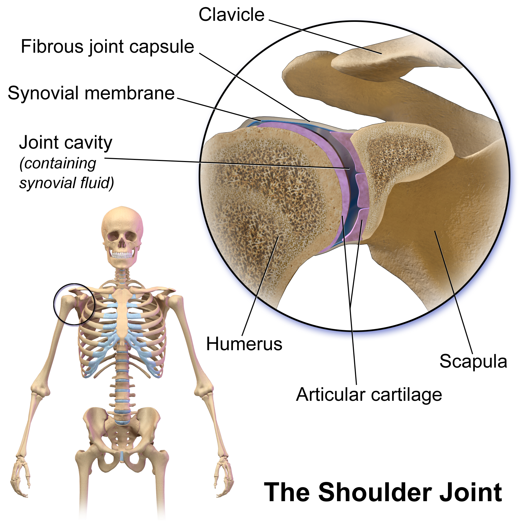
The human shoulder is made up of three bones: the clavicle (collarbone), the scapula (shoulder blade), and the humerus (upper arm bone) as well as associated muscles, ligaments and tendons. The articulations between the bones of the shoulder make up the shoulder joints. The shoulder joint, also known as the glenohumeral joint, is the major joint of the shoulder, but can more broadly include the acromioclavicular joint. In human anatomy, the shoulder joint comprises the part of the body where the humerus attaches to the scapula, and the head sits in the glenoid cavity. The shoulder is the group of structures in the region of the joint. The shoulder joint is the main joint of the shoulder. It is a ball and socket joint that allows the arm to rotate in a circular fashion or to hinge out and up away from the body. The joint capsule is a soft tissue envelope that encircles the glenohumeral joint and attaches to the scapula, humerus, and head of the biceps. It is lined by a ... [...More Info...] [...Related Items...] OR: [Wikipedia] [Google] [Baidu] [Amazon] |
Thyrohyoid Muscle
The thyrohyoid muscle is a small skeletal muscle of the neck. Above, it attaches onto the greater cornu of the hyoid bone; below, it attaches onto the oblique line of the thyroid cartilage. It is innervated by fibres derived from the cervical spinal nerve 1 that run with the Hypoglossal nerve, hypoglossal nerve (CN XII) to reach this muscle. The thyrohyoid muscle depresses the hyoid bone and elevates the larynx during swallowing. By controlling the position and shape of the larynx, it aids in making sound. Structure The thyrohyoid muscle is a small, broad and short muscle. It is quadrilateral in shape. It may be considered a superior-ward continuation of sternothyroid muscle. It belongs to the infrahyoid muscles group and the outer laryngeal muscle group. Attachments Its superior attachment is the inferior border of the greater cornu of the hyoid bone and adjacent portions of the body of hyoid bone. Its inferior attachment is the oblique line of the thyroid cartilage (alongs ... [...More Info...] [...Related Items...] OR: [Wikipedia] [Google] [Baidu] [Amazon] |
Sternothyroid Muscle
The sternothyroid muscle (or sternothyroideus) is an infrahyoid muscle of the neck. It acts to depress the hyoid bone. Structure The two muscles are in contact with each other proximally (close to their origin), but diverge distally (towards their insertions). Origin The sternothyroid arises from the posterior surface of the manubrium of the sternum from the midline to the notch for the first rib (inferior to the origin of the sternohyoid muscle), and the posterior margin of the first costal cartilage. Insertion It inserts onto the oblique line of the lamina of thyroid cartilage. Innervation The sternothyroid muscle receives motor innervation from branches of the ansa cervicalis (ultimately derived from cervical spinal nerves C1-C3). Relations The sternothyroid muscle is shorter and wider than the sternohyoid muscle and is situated deep to and partially medial to it. Variations The muscle may be absent or doubled. It may issue accessory slips to the thyrohyoid ... [...More Info...] [...Related Items...] OR: [Wikipedia] [Google] [Baidu] [Amazon] |
Muscular Triangle
The inferior carotid triangle (or muscular triangle), is bounded, in front, by the median line of the neck from the hyoid bone to the sternum; behind, by the anterior margin of the sternocleidomastoid; above, by the superior belly of the omohyoid. It is covered by the integument, superficial fascia, platysma, and deep fascia, ramifying in which are some of the branches of the supraclavicular nerves. Beneath these superficial structures are the sternohyoid and sternothyroid, which, together with the anterior margin of the sternocleidomastoid, conceal the lower part of the common carotid artery. This vessel is enclosed within its sheath, together with the internal jugular vein and vagus nerve; the vein lies lateral to the artery on the right side of the neck, but overlaps it below on the left side; the nerve lies between the artery and vein, on a plane posterior to both. In front of the sheath are a few descending filaments from the ansa cervicalis; behind the sheath are the i ... [...More Info...] [...Related Items...] OR: [Wikipedia] [Google] [Baidu] [Amazon] |
Carotid Triangle
The carotid triangle (or superior carotid triangle) is a portion of the anterior triangle of the neck. Anatomy Boundaries It is bounded: * Posteriorly by (the anterior border of) the sternocleidomastoid muscle, * Anteroinferiorly by (the superior belly of) the omohyoid muscle. * Superiorly by (the posterior belly of) the digastric muscle. Roof The roof is formed by: * Integument, * Superficial fascia, * Platysma, * Deep fascia. Floor The floor is formed by (parts of) the: * Thyrohyoid membrane, *Hyoglossus, * Constrictor pharyngis medius and constrictor pharyngis inferior muscles. Contents Arteries * Internal carotid artery * External carotid artery and some of its branches: ** Superior thyroid artery, ** Ascending pharyngeal artery, ** Lingual artery, ** Facial artery, ** Occipital artery. Veins * internal jugular vein and its tributaries (correspondng to the branches of the corresponding artery): ** Superior thyroid vein, ** Lingual veins, ** Comm ... [...More Info...] [...Related Items...] OR: [Wikipedia] [Google] [Baidu] [Amazon] |
Anterior Triangle
The anterior triangle is a region of the neck. Structure The triangle is inverted with its apex inferior to its base which is under the chin. Investing fascia covers the roof of the triangle while visceral fascia covers the floor. Anatomy Muscles: * Suprahyoid muscles - Digastric (Ant and post belly), mylohyoid, geniohyoid and stylohyoid. * Infrahyoid muscles - Omohyoid, sternohyoid, sternothyroid, and thyrohyoid. Nerve supply 2 Bellies of digastric * Anterior: Mylohyoid nerve * Posterior: Facial nerve Stylohyoid: by the facial nerve, by a branch from that to the posterior belly of digastric. Mylohyoid: by its own nerve, a branch of the inferior alveolar (from the mandibular division of trigeminal nerve), which arises just before the parent nerve enters the mandibular foramen, pierces the sphenomandibular ligament, and runs forward on the inferior surface of the mylohyoid, supplying it and the anterior belly of the digastric. Geniohyoid: by a branch from the hypoglossal n ... [...More Info...] [...Related Items...] OR: [Wikipedia] [Google] [Baidu] [Amazon] |
Posterior Triangle Of The Neck
The posterior triangle (or lateral cervical region) is a region of the neck. Boundaries The posterior triangle has the following boundaries: Apex: Union of the sternocleidomastoid and the trapezius muscles at the superior nuchal line of the occipital bone Anteriorly: Posterior border of the sternocleidomastoideus Posteriorly: Anterior border of the trapezius Inferiorly: Middle one third of the clavicle Roof: Investing layer of the deep cervical fascia Floor: (From superior to inferior) 1) M. semispinalis capitis 2) M. splenius capitis 3) M. levator scapulae 4) M. scalenus posterior 5) M. scalenus medius Divisions The posterior triangle is crossed, about 2.5 cm above the clavicle, by the inferior belly of the omohyoid muscle, which divides the space into two triangles: * an upper or occipital triangle * a lower or subclavian triangle (or supraclavicular triangle) Contents A) Nerves and plexuses: * Spinal accessory nerve (Cranial Nerve XI) * Branches of cervic ... [...More Info...] [...Related Items...] OR: [Wikipedia] [Google] [Baidu] [Amazon] |
Cervical Spinal Nerve 1
The cervical spinal nerve 1 (C1) is a spinal nerve of the cervical segment. from spinalcordinjuryzone.com. Published February 23, 2004 Archived Dec 23, 2011. Retrieved June 12, 2018. C1 carries predominantly motor fibres, but also a small meningeal branch that supplies sensation to parts of the dura around the foramen magnum (via dorsal rami). It originates from the spinal column from above the (C1). [...More Info...] [...Related Items...] OR: [Wikipedia] [Google] [Baidu] [Amazon] |

