|
Olecranon Fracture
Olecranon fracture is a fracture of the bony portion of the elbow. The injury is fairly common and often occurs following a fall or direct trauma to the elbow. The olecranon is the proximal extremity of the ulna which is articulated with the humerus bone and constitutes a part of the elbow articulation. Its location makes it vulnerable to direct trauma. Signs and symptoms People with olecranon fractures present with intense elbow pain after a direct blow or fall. Swelling over the bone site is seen and an inability to straighten the elbow is common. Due to the proximity of the olecranon to the ulnar nerve, the injury and swelling may cause numbness and tingling at the fourth and fifth fingers. Examination can bring out a palpable defect at the site of the fracture. Mechanism Olecranon fractures are common. Typically they are caused by direct blows to the elbow (e.g. motor vehicle accidents), and due to falls when the triceps are contracted. "Side-swipe" injury when driving a ... [...More Info...] [...Related Items...] OR: [Wikipedia] [Google] [Baidu] |
Bone Fracture
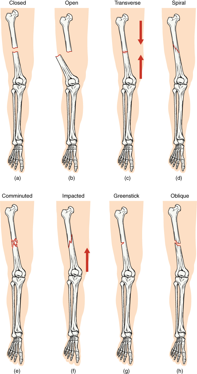
A bone fracture (abbreviated FRX or Fx, Fx, or #) is a medical condition in which there is a partial or complete break in the continuity of any bone in the body. In more severe cases, the bone may be broken into several fragments, known as a ''comminuted fracture''. A bone fracture may be the result of high force impact or stress, or a minimal trauma injury as a result of certain medical conditions that weaken the bones, such as osteoporosis, osteopenia, bone cancer, or osteogenesis imperfecta, where the fracture is then properly termed a pathologic fracture. Signs and symptoms Although bone tissue contains no pain receptors, a bone fracture is painful for several reasons: * Breaking in the continuity of the periosteum, with or without similar discontinuity in endosteum, as both contain multiple pain receptors. * Edema and hematoma of nearby soft tissues caused by ruptured bone marrow evokes pressure pain. * Involuntary muscle spasms trying to hold bone fragments in place. D ... [...More Info...] [...Related Items...] OR: [Wikipedia] [Google] [Baidu] |
X-ray
An X-ray, or, much less commonly, X-radiation, is a penetrating form of high-energy electromagnetic radiation. Most X-rays have a wavelength ranging from 10 picometers to 10 nanometers, corresponding to frequencies in the range 30 petahertz to 30 exahertz ( to ) and energies in the range 145 eV to 124 keV. X-ray wavelengths are shorter than those of UV rays and typically longer than those of gamma rays. In many languages, X-radiation is referred to as Röntgen radiation, after the German scientist Wilhelm Conrad Röntgen, who discovered it on November 8, 1895. He named it ''X-radiation'' to signify an unknown type of radiation.Novelline, Robert (1997). ''Squire's Fundamentals of Radiology''. Harvard University Press. 5th edition. . Spellings of ''X-ray(s)'' in English include the variants ''x-ray(s)'', ''xray(s)'', and ''X ray(s)''. The most familiar use of X-rays is checking for fractures (broken bones), but X-rays are also used in other ways. ... [...More Info...] [...Related Items...] OR: [Wikipedia] [Google] [Baidu] |
Elbow
The elbow is the region between the arm and the forearm that surrounds the elbow joint. The elbow includes prominent landmarks such as the olecranon, the cubital fossa (also called the chelidon, or the elbow pit), and the lateral and the medial epicondyles of the humerus. The elbow joint is a hinge joint between the arm and the forearm; more specifically between the humerus in the upper arm and the radius and ulna in the forearm which allows the forearm and hand to be moved towards and away from the body. The term ''elbow'' is specifically used for humans and other primates, and in other vertebrates forelimb plus joint is used. The name for the elbow in Latin is ''cubitus'', and so the word cubital is used in some elbow-related terms, as in ''cubital nodes'' for example. Structure Joint The elbow joint has three different portions surrounded by a common joint capsule. These are joints between the three bones of the elbow, the humerus of the upper arm, and the radius and ... [...More Info...] [...Related Items...] OR: [Wikipedia] [Google] [Baidu] |
Bone Fractures
A bone fracture (abbreviated FRX or Fx, Fx, or #) is a medical condition in which there is a partial or complete break in the continuity of any bone in the body. In more severe cases, the bone may be broken into several fragments, known as a ''comminuted fracture''. A bone fracture may be the result of high force impact or stress, or a minimal trauma injury as a result of certain medical conditions that weaken the bones, such as osteoporosis, osteopenia, bone cancer, or osteogenesis imperfecta, where the fracture is then properly termed a pathologic fracture. Signs and symptoms Although bone tissue contains no pain receptors, a bone fracture is painful for several reasons: * Breaking in the continuity of the periosteum, with or without similar discontinuity in endosteum, as both contain multiple pain receptors. * Edema and hematoma of nearby soft tissues caused by ruptured bone marrow evokes pressure pain. * Involuntary muscle spasms trying to hold bone fragments in place. ... [...More Info...] [...Related Items...] OR: [Wikipedia] [Google] [Baidu] |
Kirschner Wire
Kirschner wires or K-wires or pins are sterilized, sharpened, smooth stainless steel pins. Introduced in 1909 by Martin Kirschner, the wires are now widely used in orthopedics and other types of medical and veterinary surgery. They come in different sizes and are used to hold bone fragments together (pin fixation) or to provide an anchor for skeletal traction. The pins are often driven into the bone through the skin (percutaneous pin fixation) using a power or hand drill. They also form part of the Ilizarov apparatus. Variations * Threaded K-wires are available. Used in situations where backing out of the pin is undesirable, they are inherently weaker than smooth K-wires. * "Denham Pins" are strong, stout wires with a threaded portion at the midpoint. They are used for skeletal traction, with the threads engaging the bone. This pin was invented in 1956 by the English orthopedic surgeon Robert Arthur Denham (born 1922). Indications * K-wires are used for temporary fixation dur ... [...More Info...] [...Related Items...] OR: [Wikipedia] [Google] [Baidu] |
Fracture Of Olecranon Pre And Post Typical Surgery
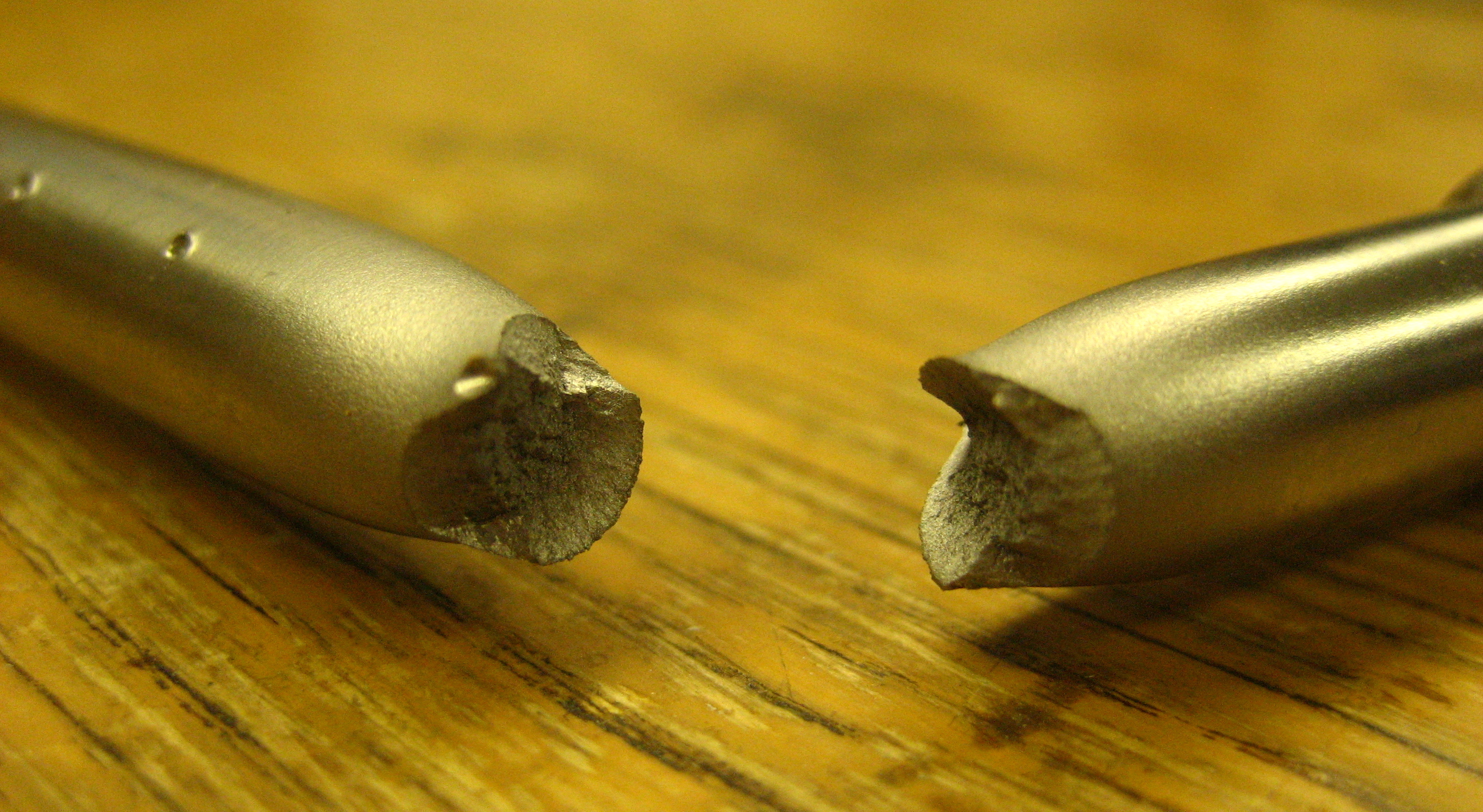
Fracture is the separation of an object or material into two or more pieces under the action of stress. The fracture of a solid usually occurs due to the development of certain displacement discontinuity surfaces within the solid. If a displacement develops perpendicular to the surface, it is called a normal tensile crack or simply a crack; if a displacement develops tangentially, it is called a shear crack, slip band or dislocation. Brittle fractures occur with no apparent deformation before fracture. Ductile fractures occur after visible deformation. Fracture strength, or breaking strength, is the stress when a specimen fails or fractures. The detailed understanding of how a fracture occurs and develops in materials is the object of fracture mechanics. Strength Fracture strength, also known as breaking strength, is the stress at which a specimen fails via fracture. This is usually determined for a given specimen by a tensile test, which charts the stress–strain cur ... [...More Info...] [...Related Items...] OR: [Wikipedia] [Google] [Baidu] |
Chris Colton
Christopher Lewis Colton (born in September 1937) is an English orthopaedic surgeon and Professor Emeritus in Orthopaedic and Accident Surgery at the University of Nottingham. He is a past president of both the British Orthopaedic Association and of the AO Foundation. Training and early career Colton qualified in medicine and surgery in 1960, studying at St Thomas's Hospital Medical School in London. He became a Fellow of the Royal College of Surgeons of England in 1963. He pursued a career as an orthopaedic surgeon, studying in Bristol, at the Royal National Orthopaedic Hospital in London, and at Dala Orthopaedic Hospital at Kano in Northern Nigeria during the Biafran civil war. Surgical career He was appointed as a Consultant Orthopaedic Surgeon in Nottingham in 1973. He was awarded an honorary title in Orthopaedic and Accident Surgery by the University of Nottingham in 1993, in recognition of his research and teaching in musculoskeletal trauma. He served as president of the Br ... [...More Info...] [...Related Items...] OR: [Wikipedia] [Google] [Baidu] |
Comminuted Fracture
A bone fracture (abbreviated FRX or Fx, Fx, or #) is a medical condition in which there is a partial or complete break in the continuity of any bone in the body. In more severe cases, the bone may be broken into several fragments, known as a ''comminuted fracture''. A bone fracture may be the result of high force impact or stress, or a minimal trauma injury as a result of certain medical conditions that weaken the bones, such as osteoporosis, osteopenia, bone cancer, or osteogenesis imperfecta, where the fracture is then properly termed a pathologic fracture. Signs and symptoms Although bone tissue contains no pain receptors, a bone fracture is painful for several reasons: * Breaking in the continuity of the periosteum, with or without similar discontinuity in endosteum, as both contain multiple pain receptors. * Edema and hematoma of nearby soft tissues caused by ruptured bone marrow evokes pressure pain. * Involuntary muscle spasms trying to hold bone fragments in place ... [...More Info...] [...Related Items...] OR: [Wikipedia] [Google] [Baidu] |
Olecranon Fracture
Olecranon fracture is a fracture of the bony portion of the elbow. The injury is fairly common and often occurs following a fall or direct trauma to the elbow. The olecranon is the proximal extremity of the ulna which is articulated with the humerus bone and constitutes a part of the elbow articulation. Its location makes it vulnerable to direct trauma. Signs and symptoms People with olecranon fractures present with intense elbow pain after a direct blow or fall. Swelling over the bone site is seen and an inability to straighten the elbow is common. Due to the proximity of the olecranon to the ulnar nerve, the injury and swelling may cause numbness and tingling at the fourth and fifth fingers. Examination can bring out a palpable defect at the site of the fracture. Mechanism Olecranon fractures are common. Typically they are caused by direct blows to the elbow (e.g. motor vehicle accidents), and due to falls when the triceps are contracted. "Side-swipe" injury when driving a ... [...More Info...] [...Related Items...] OR: [Wikipedia] [Google] [Baidu] |
Olecranon
The olecranon (, ), is a large, thick, curved bony eminence of the ulna, a long bone in the forearm that projects behind the elbow. It forms the most pointed portion of the elbow and is opposite to the cubital fossa or elbow pit. The olecranon serves as a lever for the extensor muscles that straighten the elbow joint. Structure The olecranon is situated at the proximal end of the ulna, one of the two bones in the forearm. When the hand faces forward (supination) the olecranon faces towards the back (posteriorly). It is bent forward at the summit so as to present a prominent lip which is received into the olecranon fossa of the humerus during extension of the forearm. Its base is contracted where it joins the body and the narrowest part of the upper end of the ulna. Its posterior surface, directed backward, is triangular, smooth, subcutaneous, and covered by a bursa. Its superior surface is of quadrilateral form, marked behind by a rough impression for the insertion of the Tr ... [...More Info...] [...Related Items...] OR: [Wikipedia] [Google] [Baidu] |
Avulsion Fracture
An avulsion fracture is a bone fracture which occurs when a fragment of bone tears away from the main mass of bone as a result of physical trauma. This can occur at the ligament by the application of forces external to the body (such as a fall or pull) or at the tendon by a muscular contraction that is stronger than the forces holding the bone together. Generally muscular avulsion is prevented by the neurological limitations placed on muscle contractions. Highly trained athletes can overcome this neurological inhibition of strength and produce a much greater force output capable of breaking or avulsing a bone. Types Dental avulsion Traumatic complete displacement of a tooth from its socket in alveolar bone. It is a serious dental emergency in which prompt management (within 20–40 minutes of injury) affects the prognosis of the tooth. Tuberosity avulsion of the 5th metatarsal left, Proximal fractures of 5th metatarsa The tuberosity avulsion fracture (also known as pseud ... [...More Info...] [...Related Items...] OR: [Wikipedia] [Google] [Baidu] |
Triceps Brachii Muscle
The triceps, or triceps brachii (Latin for "three-headed muscle of the arm"), is a large muscle on the back of the upper limb of many vertebrates. It consists of 3 parts: the medial, lateral, and long head. It is the muscle principally responsible for extension of the elbow joint (straightening of the arm). Structure The long head arises from the infraglenoid tubercle of the scapula. It extends distally anterior to the teres minor and posterior to the teres major. The medial head arises proximally in the humerus, just inferior to the groove of the radial nerve; from the dorsal (back) surface of the humerus; from the medial intermuscular septum; and its distal part also arises from the lateral intermuscular septum. The medial head is mostly covered by the lateral and long heads, and is only visible distally on the humerus. The lateral head arises from the dorsal surface of the humerus, lateral and proximal to the groove of the radial nerve, from the greater tubercle down to ... [...More Info...] [...Related Items...] OR: [Wikipedia] [Google] [Baidu] |


