|
Occipital Artery
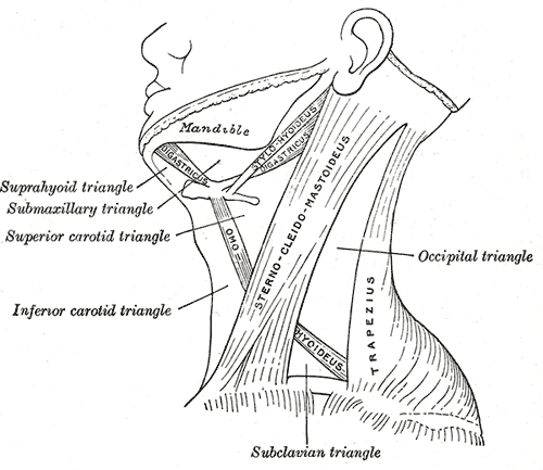
The occipital artery arises from the external carotid artery opposite the facial artery. Its path is below the posterior belly of digastric to the occipital region. This artery supplies blood to the back of the scalp and sternocleidomastoid muscles, and deep muscles in the back and neck. Structure At its origin, it is covered by the posterior belly of the digastricus and the stylohyoideus, and the hypoglossal nerve winds around it from behind forward; higher up, it crosses the internal carotid artery, the internal jugular vein, and the vagus and accessory nerves. It next ascends to the interval between the transverse process of the atlas and the mastoid process of the temporal bone, and passes horizontally backward, grooving the surface of the latter bone, being covered by the sternocleidomastoideus, splenius capitis, longissimus capitis, and digastricus, and resting upon the rectus capitis lateralis, the obliquus superior, and semispinalis capitis. It then changes its course and ... [...More Info...] [...Related Items...] OR: [Wikipedia] [Google] [Baidu] |
External Carotid Artery
The external carotid artery is a major artery of the head and neck. It arises from the common carotid artery when it splits into the external and internal carotid artery. External carotid artery supplies blood to the face and neck. Structure The external carotid artery begins at the upper border of thyroid cartilage, and curves, passing forward and upward, and then inclining backward to the space behind the neck of the mandible, where it divides into the superficial temporal and maxillary artery within the parotid gland. It rapidly diminishes in size as it travels up the neck, owing to the number and large size of its branches. At its origin, this artery is closer to the skin and more medial than the internal carotid, and is situated within the carotid triangle. Development In children, the external carotid artery is somewhat smaller than the internal carotid; but in the adult, the two vessels are of nearly equal size. Relations At the origin, external carotid artery is mo ... [...More Info...] [...Related Items...] OR: [Wikipedia] [Google] [Baidu] |
Sternocleidomastoideus
The sternocleidomastoid muscle is one of the largest and most superficial cervical muscles. The primary actions of the muscle are rotation of the head to the opposite side and flexion of the neck. The sternocleidomastoid is innervated by the accessory nerve. Etymology and location It is given the name ''sternocleidomastoid'' because it originates at the manubrium of the sternum (''sterno-'') and the clavicle (''cleido-'') and has an insertion at the mastoid process of the temporal bone of the skull. Structure The sternocleidomastoid muscle originates from two locations: the manubrium of the sternum and the clavicle. It travels obliquely across the side of the neck and inserts at the mastoid process of the temporal bone of the skull by a thin aponeurosis. The sternocleidomastoid is thick and narrow at its centre, and broader and thinner at either end. The sternal head is a round fasciculus, tendinous in front, fleshy behind, arising from the upper part of the front of the manubri ... [...More Info...] [...Related Items...] OR: [Wikipedia] [Google] [Baidu] |
Longus Capitis
The longus capitis muscle (Latin for ''long muscle of the head'', alternatively rectus capitis anticus major), is broad and thick above, narrow below, and arises by four tendinous slips, from the anterior tubercles of the transverse processes of the third, fourth, fifth, and sixth cervical vertebræ, and ascends, converging toward its fellow of the opposite side, to be inserted into the inferior surface of the basilar part of the occipital bone The occipital bone () is a neurocranium, cranial dermal bone and the main bone of the occiput (back and lower part of the skull). It is trapezoidal in shape and curved on itself like a shallow dish. The occipital bone overlies the occipital lobe .... It is innervated by a branch of cervical plexus. Longus capitis has several actions: acting unilaterally, to: *flex the head and neck laterally *rotate the head ipsilaterally acting bilaterally: *flex the head and neck Additional images File:Gray129.png, Occipital bone. Outer surface. ... [...More Info...] [...Related Items...] OR: [Wikipedia] [Google] [Baidu] |
Splenius
The splenius muscles are: *Splenius capitis muscle *Splenius cervicis muscle Their origins are in the upper thoracic and lower cervical spinous process The spinal column, a defining synapomorphy shared by nearly all vertebrates,Hagfish are believed to have secondarily lost their spinal column is a moderately flexible series of vertebrae (singular vertebra), each constituting a characteristic i ...es. Their actions are to extend and ipsilaterally rotate the head and neck. References Muscles of the torso {{Muscle-stub ... [...More Info...] [...Related Items...] OR: [Wikipedia] [Google] [Baidu] |
Stylohyoid
The stylohyoid muscle is a slender muscle, lying anterior and superior of the posterior belly of the digastric muscle. It is one of the suprahyoid muscles. It shares this muscle's innervation by the facial nerve, and functions to draw the hyoid bone backwards and elevate the tongue. Its origin is the styloid process of the temporal bone. It inserts on the body of the hyoid. Structure The stylohyoid muscle originates from the posterior and lateral surface of the styloid process of the temporal bone, near the base. Passing inferior and anterior, it inserts into the body of the hyoid bone, at its junction with the greater cornu, and just superior to the omohyoid muscle. It belongs to the group of suprahyoid muscles. It is perforated, near its insertion, by the intermediate tendon of the digastric muscle. The stylohyoid muscle has vascular supply from the lingual artery, a branch of the external carotid artery. Nerve supply A branch of the facial nerve (CN VII) innervates the st ... [...More Info...] [...Related Items...] OR: [Wikipedia] [Google] [Baidu] |
Superficial Temporal
In human anatomy, the superficial temporal artery is a major artery of the head. It arises from the external carotid artery when it splits into the superficial temporal artery and maxillary artery. Its pulse can be felt above the zygomatic arch, above and in front of the tragus of the ear. Structure The superficial temporal artery is the smaller of two end branches that split superiorly from the external carotid. Based on its direction, the superficial temporal artery appears to be a continuation of the external carotid. It begins within the parotid gland, behind the neck of the mandible, and passes superficially over the posterior root of the zygomatic process of the temporal bone; about 5 cm above this process it divides into two branches: ''a. frontal'', and ''a. parietal''. Branches The parietal branch of the superficial temporal artery (posterior temporal) is a small artery in the head. It is larger than the frontal branch and curves upward and backward on the side ... [...More Info...] [...Related Items...] OR: [Wikipedia] [Google] [Baidu] |
Posterior Auricular Artery
The posterior auricular artery is a small artery that arises from the external carotid artery, above the digastric muscle and stylohyoid muscle, opposite the apex of the styloid process. It ascends posteriorly beneath the parotid gland, along the styloid process of the temporal bone, between the cartilage of the ear and the mastoid process of the temporal bone along the lateral side of the head. The posterior auricular artery gives off the stylomastoid artery, small branches to the auricle, and supplies blood to the scalp posterior to the auricle. A person may be able to "hear" their own heart rate via this artery, under certain conditions. See also * Anterior auricular branches of superficial temporal artery The anterior auricular branches of the superficial temporal artery are distributed to the anterior portion of the auricula, the lobule, and part of the external meatus, anastomosing with the posterior auricular. They supply the external acousti ... * Posterior auric ... [...More Info...] [...Related Items...] OR: [Wikipedia] [Google] [Baidu] |
Anastomose
An anastomosis (, plural anastomoses) is a connection or opening between two things (especially cavities or passages) that are normally diverging or branching, such as between blood vessels, leaf veins, or streams. Such a connection may be normal (such as the foramen ovale in a fetus's heart) or abnormal (such as the patent foramen ovale in an adult's heart); it may be acquired (such as an arteriovenous fistula) or innate (such as the arteriovenous shunt of a metarteriole); and it may be natural (such as the aforementioned examples) or artificial (such as a surgical anastomosis). The reestablishment of an anastomosis that had become blocked is called a reanastomosis. Anastomoses that are abnormal, whether congenital or acquired, are often called fistulas. The term is used in medicine, biology, mycology, geology, and geography. Etymology Anastomosis: medical or Modern Latin, from Greek ἀναστόμωσις, anastomosis, "outlet, opening", Gr ana- "up, on, upon", stoma "mouth", ... [...More Info...] [...Related Items...] OR: [Wikipedia] [Google] [Baidu] |
Vertex (anatomy)
In arthropod and vertebrate anatomy, the vertex (or ''cranial vertex'') is the highest point of the head. In humans, the vertex is formed by four bones of the skull: the frontal bone, the two parietal bones, and the occipital bone. These bones are connected by the coronal suture between the frontal and parietal bones, the sagittal suture between the two parietal bones, and the lambdoid suture between the parietal and occipital bones. ''Vertex baldness'' refers to a form of male pattern baldness in which the baldness is limited to the vertex, resembling a tonsure. In childbirth, ''vertex birth'' refers to the common head-first presentation of the baby, as opposed to the buttocks-first position of a breech birth. In entomology, the color and shape of an insect's vertex and the structures arising from it are commonly used in identifying species. See also *Calvaria (skull) *Crown (anatomy) The crown is the top portion of the head behind the vertex. The anatomy of the crown varies ... [...More Info...] [...Related Items...] OR: [Wikipedia] [Google] [Baidu] |
Trapezius
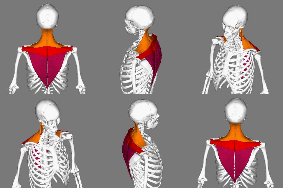
The trapezius is a large paired trapezoid-shaped surface muscle that extends longitudinally from the occipital bone to the lower thoracic vertebrae of the spine and laterally to the spine of the scapula. It moves the scapula and supports the arm. The trapezius has three functional parts: an upper (descending) part which supports the weight of the arm; a middle region (transverse), which retracts the scapula; and a lower (ascending) part which medially rotates and depresses the scapula. Name and history The trapezius muscle resembles a trapezium, also known as a trapezoid, or diamond-shaped quadrilateral. The word "spinotrapezius" refers to the human trapezius, although it is not commonly used in modern texts. In other mammals, it refers to a portion of the analogous muscle. Similarly, the term "tri-axle back plate" was historically used to describe the trapezius muscle. Structure The ''superior'' or ''upper'' (or descending) fibers of the trapezius originate from the sp ... [...More Info...] [...Related Items...] OR: [Wikipedia] [Google] [Baidu] |
Semispinalis Capitis
The semispinalis muscles are a group of three muscles belonging to the transversospinales. These are the semispinalis capitis, the semispinalis cervicis and the semispinalis thoracis. The semispinalis capitis (''complexus'') is situated at the upper and back part of the neck, deep to the splenius, and medial to the longissimus cervicis and longissimus capitis. It arises by a series of tendons from the tips of the transverse processes of the upper six or seven thoracic and the seventh cervical vertebrae, and from the articular processes of the three cervical vertebrae above this ( C4-C6). The tendons, uniting, form a broad muscle, which passes upward, and is inserted between the superior and inferior nuchal lines of the occipital bone. It lies deep to the trapezius muscle and can be palpated as a firm round muscle mass just lateral to the cervical spinous processes. The semispinalis cervicis (or ''semispinalis colli''), arises by a series of tendinous and fleshy fibers from the t ... [...More Info...] [...Related Items...] OR: [Wikipedia] [Google] [Baidu] |
Obliquus Superior
The superior oblique muscle, or obliquus oculi superior, is a fusiform muscle originating in the upper, medial side of the orbit (i.e. from beside the nose) which abducts, depresses and internally rotates the eye. It is the only extraocular muscle innervated by the trochlear nerve (the fourth cranial nerve). Structure The superior oblique muscle loops through a pulley-like structure (the trochlea of superior oblique) and inserts into the sclera on the posterotemporal surface of the eyeball. It is the pulley system that gives superior oblique its actions, causing depression of the eyeball despite being inserted on the superior surface. The superior oblique arises immediately above the margin of the optic foramen, superior and medial to the origin of the superior rectus, and, passing forward, ends in a rounded tendon, which plays in a fibrocartilaginous ring or pulley attached to the trochlear fossa of the frontal bone. The contiguous surfaces of the tendon and ring are lined by ... [...More Info...] [...Related Items...] OR: [Wikipedia] [Google] [Baidu] |


