|
Obturator Internus Muscle
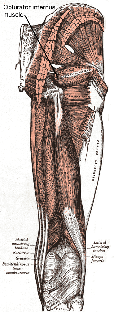
The internal obturator muscle or obturator internus muscle originates on the medial surface of the obturator membrane, the ischium near the membrane, and the rim of the pubis. It exits the pelvic cavity through the lesser sciatic foramen. The internal obturator is situated partly within the lesser pelvis, and partly at the back of the hip-joint. It functions to help laterally rotate femur with hip extension and abduct femur with hip flexion, as well as to steady the femoral head in the acetabulum. Structure Origin The internal obturator muscle arises from the inner surface of the antero-lateral wall of the pelvis. It surrounds the obturator foramen. It is attached to the inferior pubic ramus and ischium, and at the side to the inner surface of the hip bone below and behind the pelvic brim. It reaches from the upper part of the greater sciatic foramen above and behind to the obturator foramen below and in front. It also arises from the pelvic surface of the obturator membr ... [...More Info...] [...Related Items...] OR: [Wikipedia] [Google] [Baidu] |
Ischiopubic Ramus
The ischiopubic ramus is a compound structure consisting of the following two structures: * from the pubis, the inferior pubic ramus * from the ischium, the inferior ramus of the ischium It forms the inferior border of the obturator foramen and serves as part of the origin for the obturator internus The internal obturator muscle or obturator internus muscle originates on the medial surface of the obturator membrane, the ischium near the membrane, and the rim of the pubis. It exits the pelvic cavity through the lesser sciatic foramen. The i ... and externus muscles. Also, most adductors originate at the ischiopubic ramus. The fascia of Colles is attached to its margin. References External links * - "The Female Perineum" * * (, ) Bones of the pelvis {{musculoskeletal-stub ... [...More Info...] [...Related Items...] OR: [Wikipedia] [Google] [Baidu] |
Greater Sciatic Foramen
The greater sciatic foramen is an opening (foramen) in the posterior human pelvis. It is formed by the sacrotuberous and sacrospinous ligaments. The piriformis muscle passes through the foramen and occupies most of its volume. The greater sciatic foramen is wider in women than in men. Structure It is bounded as follows: * anterolaterally by the greater sciatic notch of the ilium. * posteromedially by the sacrotuberous ligament. * inferiorly by the sacrospinous ligament and the ischial spine. * superiorly by the anterior sacroiliac ligament. Function The piriformis The piriformis muscle () is a flat, pyramidally-shaped muscle in the gluteal region of the lower limbs. It is one of the six muscles in the lateral rotator group. The piriformis muscle has its origin upon the front surface of the sacrum, and in ..., which exits the pelvis through the foramen, occupies most of its volume. The following structures also exit the pelvis through the greater sciatic foramen: S ... [...More Info...] [...Related Items...] OR: [Wikipedia] [Google] [Baidu] |
Bursa (anatomy)
( grc-gre, Προῦσα, Proûsa, Latin: Prusa, ota, بورسه, Arabic:بورصة) is a city in northwestern Turkey and the administrative center of Bursa Province. The fourth-most populous city in Turkey and second-most populous in the Marmara Region, Bursa is one of the industrial centers of the country. Most of Turkey's automotive production takes place in Bursa. As of 2019, the Metropolitan Province was home to 3,056,120 inhabitants, 2,161,990 of whom lived in the 3 city urban districts (Osmangazi, Yildirim and Nilufer) plus Gursu and Kestel, largely conurbated. Bursa was the first major and second overall capital of the Ottoman State between 1335 and 1363. The city was referred to as (, meaning "God's Gift" in Ottoman Turkish, a name of Persian origin) during the Ottoman period, while a more recent nickname is ("") in reference to the parks and gardens located across its urban fabric, as well as to the vast and richly varied forests of the surrounding region ... [...More Info...] [...Related Items...] OR: [Wikipedia] [Google] [Baidu] |
Obturator Internus Muscle
The internal obturator muscle or obturator internus muscle originates on the medial surface of the obturator membrane, the ischium near the membrane, and the rim of the pubis. It exits the pelvic cavity through the lesser sciatic foramen. The internal obturator is situated partly within the lesser pelvis, and partly at the back of the hip-joint. It functions to help laterally rotate femur with hip extension and abduct femur with hip flexion, as well as to steady the femoral head in the acetabulum. Structure Origin The internal obturator muscle arises from the inner surface of the antero-lateral wall of the pelvis. It surrounds the obturator foramen. It is attached to the inferior pubic ramus and ischium, and at the side to the inner surface of the hip bone below and behind the pelvic brim. It reaches from the upper part of the greater sciatic foramen above and behind to the obturator foramen below and in front. It also arises from the pelvic surface of the obturator membr ... [...More Info...] [...Related Items...] OR: [Wikipedia] [Google] [Baidu] |
Sacral Spinal Nerve 2
The sacral spinal nerve 2 (S2) is a spinal nerve of the sacral segment. Nervous System -- Groups of Nerves It originates from the from below the 2nd body of the 
Muscles S2 supplies many muscles, either directly or through nerves originating from S2. They are not innervated with S2 as single origin, but partly by S2 and partly by other spinal nerves. They are most ...[...More Info...] [...Related Items...] OR: [Wikipedia] [Google] [Baidu] |
Sacral Spinal Nerve 1
The sacral spinal nerve 1 (S1) is a spinal nerve of the sacral segment. Nervous System -- Groups of Nerves It originates from the from below the 1st body of the 
Muscles S1 supplies many muscles, either directly or through nerves originating from S1. They are not innervated with S1 as single origin, but partly by S1 and partly by other spinal nerves. The muscles a ...[...More Info...] [...Related Items...] OR: [Wikipedia] [Google] [Baidu] |
Lumbar Spinal Nerve 5
The lumbar nerves are the five pairs of spinal nerves emerging from the lumbar vertebrae. They are divided into posterior and anterior divisions. Structure The lumbar nerves are five spinal nerves which arise from either side of the spinal cord below the thoracic spinal cord and above the sacral spinal cord. They arise from the spinal cord between each pair of lumbar spinal vertebrae and travel through the intervertebral foramina. The nerves then split into an anterior branch, which travels forward, and a posterior branch, which travels backwards and supplies the area of the back. Posterior divisions The middle divisions of the posterior branches run close to the articular processes of the vertebrae and end in the multifidus muscle. The outer branches supply the erector spinae muscles. The nerves give off branches to the skin. These pierce the aponeurosis of the greater trochanter. Anterior divisions The anterior divisions of the lumbar nerves ( la, rami anteriores) increas ... [...More Info...] [...Related Items...] OR: [Wikipedia] [Google] [Baidu] |
Obturator Internus Nerve
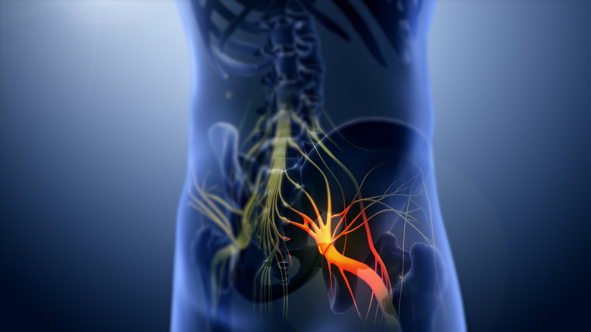
The nerve to obturator internus, also known as the obturator internus nerve, is a nerve that innervates the obturator internus and gemellus superior muscles. Structure The nerve to obturator internus originates in the lumbosacral plexus. It arises from the ventral divisions of the fifth lumbar and first and second sacral nerves. It leaves the pelvis through the greater sciatic foramen below the piriformis muscle, and gives off the branch to the gemellus superior, which enters the upper part of the posterior surface of the muscle. It then crosses the ischial spine, re-enters the pelvis through the lesser sciatic foramen, and pierces the pelvic surface of the obturator internus. See also * Obturator nerve * Nerve to quadratus femoris The nerve to quadratus femoris is a nerve that provides innervation to the quadratus femoris muscle and gemellus inferior muscle. Structure The nerve to quadratus femoris is a sacral plexus nerve. It arises from the ventral divisions of th ... [...More Info...] [...Related Items...] OR: [Wikipedia] [Google] [Baidu] |
Femur
The femur (; ), or thigh bone, is the proximal bone of the hindlimb in tetrapod vertebrates. The head of the femur articulates with the acetabulum in the pelvic bone forming the hip joint, while the distal part of the femur articulates with the tibia (shinbone) and patella (kneecap), forming the knee joint. By most measures the two (left and right) femurs are the strongest bones of the body, and in humans, the largest and thickest. Structure The femur is the only bone in the upper leg. The two femurs converge medially toward the knees, where they articulate with the proximal ends of the tibiae. The angle of convergence of the femora is a major factor in determining the femoral-tibial angle. Human females have thicker pelvic bones, causing their femora to converge more than in males. In the condition ''genu valgum'' (knock knee) the femurs converge so much that the knees touch one another. The opposite extreme is ''genu varum'' (bow-leggedness). In the general popu ... [...More Info...] [...Related Items...] OR: [Wikipedia] [Google] [Baidu] |
Sciatic Nerve
The sciatic nerve, also called the ischiadic nerve, is a large nerve in humans and other vertebrate animals which is the largest branch of the sacral plexus and runs alongside the hip joint and down the lower limb. It is the longest and widest single nerve in the human body, going from the top of the leg to the foot on the posterior aspect. The sciatic nerve has no cutaneous branches for the thigh. This nerve provides the connection to the nervous system for the skin of the lateral leg and the whole foot, the muscles of the back of the thigh, and those of the leg and foot. It is derived from spinal nerves L4 to S3. It contains fibers from both the anterior and posterior divisions of the lumbosacral plexus. Structure In humans, the sciatic nerve is formed from the L4 to S3 segments of the sacral plexus, a collection of nerve fibres that emerge from the sacral part of the spinal cord. The lumbosacral trunk from the L4 and L5 roots descends between the sacral promontory and al ... [...More Info...] [...Related Items...] OR: [Wikipedia] [Google] [Baidu] |
Coccygeus Muscle
The coccygeus muscle or ischiococcygeus is a muscle of the pelvic floor, located posterior to levator ani and anterior to the sacrospinous ligament. Structure The coccygeus muscle is posterior to levator ani and anterior to the sacrospinous ligament in the pelvic floor. It is a triangular plane of muscular and tendinous fibers. It arises by its apex from the spine of the ischium and sacrospinous ligament. It is inserted by its base into the margin of the coccyx and into the side of the lowest piece of the sacrum. In combination with the levator ani, it forms the pelvic diaphragm. The pudendal nerve runs between the coccygeus muscle and the piriformis muscle, superficial to the coccygeus muscle. Nerve supply The coccygeus muscle is innervated by the pudendal nerve, which runs between it and the piriformis muscle. Function The coccygeus muscle assists the levator ani and piriformis muscle in closing in the back part of the outlet of the pelvis. This helps to ... [...More Info...] [...Related Items...] OR: [Wikipedia] [Google] [Baidu] |
Pudendal Nerve
The pudendal nerve is the main nerve of the perineum. It carries sensation from the external genitalia of both sexes and the skin around the anus and perineum, as well as the motor supply to various pelvic muscles, including the male or female external urethral sphincter and the external anal sphincter. If damaged, most commonly by childbirth, lesions may cause sensory loss or fecal incontinence. The nerve may be temporarily blocked as part of an anaesthetic procedure. The pudendal canal that carries the pudendal nerve is also known by the eponymous term "Alcock's canal", after Benjamin Alcock, an Irish anatomist who documented the canal in 1836. Structure The pudendal nerve is paired, meaning there are two nerves, one on the left and one on the right side of the body. Each is formed as three roots immediately converge above the upper border of the sacrotuberous ligament and the coccygeus muscle. The three roots become two cords when the middle and lower root join to ... [...More Info...] [...Related Items...] OR: [Wikipedia] [Google] [Baidu] |


