|
Ortolani Maneuver
The Ortolani test is part of the physical examination for developmental dysplasia of the hip, along with the Barlow maneuver. Specifically, the Ortolani test is positive when a posterior dislocation of the hip is reducible with this maneuver. This is part of the standard infant exam performed preferably in early infancy.The Ortolani test is named after Marino Ortolani, who developed it in 1937. Procedure The Ortolani test is performed with the Barlow maneuver and inspection of the hip joint and legs. It relocates the dislocation of the hip joint that has just been elicited by the Barlow maneuver. The Ortolani test is performed by an examiner first flexing the hips and knees of a supine infant to 90°, then with the examiner's index fingers placing anterior pressure on the greater trochanters, gently and smoothly abducting the infant's legs using the examiner's thumbs. Interpretation A positive sign is a distinctive 'clunk' which can be heard and felt as the femoral head relo ... [...More Info...] [...Related Items...] OR: [Wikipedia] [Google] [Baidu] |
Physical Examination
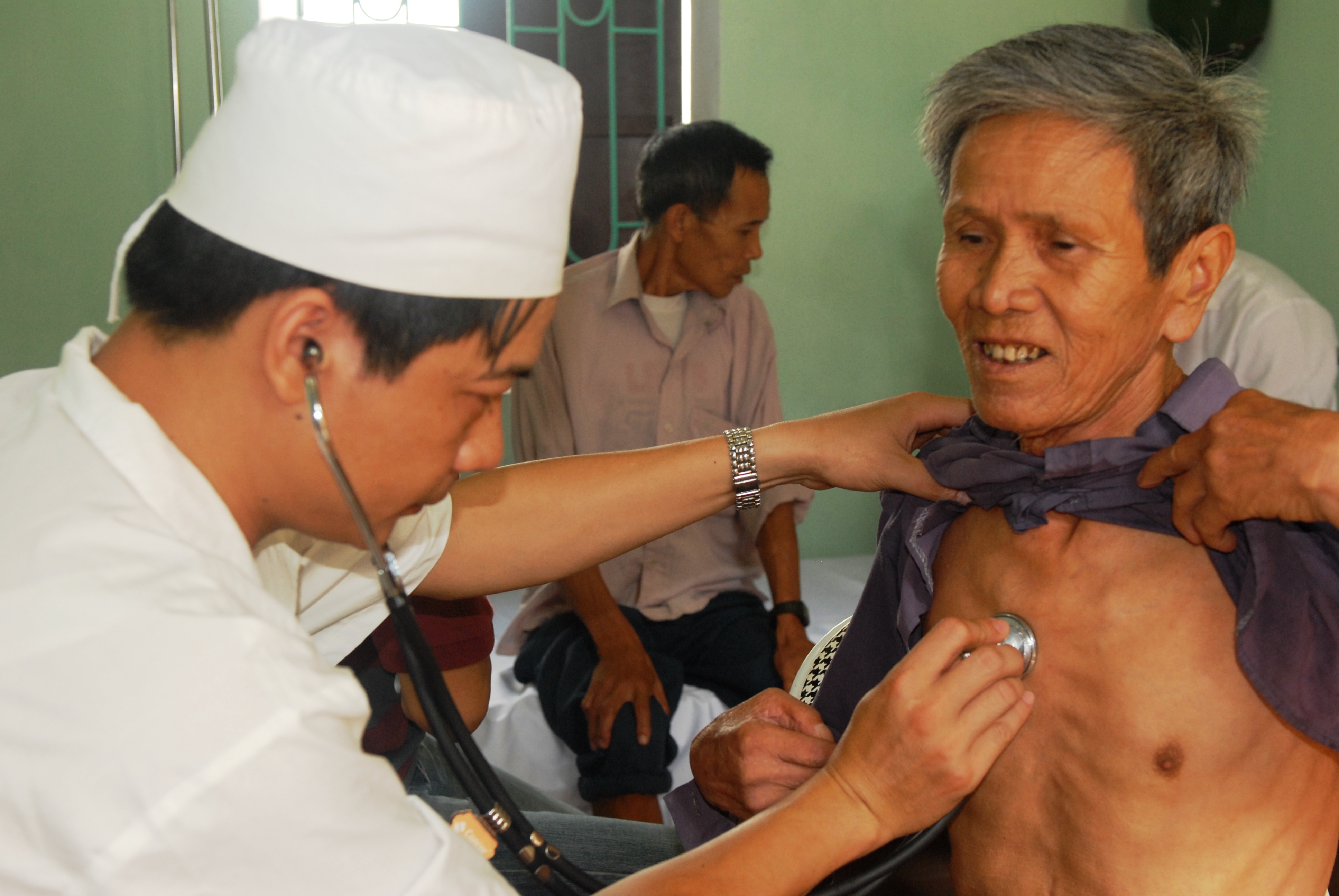
In a physical examination, medical examination, or clinical examination, a medical practitioner examines a patient for any possible medical signs or symptoms of a medical condition. It generally consists of a series of questions about the patient's medical history followed by an examination based on the reported symptoms. Together, the medical history and the physical examination help to determine a diagnosis and devise the treatment plan. These data then become part of the medical record. Types Routine The ''routine physical'', also known as ''general medical examination'', ''periodic health evaluation'', ''annual physical'', ''comprehensive medical exam'', ''general health check'', ''preventive health examination'', ''medical check-up'', or simply ''medical'', is a physical examination performed on an asymptomatic patient for medical screening purposes. These are normally performed by a pediatrician, family practice physician, physician assistant, a certified nurse ... [...More Info...] [...Related Items...] OR: [Wikipedia] [Google] [Baidu] |
Hip Dysplasia (human)
Hip dysplasia is an abnormality of the hip joint where the socket portion does not fully cover the ball portion, resulting in an increased risk for joint dislocation. Hip dysplasia may occur at birth or develop in early life. Regardless, it does not typically produce symptoms in babies less than a year old. Occasionally one leg may be shorter than the other. The left hip is more often affected than the right. Complications without treatment can include arthritis, limping, and low back pain. Females are affected more often than males. Hip dysplasia was described at least as early as the 300s BC by Hippocrates. Risk factors for hip dysplasia include female sex, family history, certain swaddling practices, and breech presentation whether an infant is delivered vaginally or by cesarean section. If one identical twin is affected, there is a 40% risk the other will also be affected. Screening all babies for the condition by physical examination is recommended. Ultrasonography ... [...More Info...] [...Related Items...] OR: [Wikipedia] [Google] [Baidu] |
Barlow Maneuver
The Barlow maneuver is a physical examination performed on infants to screen for developmental dysplasia of the hip. It is named for Dr. Thomas Geoffrey Barlow (September 25, 1915 – May 25, 1975), an English orthopedic surgeon, who devised this test. It was clinically tested during 1957–1962 at Hope Hospital, Salford, Lancashire Lancashire ( , ; abbreviated Lancs) is the name of a Historic counties of England, historic county, Ceremonial County, ceremonial county, and non-metropolitan county in North West England. The boundaries of these three areas differ significa .... Procedure The maneuver is easily performed by adducting the hip (bringing the thigh towards the midline) while applying pressure on the knee, directing the force posteriorly. Interpretation If the hip is dislocatable — that is, if the hip can be popped out of socket with this maneuver — the test is considered positive. The Ortolani maneuver is then used, to confirm the positive finding ( ... [...More Info...] [...Related Items...] OR: [Wikipedia] [Google] [Baidu] |
Posterior (anatomy)
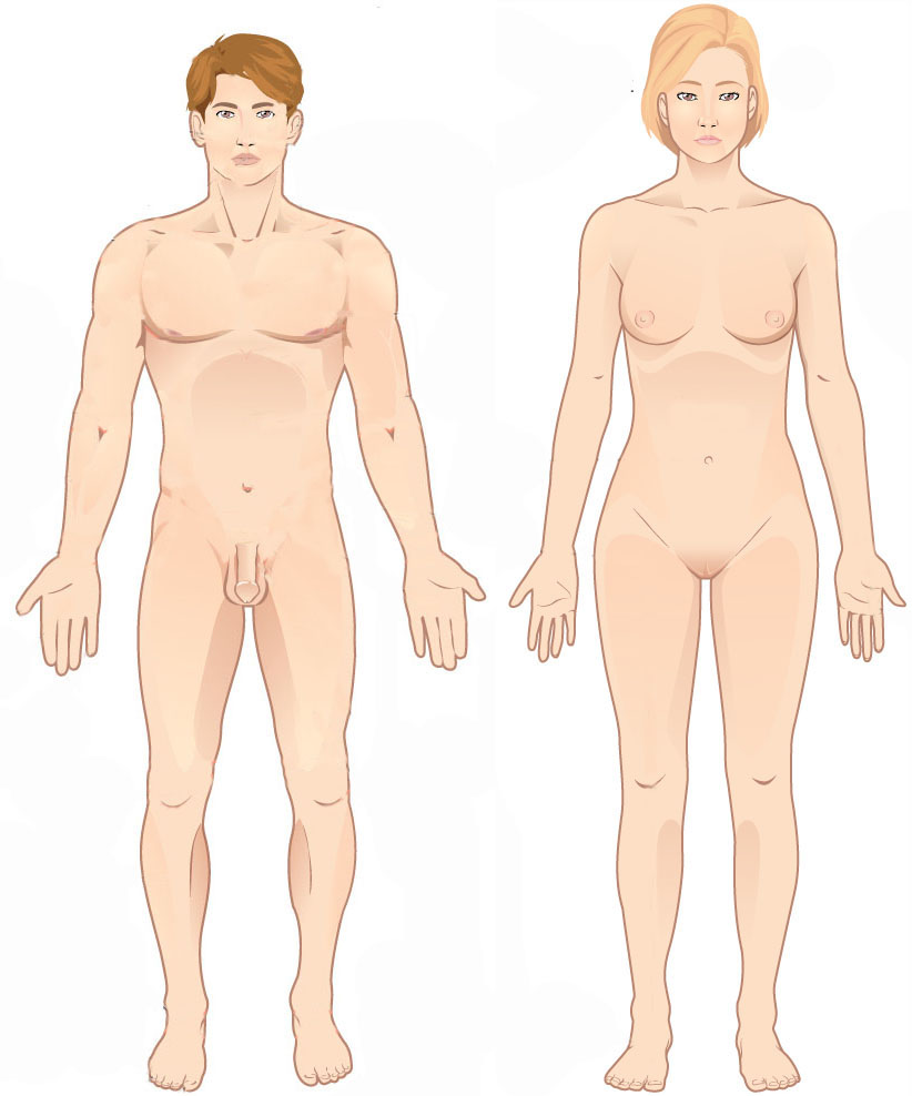
Standard anatomical terms of location are used to unambiguously describe the anatomy of animals, including humans. The terms, typically derived from Latin or Greek roots, describe something in its standard anatomical position. This position provides a definition of what is at the front ("anterior"), behind ("posterior") and so on. As part of defining and describing terms, the body is described through the use of anatomical planes and anatomical axes. The meaning of terms that are used can change depending on whether an organism is bipedal or quadrupedal. Additionally, for some animals such as invertebrates, some terms may not have any meaning at all; for example, an animal that is radially symmetrical will have no anterior surface, but can still have a description that a part is close to the middle ("proximal") or further from the middle ("distal"). International organisations have determined vocabularies that are often used as standard vocabularies for subdisciplines of anatomy ... [...More Info...] [...Related Items...] OR: [Wikipedia] [Google] [Baidu] |
Dislocation (medicine)
A joint dislocation, also called luxation, occurs when there is an abnormal separation in the joint, where two or more bones meet.Dislocations. Lucile Packard Children’s Hospital at Stanford. Retrieved 3 March 2013 A partial dislocation is referred to as a subluxation. Dislocations are often caused by sudden trauma on the joint like an impact or fall. A joint dislocation can cause damage to the surrounding ligaments, tendons, muscles, and nerves. Dislocations can occur in any major joint (shoulder, knees, etc.) or minor joint (toes, fingers, etc.). The most common joint dislocation is a shoulder dislocation. Treatment for joint dislocation is usually by closed reduction, that is, skilled manipulation to return the bones to their normal position. Reduction should only be performed by trained medical professionals, because it can cause injury to soft tissue and/or the nerves and vascular structures around the dislocation. Symptoms and signs The following symptoms are common wit ... [...More Info...] [...Related Items...] OR: [Wikipedia] [Google] [Baidu] |
Marino Ortolani
Marino Ortolani (26 July 1904 in Altedo, Malalbergo, Province of Bologna, Italy; † 1983) was an Italian pediatrician who developed a clinical test for the recognition of hip dysplasia called the Ortolani test The Ortolani test is part of the physical examination for developmental dysplasia of the hip, along with the Barlow maneuver. Specifically, the Ortolani test is positive when a posterior dislocation of the hip is reducible with this maneuver. .... References *Italo Farnetani, Francesca Farnetani, ''La top twelve della ricerca italiana'', « Minerva Pediatrica» 2015; 67 (5): pp.437-450 *Italo Farnetani, ''Qualche notazione di storia della pediatria, in margine alla V edizione di Pediatria Essenziale'', Postfazione. In Burgio G.R.( (a cura di). Pediatria Essenziale. 5a Ed. Milano: Edi-Ermes; 2012. . vol. 2°, pp. 1757-1764. External links Who's Who in Orthopedics: Marino Ortolani- Seyed Behrooz Mostofi *Italo Farnetani, ''Ortolani, Marino'', Dizionario Biografico deg ... [...More Info...] [...Related Items...] OR: [Wikipedia] [Google] [Baidu] |
Inspection (medicine)
In a physical examination, medical examination, or clinical examination, a medical practitioner examines a patient for any possible medical signs or symptoms of a medical condition. It generally consists of a series of questions about the patient's medical history followed by an examination based on the reported symptoms. Together, the medical history and the physical examination help to determine a diagnosis and devise the treatment plan. These data then become part of the medical record. Types Routine The ''routine physical'', also known as ''general medical examination'', ''periodic health evaluation'', ''annual physical'', ''comprehensive medical exam'', ''general health check'', ''preventive health examination'', ''medical check-up'', or simply ''medical'', is a physical examination performed on an asymptomatic patient for medical screening purposes. These are normally performed by a pediatrician, family practice physician, physician assistant, a certified nurse ... [...More Info...] [...Related Items...] OR: [Wikipedia] [Google] [Baidu] |
Anatomical Terms Of Motion
Motion, the process of movement, is described using specific anatomical terms. Motion includes movement of organs, joints, limbs, and specific sections of the body. The terminology used describes this motion according to its direction relative to the anatomical position of the body parts involved. Anatomists and others use a unified set of terms to describe most of the movements, although other, more specialized terms are necessary for describing unique movements such as those of the hands, feet, and eyes. In general, motion is classified according to the anatomical plane it occurs in. ''Flexion'' and ''extension'' are examples of ''angular'' motions, in which two axes of a joint are brought closer together or moved further apart. ''Rotational'' motion may occur at other joints, for example the shoulder, and are described as ''internal'' or ''external''. Other terms, such as ''elevation'' and ''depression'', describe movement above or below the horizontal plane. Many ana ... [...More Info...] [...Related Items...] OR: [Wikipedia] [Google] [Baidu] |
Anterior
Standard anatomical terms of location are used to unambiguously describe the anatomy of animals, including humans. The terms, typically derived from Latin or Greek roots, describe something in its standard anatomical position. This position provides a definition of what is at the front ("anterior"), behind ("posterior") and so on. As part of defining and describing terms, the body is described through the use of anatomical planes and anatomical axes. The meaning of terms that are used can change depending on whether an organism is bipedal or quadrupedal. Additionally, for some animals such as invertebrates, some terms may not have any meaning at all; for example, an animal that is radially symmetrical will have no anterior surface, but can still have a description that a part is close to the middle ("proximal") or further from the middle ("distal"). International organisations have determined vocabularies that are often used as standard vocabularies for subdisciplines of anatomy ... [...More Info...] [...Related Items...] OR: [Wikipedia] [Google] [Baidu] |
Greater Trochanter
The greater trochanter of the femur is a large, irregular, quadrilateral eminence and a part of the skeletal system. It is directed lateral and medially and slightly posterior. In the adult it is about 2–4 cm lower than the femoral head.Standring, Susan, editor. ''Gray’s Anatomy: The Anatomical Basis of Clinical Practice''. Forty-First edition, Elsevier Limited, 2016, p. 1327. Because the pelvic outlet in the female is larger than in the male, there is a greater distance between the greater trochanters in the female. It has two surfaces and four borders. It is a traction epiphysis. Surfaces The ''lateral surface'', quadrilateral in form, is broad, rough, convex, and marked by a diagonal impression, which extends from the postero-superior to the antero-inferior angle, and serves for the insertion of the tendon of the gluteus medius. Above the impression is a triangular surface, sometimes rough for part of the tendon of the same muscle, sometimes smooth for the interpo ... [...More Info...] [...Related Items...] OR: [Wikipedia] [Google] [Baidu] |
Femur Head
The femoral head (femur head or head of the femur) is the highest part of the thigh bone (femur). It is supported by the femoral neck. Structure The head is globular and forms rather more than a hemisphere, is directed upward, medialward, and a little forward, the greater part of its convexity being above and in front. The femoral head's surface is smooth. It is coated with cartilage in the fresh state, except over an ovoid depression, the fovea capitis, which is situated a little below and behind the center of the femoral head, and gives attachment to the ligament of head of femur. The thickest region of the articular cartilage is at the centre of the femoral head, measuring up to 2.8 mm. The diameter of the femoral head is usually larger in men than in women. Fovea capitis The fovea capitis is a small, concave depression within the head of the femur that serves as an attachment point for the ligamentum teres (Saladin). It is slightly ovoid in shape and is oriented "superior ... [...More Info...] [...Related Items...] OR: [Wikipedia] [Google] [Baidu] |


