|
Occipital Nerve Block
Occipital nerve block is a procedure involving injection of steroids or anesthetics into regions of the greater occipital nerve and the lesser occipital nerve used to treat chronic headaches. These nerves are located in the back of the head near in the suboccipital triangle along the line between the inion and the mastoid process. They innervate muscles in the suboccipital and posterior scalp regions. The injection will either block pain signals or reduce swelling and inflammation in these regions depending on the choice of injection. The procedure is helpful in treating occipital neuralgia and chronic headaches that arise from the neck. __TOC__ Procedure The patient is kept conscious for the duration of the procedure. A small gauge needle is inserted at points of the greater and lesser occipital nerves down to the periosteum of the occiput. Some pain may be felt during the insertion of the needle through the skin. Injected anesthetics give pain relief almost immediately whil ... [...More Info...] [...Related Items...] OR: [Wikipedia] [Google] [Baidu] |
Steroids
A steroid is a biologically active organic compound with four rings arranged in a specific molecular configuration. Steroids have two principal biological functions: as important components of cell membranes that alter membrane fluidity; and as signaling molecules. Hundreds of steroids are found in plants, animals and fungi. All steroids are manufactured in cells from the sterols lanosterol (opisthokonts) or cycloartenol (plants). Lanosterol and cycloartenol are derived from the cyclization of the triterpene squalene. The steroid core structure is typically composed of seventeen carbon atoms, bonded in four " fused" rings: three six-member cyclohexane rings (rings A, B and C in the first illustration) and one five-member cyclopentane ring (the D ring). Steroids vary by the functional groups attached to this four-ring core and by the oxidation state of the rings. Sterols are forms of steroids with a hydroxy group at position three and a skeleton derived from cholestane. '' ... [...More Info...] [...Related Items...] OR: [Wikipedia] [Google] [Baidu] |
Anesthetics
An anesthetic (American English) or anaesthetic (British English; see spelling differences) is a drug used to induce anesthesia — in other words, to result in a temporary loss of sensation or awareness. They may be divided into two broad classes: general anesthetics, which result in a reversible loss of consciousness, and local anesthetics, which cause a reversible loss of sensation for a limited region of the body without necessarily affecting consciousness. A wide variety of drugs are used in modern anesthetic practice. Many are rarely used outside anesthesiology, but others are used commonly in various fields of healthcare. Combinations of anesthetics are sometimes used for their synergistic and additive therapeutic effects. Adverse effects, however, may also be increased. Anesthetics are distinct from analgesics, which block only sensation of painful stimuli. Local anesthetics Local anesthetic agents prevent the transmission of nerve impulses without causi ... [...More Info...] [...Related Items...] OR: [Wikipedia] [Google] [Baidu] |
Greater Occipital Nerve
The greater occipital nerve is a nerve of the head. It is a spinal nerve, specifically the medial branch of the dorsal primary ramus of cervical spinal nerve 2. It arises from between the first and second cervical vertebrae, ascends, and then passes through the semispinalis muscle. It ascends further to supply the skin along the posterior part of the scalp to the vertex. It supplies sensation the scalp at the top of the head, over the ear and over the parotid glands. Structure The greater occipital nerve is the medial branch of the dorsal primary ramus of cervical spinal nerve 2. It may also involve fibres from cervical spinal nerve 3. It arises from between the first and second cervical vertebrae, along with the lesser occipital nerve. It ascends after emerging from below the suboccipital triangle beneath the obliquus capitis inferior muscle. Just below the superior nuchal ridge, it pierces the fascia. It ascends further to supply the skin along the posterior part of t ... [...More Info...] [...Related Items...] OR: [Wikipedia] [Google] [Baidu] |
Lesser Occipital Nerve
The lesser occipital nerve or small occipital nerve is a cutaneous spinal nerve. It arises from cervical spinal nerve 2, second cervical (spinal) nerve (along with the greater occipital nerve). It innervates the scalp in the lateral area of the head posterior to the ear. Structure The lesser occipital nerve is one of the four cutaneous branches of the cervical plexus. Origin It arises from the (lateral branch of the ventral ramus) of cervical spinal nerve 2, cervical spinal nerve C2; it may also receive fibres from cervical spinal nerve 3, cervical spinal nerve C3. It originates between the Atlas (anatomy), atlas, and Axis (anatomy), axis. Course and relations It curves around the Accessory nerve, accessory nerve (CN XI) to come to course anterior to it. It then curves around and ascends along the posterior border of the sternocleidomastoid muscle; rarely, it may pierce the muscle. Near the cranium, it perforates the deep fascia. It is continues upwards along the scalp po ... [...More Info...] [...Related Items...] OR: [Wikipedia] [Google] [Baidu] |
Chronic Headache
Headache is the symptom of pain in the face, head, or neck. It can occur as a migraine, tension-type headache, or cluster headache. There is an increased risk of depression in those with severe headaches. Headaches can occur as a result of many conditions. There are a number of different classification systems for headaches. The most well-recognized is that of the International Headache Society, which classifies it into more than 150 types of primary and secondary headaches. Causes of headaches may include dehydration; fatigue; sleep deprivation; stress; the effects of medications (overuse) and recreational drugs, including withdrawal; viral infections; loud noises; head injury; rapid ingestion of a very cold food or beverage; and dental or sinus issues (such as sinusitis). Treatment of a headache depends on the underlying cause, but commonly involves pain medication (especially in case of migraine or cluster headache). A headache is one of the most commonly experienced ... [...More Info...] [...Related Items...] OR: [Wikipedia] [Google] [Baidu] |
Suboccipital Triangle
The suboccipital triangle is a region of the neck bounded by the following three muscles of the suboccipital group of muscles: * Rectus capitis posterior major - above and medially * Obliquus capitis superior - above and laterally * Obliquus capitis inferior - below and laterally (Rectus capitis posterior minor is also in this region but does not form part of the triangle) It is covered by a layer of dense fibro-fatty tissue, situated beneath the semispinalis capitis. The floor is formed by the posterior atlantooccipital membrane, and the posterior arch of the atlas. In the deep groove on the upper surface of the posterior arch of the atlas are the vertebral artery and the first cervical or suboccipital nerve. In the past, the vertebral artery was accessed here in order to conduct angiography of the circle of Willis. Presently, formal angiography of the circle of Willis is performed via catheter angiography, with access usually being acquired at the common femoral artery. Alter ... [...More Info...] [...Related Items...] OR: [Wikipedia] [Google] [Baidu] |
Inion
Near the middle of the squamous part of occipital bone is the external occipital protuberance, the highest point of which is referred to as the inion. The inion is the most prominent projection of the protuberance which is located at the posterioinferior (rear lower) part of the human skull. The nuchal ligament and trapezius muscle attach to it. The inion (ἰνίον, iníon, Greek for the occipital bone) is used as a landmark in the 10-20 system in electroencephalography (EEG) recording. Extending laterally from it on either side is the superior nuchal line, and above it is the faintly marked highest nuchal line. A study of 16th-century Anatolian remains showed that the external occipital protuberance statistically tends to be less pronounced in female remains. Additional images See also * Internal occipital protuberance * Occipital bun The occipital bone () is a cranial dermal bone and the main bone of the occiput (back and lower part of the skull). It is trapezoidal in ... [...More Info...] [...Related Items...] OR: [Wikipedia] [Google] [Baidu] |
Mastoid Process
The mastoid part of the temporal bone is the posterior (back) part of the temporal bone, one of the bones of the skull. Its rough surface gives attachment to various muscles (via tendons) and it has openings for blood vessels. From its borders, the mastoid part articulates with two other bones. Etymology The word "mastoid" is derived from the Greek word for "breast", a reference to the shape of this bone. Surfaces Outer surface Its outer surface is rough and gives attachment to the occipitalis and posterior auricular muscles. It is perforated by numerous foramina (holes); for example, the mastoid foramen is situated near the posterior border and transmits a vein to the transverse sinus and a small branch of the occipital artery to the dura mater. The position and size of this foramen are very variable; it is not always present; sometimes it is situated in the occipital bone, or in the suture between the temporal and the occipital. Mastoid process The mastoid process is ... [...More Info...] [...Related Items...] OR: [Wikipedia] [Google] [Baidu] |
Scalp
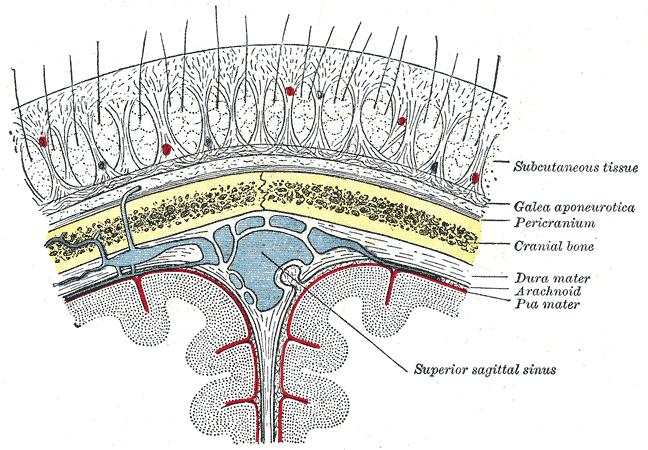
The scalp is the anatomical area bordered by the human face at the front, and by the neck at the sides and back. Structure The scalp is usually described as having five layers, which can conveniently be remembered as a mnemonic: * S: The skin on the head from which head hair grows. It contains numerous sebaceous glands and hair follicles. * C: Connective tissue. A dense subcutaneous layer of fat and fibrous tissue that lies beneath the skin, containing the nerves and vessels of the scalp. * A: The aponeurosis called epicranial aponeurosis (or galea aponeurotica) is the next layer. It is a tough layer of dense fibrous tissue which runs from the frontalis muscle anteriorly to the occipitalis posteriorly. * L: The loose areolar connective tissue layer provides an easy plane of separation between the upper three layers and the pericranium. In scalping the scalp is torn off through this layer. It also provides a plane of access in craniofacial surgery and neurosurgery. This layer i ... [...More Info...] [...Related Items...] OR: [Wikipedia] [Google] [Baidu] |
Occipital Neuralgia
Occipital neuralgia (ON) is a painful condition affecting the posterior head in the distributions of the greater occipital nerve (GON), lesser occipital nerve (LON), third occipital nerve (TON), or a combination of the three. It is paroxysmal, lasting from seconds to minutes, and often consists of lancinating pain that directly results from the pathology of one of these nerves. It is paramount that physicians understand the differential diagnosis for this condition and specific diagnostic criteria. There are multiple treatment modalities, several of which have well-established efficacy in treating this condition. Text was copied from this source, which is available under Creative Commons Attribution 4.0 International License Signs and symptoms Patients presenting with a headache originating at the posterior skull base should be evaluated for ON. This condition typically presents as a paroxysmal, lancinating or stabbing pain lasting from seconds to minutes, and therefore a continuo ... [...More Info...] [...Related Items...] OR: [Wikipedia] [Google] [Baidu] |
Periosteum
The periosteum is a membrane that covers the outer surface of all bones, except at the articular surfaces (i.e. the parts within a joint space) of long bones. Endosteum lines the inner surface of the medullary cavity of all long bones. Structure The periosteum consists of an outer fibrous layer, and an inner cambium layer (or osteogenic layer). The fibrous layer is of dense irregular connective tissue, containing fibroblasts, while the cambium layer is highly cellular containing progenitor cells that develop into osteoblasts. These osteoblasts are responsible for increasing the width of a long bone and the overall size of the other bone types. After a bone fracture, the progenitor cells develop into osteoblasts and chondroblasts, which are essential to the healing process. The outer fibrous layer and the inner cambium layer is differentiated under electron micrography. As opposed to osseous tissue, the periosteum has nociceptors, sensory neurons that make it very sensitive to ... [...More Info...] [...Related Items...] OR: [Wikipedia] [Google] [Baidu] |
Journal Of The American Board Of Family Medicine
The ''Journal of the American Board of Family Medicine'' is the official publication of the American Board of Family Medicine. It was formerly published as ''The Journal of the American Board of Family Practice'' by the Board of the Massachusetts Medical Society The Massachusetts Medical Society (MMS) is the oldest continuously operating state medical association in the United States. Incorporated on November 1, 1781, by an act of the Massachusetts General Court, the MMS is a non-profit organization th .... Publications established in 1988 Family medicine journals Bimonthly journals English-language journals Academic journals published by learned and professional societies of the United States {{med-journal-stub ... [...More Info...] [...Related Items...] OR: [Wikipedia] [Google] [Baidu] |

.jpg)


