|
Nucleus Ventralis Posterior Lateralis Pars Oralis
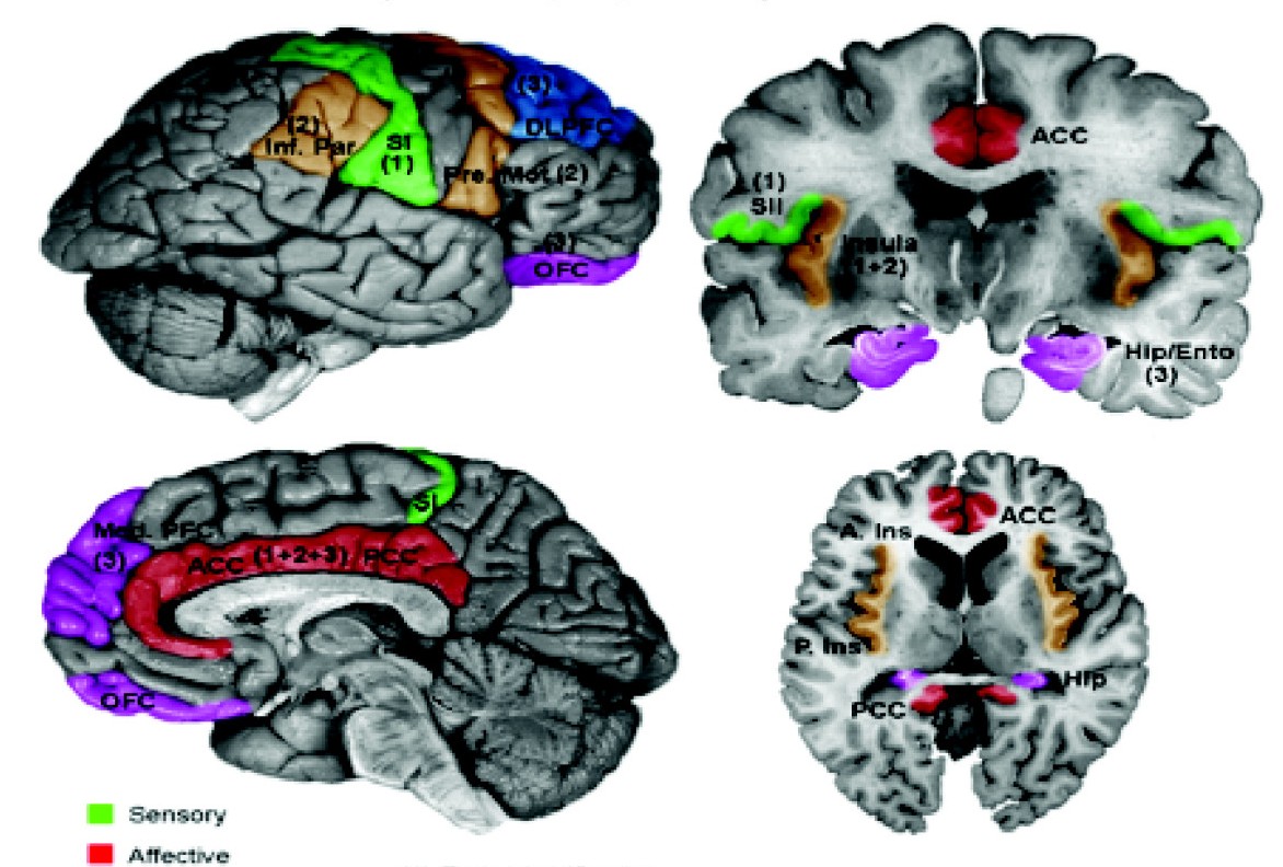
The ventral posterolateral nucleus (VPL) is a nucleus of the thalamus. Together with the ventral posteromedial nucleus (VPM), ventral posterior inferior nucleus (VPI) and ventromedial posterior nucleus (VMpo), it constitutes the ventral posterior nucleus. There is uncertainty in the location of VMpo, as determined by spinothalamic tract (STT) terminations and staining for calcium-binding proteins, and several authorities do not consider its existence as being proved. The nucleus ventralis posterior lateralis pars oralis (VPLo) is a subdivision of the ventral posterolateral thalamus which has substantial projections to the motor cortex. Input and output The VPL receives information from the neospinothalamic tract and the medial lemniscus of the posterior column-medial lemniscus pathway. It then projects this sensory information to Brodmann's Areas 3, 1 and 2 in the postcentral gyrus. Collectively, Brodmann areas 3, 1 and 2 make up the primary somatosensory In physiology, t ... [...More Info...] [...Related Items...] OR: [Wikipedia] [Google] [Baidu] |
Midline Nuclear Group
The midline nuclear group (or midline thalamic nuclei) is a region of the thalamus consisting of the following nuclei: * paraventricular nucleus of thalamus (''nucleus paraventricularis thalami'') - not to be confused with paraventricular nucleus of hypothalamus * paratenial nucleus The paratenial nucleus, or parataenial nucleus ( la, nucleus parataenialis), is a component of the midline nuclear group in the thalamus. It is sometimes subdivided into the nucleus parataenialis interstitialis and nucleus parataenialis parvocellu ... (''nucleus parataenialis'') * nucleus reuniens * rhomboid nucleus (''nucleus commissuralis rhomboidalis'') * subfascicular nucleus (''nucleus subfascicularis'') The midline nuclei are often called "nonspecific" in that they project widely to the cortex and elsewhere. This has led to the assumption that they may be involved in general functions such as alerting. However, anatomical connections might suggest more specific functions, with the paraventr ... [...More Info...] [...Related Items...] OR: [Wikipedia] [Google] [Baidu] |
Thalamus
The thalamus (from Greek θάλαμος, "chamber") is a large mass of gray matter located in the dorsal part of the diencephalon (a division of the forebrain). Nerve fibers project out of the thalamus to the cerebral cortex in all directions, allowing hub-like exchanges of information. It has several functions, such as the relaying of sensory signals, including motor signals to the cerebral cortex and the regulation of consciousness, sleep, and alertness. Anatomically, it is a paramedian symmetrical structure of two halves (left and right), within the vertebrate brain, situated between the cerebral cortex and the midbrain. It forms during embryonic development as the main product of the diencephalon, as first recognized by the Swiss embryologist and anatomist Wilhelm His Sr. in 1893. Anatomy The thalamus is a paired structure of gray matter located in the forebrain which is superior to the midbrain, near the center of the brain, with nerve fibers projecting out to the ... [...More Info...] [...Related Items...] OR: [Wikipedia] [Google] [Baidu] |
Somatosensory System
In physiology, the somatosensory system is the network of neural structures in the brain and body that produce the perception of touch (haptic perception), as well as temperature (thermoception), body position (proprioception), and pain. It is a subset of the sensory nervous system, which also represents visual, auditory, olfactory, and gustatory stimuli. Somatosensation begins when mechano- and thermosensitive structures in the skin or internal organs sense physical stimuli such as pressure on the skin (see mechanotransduction, nociception). Activation of these structures, or receptors, leads to activation of peripheral sensory neurons that convey signals to the spinal cord as patterns of action potentials. Sensory information is then processed locally in the spinal cord to drive reflexes, and is also conveyed to the brain for conscious perception of touch and proprioception. Note, somatosensory information from the face and head enters the brain through periphera ... [...More Info...] [...Related Items...] OR: [Wikipedia] [Google] [Baidu] |
Postcentral Gyrus
In neuroanatomy, the postcentral gyrus is a prominent gyrus in the lateral parietal lobe of the human brain. It is the location of the primary somatosensory cortex, the main sensory receptive area for the somatosensory system, sense of touch. Like other sensory areas, there is a map of sensory space in this location, called the ''sensory homunculus''. The primary somatosensory cortex was initially defined from surface stimulation studies of Wilder Penfield, and parallel surface potential studies of Bard, Woolsey, and Marshall. Although initially defined to be roughly the same as Brodmann areas Brodmann area 3, 3, Brodmann area 1, 1 and Brodmann area 2, 2, more recent work by Jon Kaas, Kaas has suggested that for homogeny with other sensory fields only area 3 should be referred to as "primary somatosensory cortex", as it receives the bulk of the Thalamocortical radiations, thalamocortical projections from the sensory input fields. Structure The lateral postcentral gyrus is bounded ... [...More Info...] [...Related Items...] OR: [Wikipedia] [Google] [Baidu] |
Brodmann Areas 3, 1 And 2
In neuroanatomy, the primary somatosensory cortex is located in the postcentral gyrus of the brain's parietal lobe, and is part of the somatosensory system. It was initially defined from surface stimulation studies of Wilder Penfield, and parallel surface potential studies of Bard, Woolsey, and Marshall. Although initially defined to be roughly the same as Brodmann areas 3, 1 and 2, more recent work by Kaas has suggested that for homogeny with other sensory fields only area 3 should be referred to as "primary somatosensory cortex", as it receives the bulk of the thalamocortical projections from the sensory input fields. At the primary somatosensory cortex, tactile representation is orderly arranged (in an inverted fashion) from the toe (at the top of the cerebral hemisphere) to mouth (at the bottom). However, some body parts may be controlled by partially overlapping regions of cortex. Each cerebral hemisphere of the primary somatosensory cortex only contains a tactile represen ... [...More Info...] [...Related Items...] OR: [Wikipedia] [Google] [Baidu] |
Posterior Column-medial Lemniscus Pathway
Posterior may refer to: * Posterior (anatomy), the end of an organism opposite to its head ** Buttocks, as a euphemism * Posterior horn (other) * Posterior probability The posterior probability is a type of conditional probability that results from updating the prior probability with information summarized by the likelihood via an application of Bayes' rule. From an epistemological perspective, the posterior p ..., the conditional probability that is assigned when the relevant evidence is taken into account * Posterior tense, a relative future tense {{disambiguation ... [...More Info...] [...Related Items...] OR: [Wikipedia] [Google] [Baidu] |
Medial Lemniscus
In neuroanatomy, the medial lemniscus, also known as Reil's band or Reil's ribbon (for German anatomist Johann Christian Reil), is a large ascending bundle of heavily myelinated axons that decussate (cross) in the brainstem, specifically in the medulla oblongata. The medial lemniscus is formed by the crossings of the internal arcuate fibers. The internal arcuate fibers are composed of axons of nucleus gracilis and nucleus cuneatus. The axons of the nucleus gracilis and nucleus cuneatus in the medial lemniscus have cell bodies that lie contralaterally. The medial lemniscus is part of the dorsal column–medial lemniscus pathway, which ascends from the skin to the thalamus, which is important for somatosensation from the skin and joints, therefore, lesion of the medial lemnisci causes an impairment of vibratory and touch-pressure sense. Etymology Lemniscus means "ribbon", so named because the medial lemniscus "spirals" or "turns" as it ascends. Path After neurons carrying propri ... [...More Info...] [...Related Items...] OR: [Wikipedia] [Google] [Baidu] |
Neospinothalamic Tract
Nociception (also nocioception, from Latin ''nocere'' 'to harm or hurt') is the sensory nervous system's process of encoding noxious stimuli. It deals with a series of events and processes required for an organism to receive a painful stimulus, convert it to a molecular signal, and recognize and characterize the signal in order to trigger an appropriate defense response. In nociception, intense chemical (e.g., capsaicin present in Chili pepper or Cayenne pepper), mechanical (e.g., cutting, crushing), or thermal (heat and cold) stimulation of sensory neurons called nociceptors produces a signal that travels along a chain of nerve fibers via the spinal cord to the brain. Nociception triggers a variety of physiological and behavioral responses to protect the organism against an aggression and usually results in a subjective experience, or perception, of pain in sentient beings. Detection of noxious stimuli Potentially damaging mechanical, thermal, and chemical stimuli are detected ... [...More Info...] [...Related Items...] OR: [Wikipedia] [Google] [Baidu] |
Brain Research
''Brain Research'' is a peer-reviewed scientific journal focusing on several aspects of neuroscience. It publishes research reports and " minireviews". The editor-in-chief is Matthew J. LaVoie (University of Florida). Until 2011, full reviews were published in ''Brain Research Reviews'', which is now integrated into the main section, albeit with independent volume numbering. In 2006, four other previously established semi-independent journal sections ('' Cognitive Brain Research, Developmental Brain Research, Molecular Brain Research,'' and '' Brain Research Protocols'') were merged with ''Brain Research''. The journal has nine main subsections: * ''Cellular and Molecular Systems'' * ''Nervous System Development, Regeneration and Aging'' * ''Neurophysiology, Neuropharmacology and other forms of Intercellular Communication'' * ''Structural Organization of the Brain'' * ''Sensory and Motor Systems'' * ''Regulatory Systems'' * ''Cognitive and Behavioral Neuroscience'' * ''Disease-Re ... [...More Info...] [...Related Items...] OR: [Wikipedia] [Google] [Baidu] |
Ventral Posterior Nucleus
The ventral posterior nucleus is the somato-sensory relay nucleus in thalamus of the brain. Input and output The ventral posterior nucleus receives neuronal input from the medial lemniscus, spinothalamic tracts, and trigeminothalamic tract. It projects to the somatosensory cortex and the ascending reticuloactivation system. Subdivisions The ventral posterior nucleus is divided into: *Ventral posterolateral nucleus, which receives sensory information from the body. *Ventral posteromedial nucleus, which receives sensory information from the head and face via the trigeminal nerve. *Ventral intermediate nucleus, implicated in oscillatory tremor generation in Parkinson's disease and essential tremor. Function Functions in touch, body position, pain, temperature, itch, taste, and arousal Arousal is the physiological and psychological state of being awoken or of sense organs stimulated to a point of perception. It involves activation of the ascending reticular activating system (A ... [...More Info...] [...Related Items...] OR: [Wikipedia] [Google] [Baidu] |
Ventral Posterior Inferior Nucleus
Standard anatomical terms of location are used to unambiguously describe the anatomy of animals, including humans. The terms, typically derived from Latin or Greek roots, describe something in its standard anatomical position. This position provides a definition of what is at the front ("anterior"), behind ("posterior") and so on. As part of defining and describing terms, the body is described through the use of anatomical planes and anatomical axes. The meaning of terms that are used can change depending on whether an organism is bipedal or quadrupedal. Additionally, for some animals such as invertebrates, some terms may not have any meaning at all; for example, an animal that is radially symmetrical will have no anterior surface, but can still have a description that a part is close to the middle ("proximal") or further from the middle ("distal"). International organisations have determined vocabularies that are often used as standard vocabularies for subdisciplines of anatomy ... [...More Info...] [...Related Items...] OR: [Wikipedia] [Google] [Baidu] |
Ventral Posteromedial Nucleus
The ventral posteromedial nucleus (VPM) is a nucleus of the thalamus. Inputs and outputs The VPM contains synapses between second and third order neurons from the anterior (ventral) trigeminothalamic tract and posterior (dorsal) trigeminothalamic tract. These neurons convey sensory information from the face and oral cavity. Third order neurons in the trigeminothalamic systems project to the postcentral gyrus. The VPM also receives taste afferent information from the solitary tract The solitary tract (tractus solitarius, or fasciculus solitarius), is a compact fiber bundle that extends longitudinally through the posterolateral region of the medulla oblongata. The solitary tract is surrounded by the solitary nucleus, and des ... and projects to the cortical gustatory area. Subareas VPMpc The parvicellular part of the ventroposterior medial nucleus (VPMpc) is argued by some as not an actually part of the VPM, because it does not project to the somatosensory cortex as the r ... [...More Info...] [...Related Items...] OR: [Wikipedia] [Google] [Baidu] |


