|
Nuclear Chain Fiber
A nuclear chain fiber is a specialized sensory organ contained within a muscle. Nuclear chain fibers are intrafusal fibers that, along with nuclear bag fibers, make up the muscle spindle responsible for the detection of changes in muscle length. There are 3–9 nuclear chain fibers per muscle spindle that are half the size of the nuclear bag fibers. Their nuclei are aligned in a chain and they excite the secondary nerve. They are static, whereas the nuclear bag fibers are dynamic in comparison. The name "nuclear chain" refers to the structure of the central region of the fiber, where the sensory axons wrap around the intrafusal fibers. The secondary nerve association involves an efferent and afferent pathway that measure the stress and strain placed on the muscle (usually the extrafusal fibers connected from the muscle portion to a bone). The afferent pathway resembles a spring wrapping around the nuclear chain fiber and connecting to one of its ends away from the bone. Agai ... [...More Info...] [...Related Items...] OR: [Wikipedia] [Google] [Baidu] |
Muscle Spindle
Muscle spindles are stretch receptors within the body of a skeletal muscle that primarily detect changes in the length of the muscle. They convey length information to the central nervous system via afferent nerve fibers. This information can be processed by the brain as proprioception. The responses of muscle spindles to changes in length also play an important role in regulating the contraction of muscles, for example, by activating motor neurons via the stretch reflex to resist muscle stretch. The muscle spindle has both sensory and motor components. * Sensory information conveyed by primary type Ia sensory fibers which spiral around muscle fibres within the spindle, and secondary type II sensory fibers * Activation of muscle fibres within the spindle by up to a dozen gamma motor neurons and to a lesser extent by one or two beta motor neurons Structure Muscle spindles are found within the belly of a skeletal muscle. Muscle spindles are fusiform (spindle-shaped), and the spec ... [...More Info...] [...Related Items...] OR: [Wikipedia] [Google] [Baidu] |
Series And Parallel Circuits
Two-terminal components and electrical networks can be connected in series or parallel. The resulting electrical network will have two terminals, and itself can participate in a series or parallel topology. Whether a two-terminal "object" is an electrical component (e.g. a resistor) or an electrical network (e.g. resistors in series) is a matter of perspective. This article will use "component" to refer to a two-terminal "object" that participate in the series/parallel networks. Components connected in series are connected along a single "electrical path", and each component has the same current through it, equal to the current through the network. The voltage across the network is equal to the sum of the voltages across each component. Components connected in parallel are connected along multiple paths, and each component has the same voltage across it, equal to the voltage across the network. The current through the network is equal to the sum of the currents through each com ... [...More Info...] [...Related Items...] OR: [Wikipedia] [Google] [Baidu] |
Gamma Motor Neurons
A gamma motor neuron (γ motor neuron), also called gamma motoneuron, or fusimotor neuron, is a type of lower motor neuron that takes part in the process of muscle contraction, and represents about 30% of ( Aγ) fibers going to the muscle. Like alpha motor neurons, their cell bodies are located in the anterior grey column of the spinal cord. They receive input from the reticular formation of the pons in the brainstem. Their axons are smaller than those of the alpha motor neurons, with a diameter of only 5 μm. Unlike the alpha motor neurons, gamma motor neurons do not directly adjust the lengthening or shortening of muscles. However, their role is important in keeping muscle spindles taut, thereby allowing the continued firing of alpha neurons, leading to muscle contraction. These neurons also play a role in adjusting the sensitivity of muscle spindles. The presence of myelination in gamma motor neurons allows a conduction velocity of 4 to 24 meters per second, significantl ... [...More Info...] [...Related Items...] OR: [Wikipedia] [Google] [Baidu] |
Spinal Cord
The spinal cord is a long, thin, tubular structure made up of nervous tissue, which extends from the medulla oblongata in the brainstem to the lumbar region of the vertebral column (backbone). The backbone encloses the central canal of the spinal cord, which contains cerebrospinal fluid. The brain and spinal cord together make up the central nervous system (CNS). In humans, the spinal cord begins at the occipital bone, passing through the foramen magnum and then enters the spinal canal at the beginning of the cervical vertebrae. The spinal cord extends down to between the first and second lumbar vertebrae, where it ends. The enclosing bony vertebral column protects the relatively shorter spinal cord. It is around long in adult men and around long in adult women. The diameter of the spinal cord ranges from in the cervical and lumbar regions to in the thoracic area. The spinal cord functions primarily in the transmission of nerve signals from the motor cortex to the body, ... [...More Info...] [...Related Items...] OR: [Wikipedia] [Google] [Baidu] |
Posterior Horn Of Spinal Cord
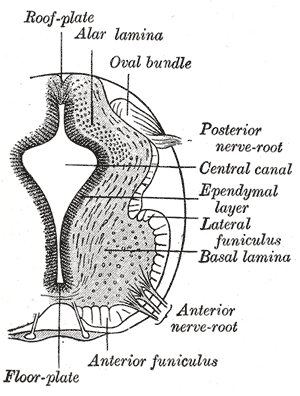
The posterior grey column (posterior cornu, dorsal horn, spinal dorsal horn, posterior horn, sensory horn) of the spinal cord is one of the three grey columns of the spinal cord. It receives several types of sensory information from the body, including fine touch, proprioception, and vibration. This information is sent from receptors of the skin, bones, and joints through sensory neurons whose cell bodies lie in the dorsal root ganglion. Anatomy The posterior grey column is subdivided into six layers termed Rexed laminae I-VI *Marginal nucleus of spinal cord (lamina I) *Substantia gelatinosa of Rolando (lamina II) *Nucleus proprius (laminae III, IV) *Spinal lamina V, the neck of the posterior horn *Spinal lamina VI, the base of the posterior horn. The other four Rexed laminae are located in the other two grey columns in the spinal cord. Additional images File:Gray687.png, Section of the medulla oblongata through the lower part of the decussation of the pyramids See also * ... [...More Info...] [...Related Items...] OR: [Wikipedia] [Google] [Baidu] |
Nucleus Proprius
The nucleus proprius is a layer of the spinal cord adjacent to the substantia gelatinosa. The nucleus proprius can be found in the gray matter in all levels of the spinal cord. It constitutes the first synapse of the spinothalamic tract carrying pain and temperature sensations from peripheral nerves. Cells in this nucleus project to deeper laminae of the spinal cord, to the posterior column nuclei, and to other supraspinal relay centers including the midbrain, thalamus, and hypothalamus. Rexed laminae III and IV make up the nucleus proprius. The nucleus proprius (NP), along with the substantia gelatinosa of Rolando are involved in sensing pain and temperature. See also * Rexed laminae The Rexed laminae comprise a system of ten layers of grey matter (I–X), identified in the early 1950s by Bror Rexed to label portions of the grey columns of the spinal cord. Similar to Brodmann areas, they are defined by their cellular struct ... References External links * Diagram at pixel ... [...More Info...] [...Related Items...] OR: [Wikipedia] [Google] [Baidu] |
Type II Sensory Fibers
Type II sensory fiber (group Aβ) is a type of sensory fiber, the second of the two main groups of touch receptors. The responses of different type Aβ fibers to these stimuli can be subdivided based on their adaptation properties, traditionally into rapidly adapting (RA) or slowly adapting (SA) neurons. Type II sensory fibers are slowly-adapting (SA), meaning that even when there is no change in touch, they keep respond to stimuli and fire action potentials. In the body, Type II sensory fibers belong to pseudounipolar neurons. The most notable example are neurons with Merkel cell-neurite complexes on their dendrites (sense static touch) and Ruffini endings (sense stretch on the skin and over-extension inside joints). Under pathological conditions they may become hyper-excitable leading to stimuli that would usually elicit sensations of tactile touch causing pain. These changes are in part induced by PGE2 which is produced by COX1, and type II fibers with free nerve endings ... [...More Info...] [...Related Items...] OR: [Wikipedia] [Google] [Baidu] |
Type Ia Sensory Fibers
A type Ia sensory fiber, or a primary afferent fiber is a type of afferent nerve fiber. It is the sensory fiber of a stretch receptor called the muscle spindle found in muscles, which constantly monitors the rate at which a muscle stretch changes. The information carried by type Ia fibers contributes to the sense of proprioception. Function of muscle spindles For the body to keep moving properly and with finesse, the nervous system has to have a constant input of sensory data coming from areas such as the muscles and joints. In order to receive a continuous stream of sensory data, the body has developed special sensory receptors called proprioceptors. Muscle spindles are a type of proprioceptor, and they are found inside the muscle itself. They lie parallel with the contractile fibers. This gives them the ability to monitor muscle length with precision. Types of sensory fibers This change in length of the spindle is transduced (transformed into electric membrane potentials) b ... [...More Info...] [...Related Items...] OR: [Wikipedia] [Google] [Baidu] |
Efferent Nerve Fiber
Efferent nerve fibers refer to axonal projections that ''exit'' a particular region; as opposed to afferent projections that ''arrive'' at the region. These terms have a slightly different meaning in the context of the peripheral nervous system (PNS) and central nervous system (CNS). The efferent fiber is a long process projecting far from the neuron's body that carries nerve impulses away from the central nervous system toward the peripheral effector organs (mainly muscles and glands). A bundle of these fibers is called an efferent nerve (if it connects to muscles, then it is a motor nerve). The opposite direction of neural activity is afferent conduction, which carries impulses by way of the afferent nerve fibers of sensory neurons. In the nervous system there is a "closed loop" system of sensation, decision, and reactions. This process is carried out through the activity of sensory neurons, interneurons, and motor neurons. In the CNS, afferent and efferent projections ... [...More Info...] [...Related Items...] OR: [Wikipedia] [Google] [Baidu] |
Afferent Nerve Fiber
Afferent nerve fibers are the axons (nerve fibers) carried by a sensory nerve that relay sensory information from sensory receptors to regions of the brain. Afferent projections ''arrive'' at a particular brain region. Efferent nerve fibers are carried by efferent nerves and ''exit'' a region to act on muscles and glands. In the peripheral nervous system afferent and efferent nerve fibers are part of the somatic nervous system and arise from outside of the spinal cord. Sensory nerves carry the afferent fibers to enter into the spinal cord, and motor nerves carry the efferent fibers out of the spinal cord to act on skeletal muscles. In the central nervous system non-motor efferents are carried in efferent nerves to act on glands. Structure Afferent neurons are pseudounipolar neurons that have a single process leaving the cell body dividing into two branches: the long one towards the sensory organ, and the short one toward the central nervous system (e.g. spinal cord). The ... [...More Info...] [...Related Items...] OR: [Wikipedia] [Google] [Baidu] |
Nuclear Bag Fibers
A nuclear bag fiber is a type of intrafusal muscle fiber that lies in the center of a muscle spindle. Each has many nuclei concentrated in bags and they cause excitation of the primary sensory fibers. There are two kinds of bag fibers based upon contraction speed and motor innervation. #BAG2 fibers are the largest. They have no striations in middle region and swell to enclose nuclei, hence their name. #BAG1 fibers, smaller than BAG2. Both bag types extend beyond the spindle capsule. These sense dynamic length of the muscle. They are sensitive to length and velocity. See also * Nuclear chain fiber A nuclear chain fiber is a specialized sensory organ contained within a muscle. Nuclear chain fibers are intrafusal fibers that, along with nuclear bag fibers, make up the muscle spindle responsible for the detection of changes in muscle len ... References External links * http://www.unmc.edu/Physiology/Mann/mann11.html Nervous system Muscular system {{neuros ... [...More Info...] [...Related Items...] OR: [Wikipedia] [Google] [Baidu] |
Sensory Receptor
Sensory neurons, also known as afferent neurons, are neurons in the nervous system, that convert a specific type of stimulus, via their receptors, into action potentials or graded potentials. This process is called sensory transduction. The cell bodies of the sensory neurons are located in the dorsal ganglia of the spinal cord. The sensory information travels on the afferent nerve fibers in a sensory nerve, to the brain via the spinal cord. The stimulus can come from ''exteroreceptors'' outside the body, for example those that detect light and sound, or from ''interoreceptors'' inside the body, for example those that are responsive to blood pressure or the sense of body position. Types and function Different types of sensory neurons have different sensory receptors that respond to different kinds of stimuli. There are at least six external and two internal sensory receptors: External receptors External receptors that respond to stimuli from outside the body are called ex ... [...More Info...] [...Related Items...] OR: [Wikipedia] [Google] [Baidu] |

