|
Neuromuscular Monitoring
In anesthesia, neuromuscular blocking agents may be required to facilitate Tracheal intubation, endotracheal intubation and provide optimal surgical conditions. When neuromuscular blocking agents are administered, neuromuscular function of the patient must be monitored. Neuromuscular function monitoring is a technique that involves the electrical stimulation of a motor nerve and monitoring the response of the muscle supplied by that nerve. It may be used from the induction of to recovery from neuromuscular blockade. Importantly, it is used to confirm adequacy of recovery after the administration of neuromuscular blocking agents. The response of the muscles to electrical stimulation of the nerves can be recorded subjectively (qualitative) or objectively (quantitatively). Quantitative techniques include electromyography, acceleromyography, Piezoelectric sensor, kinemyography, Phonomyography, phonomygraphy and mechanomyography. Neuromuscular monitoring is recommended when neuromuscular ... [...More Info...] [...Related Items...] OR: [Wikipedia] [Google] [Baidu] |
Muscle Tone
In physiology, medicine, and anatomy, muscle tone (residual muscle tension or tonus) is the continuous and passive partial muscle contraction, contraction of the muscles, or the muscle's resistance to passive stretch during resting state.O’Sullivan, S. B. (2007). Examination of motor function: Motor control and motor learning. In S. B. O’Sullivan, & T. J. Schmitz (Eds), Physical rehabilitation (5th ed.) (pp. 233-234). Philadelphia, Pennsylvania: F. A. Davis Company. It helps to maintain neutral spine, posture and declines during REM sleep. Purpose If a sudden muscle pull, pull or stretch occurs, the body responds by automatically increasing the muscle's tension, a reflex which helps guard against danger as well as helping maintain balance disorder, balance. Such near-continuous innervation can be thought of as a "default" or "steady state" condition for muscles. Both the Extension (kinesiology), extensor and flexion, flexor muscles are involved in the maintenance of a consta ... [...More Info...] [...Related Items...] OR: [Wikipedia] [Google] [Baidu] |
Neuromuscular Blockade
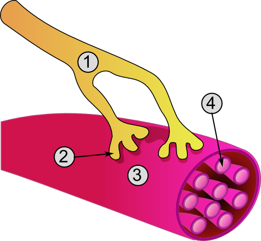
Neuromuscular-blocking drugs block neuromuscular transmission at the neuromuscular junction, causing paralysis of the affected skeletal muscles. This is accomplished via their action on the post-synaptic acetylcholine (Nm) receptors. In clinical use, neuromuscular block is used adjunctively to anesthesia to produce paralysis, firstly to paralyze the vocal cords, and permit intubation of the trachea, and secondly to optimize the surgical field by inhibiting spontaneous ventilation, and causing relaxation of skeletal muscles. Because the appropriate dose of neuromuscular-blocking drug may paralyze muscles required for breathing (i.e., the diaphragm), mechanical ventilation should be available to maintain adequate respiration. Patients are still aware of pain even after full conduction block has occurred; hence, general anesthetics and/or analgesics must also be given to prevent anesthesia awareness. Nomenclature Neuromuscular blocking drugs are often classified into two broad ... [...More Info...] [...Related Items...] OR: [Wikipedia] [Google] [Baidu] |
Extubation
Tracheal intubation, usually simply referred to as intubation, is the placement of a flexible plastic tube into the trachea (windpipe) to maintain an open airway or to serve as a conduit through which to administer certain drugs. It is frequently performed in critically injured, ill, or anesthetized patients to facilitate ventilation of the lungs, including mechanical ventilation, and to prevent the possibility of asphyxiation or airway obstruction. The most widely used route is orotracheal, in which an endotracheal tube is passed through the mouth and vocal apparatus into the trachea. In a nasotracheal procedure, an endotracheal tube is passed through the nose and vocal apparatus into the trachea. Other methods of intubation involve surgery and include the cricothyrotomy (used almost exclusively in emergency circumstances) and the tracheotomy, used primarily in situations where a prolonged need for airway support is anticipated. Because it is an invasive and uncomfortable medica ... [...More Info...] [...Related Items...] OR: [Wikipedia] [Google] [Baidu] |
The Australian And New Zealand College Of Anaesthetists
''The'' () is a grammatical article in English, denoting persons or things that are already or about to be mentioned, under discussion, implied or otherwise presumed familiar to listeners, readers, or speakers. It is the definite article in English. ''The'' is the most frequently used word in the English language; studies and analyses of texts have found it to account for seven percent of all printed English-language words. It is derived from gendered articles in Old English which combined in Middle English and now has a single form used with nouns of any gender. The word can be used with both singular and plural nouns, and with a noun that starts with any letter. This is different from many other languages, which have different forms of the definite article for different genders or numbers. Pronunciation In most dialects, "the" is pronounced as (with the voiced dental fricative followed by a schwa) when followed by a consonant sound, and as (homophone of the archaic pron ... [...More Info...] [...Related Items...] OR: [Wikipedia] [Google] [Baidu] |
Association Of Anaesthetists Of Great Britain And Ireland
The Association of Anaesthetists, in full the Association of Anaesthetists of Great Britain and Ireland (AAGBI), is a professional association for anaesthetists in the United Kingdom and Ireland. It was founded by Dr Henry Featherstone in 1932, when GPs gave most anaesthetics in the UK and Ireland as a sideline. Anaesthetists were not respected by other specialists and were poorly paid. Surgeons provided referrals and collected and paid their fees. The AAGBI's negotiations before the NHS was established in 1948 ensured anaesthetists received consultant status. It instigated the founding of the Faculty of Anaesthetists of the Royal College of Surgeons of England (now the Royal College of Anaesthetists) in 1947 and supported the foundation of the equivalent Faculty of the Royal College of Surgeons in Ireland (now the College of Anaesthesiologists of Ireland in 1959. The AAGBI adopted the motto in somno securitas (safe in sleep) when it was granted the right to bear arms by King ... [...More Info...] [...Related Items...] OR: [Wikipedia] [Google] [Baidu] |
Neuromuscular Blocking Drug
Neuromuscular-blocking drugs block neuromuscular transmission at the neuromuscular junction, causing paralysis of the affected skeletal muscles. This is accomplished via their action on the post-synaptic acetylcholine (Nm) receptors. In clinical use, neuromuscular block is used adjunctively to anesthesia to produce paralysis, firstly to paralyze the vocal cords, and permit intubation of the trachea, and secondly to optimize the surgical field by inhibiting spontaneous ventilation, and causing relaxation of skeletal muscles. Because the appropriate dose of neuromuscular-blocking drug may paralyze muscles required for breathing (i.e., the diaphragm), mechanical ventilation should be available to maintain adequate respiration. Patients are still aware of pain even after full conduction block has occurred; hence, general anesthetics and/or analgesics must also be given to prevent anesthesia awareness. Nomenclature Neuromuscular blocking drugs are often classified into two broad ... [...More Info...] [...Related Items...] OR: [Wikipedia] [Google] [Baidu] |
Acceleromyography Monitoring With Preload Hand Adapter
An acceleromyograph is a piezoelectric myograph, used to measure the force produced by a muscle after it has undergone nerve stimulation. Acceleromyographs may be used, during anaesthesia when muscle relaxants are administered, to measure the depth of neuromuscular blockade and to assess adequacy of recovery from these agents at the end of surgery. Acceleromyography is classified as quantitative neuromuscular monitoring. Rationale Patients who undergo anesthesia may receive a drug that paralyzes muscles, facilitating endotracheal intubation and improving operating conditions for the surgeon. Longer-acting drugs have higher prevalence of residual blockade in the PACU or ICU than shorter acting drugs. Different clinical tests to measure or exclude evidence of residual muscle weakness have been described but cannot exclude postoperative residual curarization. Small degrees of muscle blockade can only accurately be measured by the use of quantitative neuromuscular monitoring. Spec ... [...More Info...] [...Related Items...] OR: [Wikipedia] [Google] [Baidu] |
Mechanomyography
The mechanomyogram (MMG) is the mechanical signal observable from the surface of a muscle when the muscle is contracted. At the onset of muscle contraction, gross changes in the muscle shape cause a large peak in the MMG. Subsequent vibrations are due to oscillations of the muscle fibres at the resonance frequency of the muscle. The mechanomyogram is also known as the phonomyogram, acoustic myogram, sound myogram, vibromyogram or muscle sound. Signal characteristics The MMG is a low frequency vibration that may be observed when a muscle is contracted using suitable measuring techniques. Measurement techniques It can be measured using an accelerometer or a microphone placed on the skin over the belly of the muscle. When measured using a microphone is may be termed the acoustic myogram. Uses The MMG may provide a useful alternative to the electromyogram (EMG) for monitoring muscle activity. It has a higher signal-to-noise ratio than the surface EMG and thus can be used to m ... [...More Info...] [...Related Items...] OR: [Wikipedia] [Google] [Baidu] |
Action Potential
An action potential occurs when the membrane potential of a specific cell location rapidly rises and falls. This depolarization then causes adjacent locations to similarly depolarize. Action potentials occur in several types of animal cells, called excitable cells, which include neurons, muscle cells, and in some plant cells. Certain endocrine cells such as pancreatic beta cells, and certain cells of the anterior pituitary gland are also excitable cells. In neurons, action potentials play a central role in cell-cell communication by providing for—or with regard to saltatory conduction, assisting—the propagation of signals along the neuron's axon toward synaptic boutons situated at the ends of an axon; these signals can then connect with other neurons at synapses, or to motor cells or glands. In other types of cells, their main function is to activate intracellular processes. In muscle cells, for example, an action potential is the first step in the chain of events l ... [...More Info...] [...Related Items...] OR: [Wikipedia] [Google] [Baidu] |


.png)


